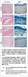
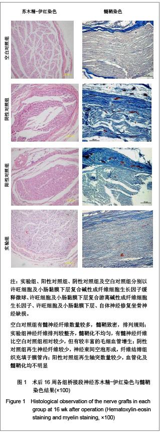
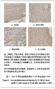
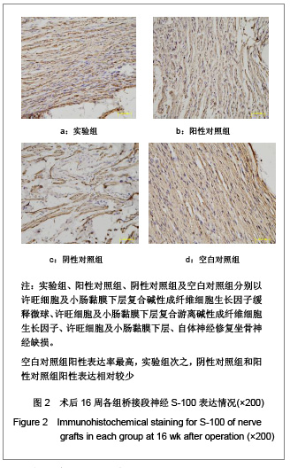
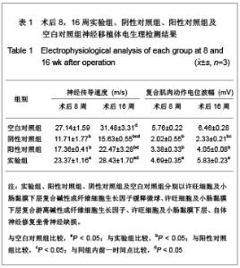
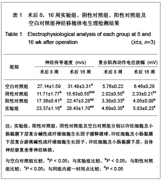
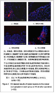
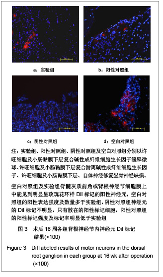
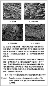
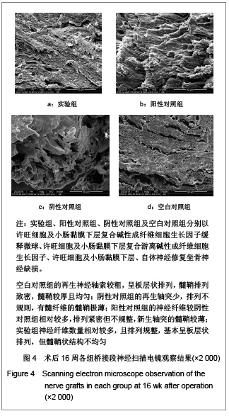
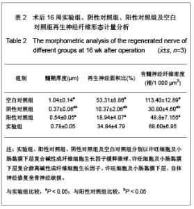
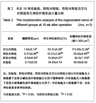
| [1] Hudson TW,Evans GR,Schmidt CE. Engineering strategies for peripheral nerve repair. Clin Plast Surg.1999; 26(4): 617-628.[2] Battiston B,Geuna S,Ferrero M,et al. Nerve repair by means of tubulization: literature review and personal clinical experience comparing biological and synthetic conduits for sensory nerve repair.J Microsurgery.2005;25(4):258-267.[3] Zhang KW,Xiao R,Duan H,et al. Zhongguo Zuzhi Gongcheng Yanjiu yu Linchuang Kangfu. 2010;14(21):3827-3831. 张开伟,肖睿,段宏,等. 碱性成纤维细胞生长因子对许旺细胞在小肠黏膜下层支架上黏附及增殖的影响[J].中国组织工程研究与临床康复,2010,14(21):3827-3831.[4] The Ministry of Science and Technology of the People’s Republic of China. Guidance suggestion of caring laboratory animals.2006-09-30. 中华人民共和国科学技术部. 关于善待实验动物的指导性意见. 2006-09-30.[5] Fansa H,Keihoff G,Plogmeier K,et al.Successful implantation of Schwann cells in acellular muscles.J Reconstr Microsurg. 1999;15(1):61-65.[6] Jones NC,Trainor PA. Role of morphogens in neural crest cell determination.J Neurobiol.2005;64(4):388-404.[7] Tada H,Hatoko M,Tanaka A,et al. Detection of E-cadherin expression after nerve repair in a rat sciatic nerve model.Ann Plast Surg.2001;47(2):178-182.[8] Torigoe K,Tanaka HF,Takahashi A,et al.Basic behavior of migratory Schwanns cells in peripheral nerve regeneration. Exp Neurol.1996;137(2):301-308.[9] Su Y,Zhang CQ,Sun LY,et al.Zhonghua Chuangshang Guke Zazhi. 2004;6(10):1136-1139. 苏琰,张长青,孙鲁源,等.小肠粘膜下层(SIS)桥接周围神经缺损的实验研究[J].中华创伤骨科杂志,2004,6(10):1136-1139. [10] Voytik-Harbin SL,Brightman AO,Kraine MR,et al.Identification of extractable growth factors from small intestinal submucosa. J Cell Biochem.1997; 67(4): 478-483.[11] Han TK,Dou QY,Chen F,et al. Zhongguo Zuzhi Gongcheng Yanjiu yu Linchuang Kangfu. 2008;12(49):9635-9638. 韩同坤,窦庆寅,陈峰,等.复合许旺细胞的猪肠黏膜下层桥接修复周围神经缺损[J].中国组织工程研究与临床康复,2008,12(49): 9635-9638.[12] Smith RM,Wiedl C,Chubb P,et al.Role of small intestine submucosa (SIS) as a nerve conduit: preliminary report.J Invest Surg.2004;17(6): 339-344.[13] Navarro X,Rodriguez FJ,Ceballos D,et al.Engineering an artificial nerve graft for the repair of severe nerve injuries.Med Biol Eng Comput.2003; 41(2):220-226.[14] Novikova LN,Mosahebi A,Wiberg M.Alginate hydrogel and matrigel as potential cell carriers for neurotransplantation.J Biomed Mater Res A.2006; 77A(2):242-252[15] Brandt J,Nilsson A,Kanje M,et al. Acutely-dissociated Schwann cells used in tendon autografts for bridging nerve defects in rats: a new principle for tissue engineering in nerve reconstruction. Seand J Plast Reconstr Surg Hand Surg.2005;39(6):321-325.[16] Bryan DJ,Holway AH,Wang KK,et al.Influence of glial growth factor and Schwann cells in a bioresorbable guidance channel on peripheral nerve regeneration.J Tissue Eng.2000; 6(2):129-138.[17] Mohanna PN,Young RC,Wiberg M,et al. A composite poly-hydroxybutyrate–glial growth factor conduit for long nerve gap repairs.J Anat.2003;203(6):553-565.[18] Wang GL,Lin W,Gao WQ,et al.Zhongguo Xiufu Chongjian Waike Zazhi. 2008;22(12):1485-1490. 王光林,林卫,高伟强,等. bFGF-PLGA缓释微球生物活性组织工程神经的构建和效果评价研究[J].中国修复重建外科杂志,2008, 22(12):1485-1490.[19] Mersa B,Agir H,Aydin A,et al.Comparison of expanded polytetrafluoroethylene (ePTFE) with autogenous vein as a nerve conduit in rat sciatic nerve defects.Kulak Burun Bogaz Ihtis Derg.2004;13(526): 103-111.[20] Mohanna PN,Terenghi G,Wiberg M.Composite PHB-GGF conduit for long nerve gap repair: a long-term evaluation.Scand J Plast Reconstr Surg Hand Surg.2005; 39(3):129-137.[21] Jneg CB,Coggeshall RE. Permeable tubes increase the length of the gap that regenerating axons can span. Barin Res.1987;408(1-2):239-242.[22] Rutkowski GE,Heath CA.Development of a bioartificial nerve graft. I. Design based on a reaction-diffusion model.Biotechnol Prog.2002;18(2):362-372.[23] Burnett MG,Zager EL. Pathophysiology of peripheral nerve injury:a brief review. Neurosurg Focus.2004;16(5):E1.[24] Chamberlain LJ,Yannas IV,Arrsabalaga A,et al.Early peripheral nerve healing in collagen and silicone tube implants:myofibroblasts and the cellular response. Biomaterials.1998;19(15): 1393-1403.[25] Lundborg G,Longo FM,Varon S. Nerve regeneration model and trophic factors in vivo.Brain Res.1982;232(1):157-164.[26] Zhang W,Ochi M,Takata H,et al.Influence of distal nerve segment volumen on nerve regeneration in silicone tubes. Exp Neurol.1997;146(2):600-603.[27] Grabb WC.Median and ulnr nerve sucture.An experimental study comparing primary and secondary repair in monkeys. J Bone Joint Surg.1998;50(5):964-972.[28] Yin ZS,Gu YD,Zhu JH,et al.Zhonghua Xianwei Waike Zazhi. 2002;25(4): 285-287. 尹宗生,顾玉东,朱建华,等.125碘-辣根过氧化物酶评价手术后神经再生的实验[J].中华显微外科杂志,2002,25(4): 285-287.[29] Xu WD,Deng SZ,Xu JG,et al.Zhonghua Shouwaike Zazhi. 1998;14(4):239-241. 徐文东,邓守真,徐建光,等.鼠坐骨神经再生早期轴浆流改变的实验研究[J].中华手外科杂志,1998,14(4):239-241.[30] Mandado M,Molist P,Anadón R,et al.A DiI-tracing study of the neural connections of the pineal organ in two elasmobranchs (Scyliorhinus canicula and Raja montagui) suggests a pineal projection to the midbrain GnRH-immunoreactive nucleus.Cell Tissue Res.2001;303(3): 391-401.[31] Fernández-Borja M,Bellido D,Vilella E,et al.Lipoprotein lipase-mediated uptake of lipoprotein in human fibroblasts: evidence for an LDL receptor-independent internalization pathway.J Lipid Res.1996;37(3): 464-481.[32] Hagg T. Neuronal cell death: Retraction. Science.1999;185: 340-342. |