Chinese Journal of Tissue Engineering Research ›› 2023, Vol. 27 ›› Issue (15): 2363-2370.doi: 10.12307/2023.371
Previous Articles Next Articles
Effects of neural stem cell transplantation in different ways on neuroinflammation in amyotrophic lateral sclerosis
Lin Yanchen1, Li Jingjing2, Lu Guangxu3, Dai Erqing1, Chen Jing4, Zhong Shijiang4
- 1Department of Rehabilitation Medicine, 4Department of Neurology, Characteristic Medical Center of the Armed Police, Tianjin 300162, China; 2Department of Pharmacy, Tianjin Fourth Central Hospital, Tianjin 314000, China; 3The Second Mobile Corps Hospital of the Armed Police, Wuxi 214000, Jiangsu Province, China
-
Received:2022-05-09Accepted:2022-06-27Online:2023-05-28Published:2022-10-18 -
Contact:Chen Jing, Master, Associate chief physician, Department of Neurology, Characteristic Medical Center of the Armed Police, Tianjin 300162, China Zhong Shijiang, MD, Chief physician, Department of Neurology, Characteristic Medical Center of the Armed Police, Tianjin 300162, China -
About author:Lin Yanchen, Master, Attending physician, Department of Rehabilitation Medicine, Characteristic Medical Center of the Armed Police, Tianjin 300162, China Li Jingjing, Pharmacist, Department of Pharmacy, Tianjin Fourth Central Hospital, Tianjin 314000, China -
Supported by:Tianjin Natural Science Foundation Project, No. 13JCBJC24100 (to ZSJ); Tianjin Key Research Project of Health Industry, No. 16KG136 (to ZSJ)
CLC Number:
Cite this article
Lin Yanchen, Li Jingjing, Lu Guangxu, Dai Erqing, Chen Jing, Zhong Shijiang. Effects of neural stem cell transplantation in different ways on neuroinflammation in amyotrophic lateral sclerosis[J]. Chinese Journal of Tissue Engineering Research, 2023, 27(15): 2363-2370.
share this article
Add to citation manager EndNote|Reference Manager|ProCite|BibTeX|RefWorks
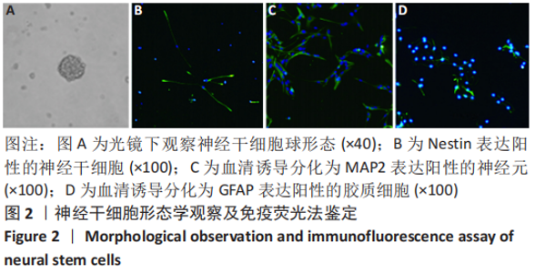
2.1 神经干细胞形态学观察及鉴定结果 分离后24 h可见单个悬浮细胞、不规则悬浮细胞团及部分贴壁细胞混合存在,细胞均为透亮,呈圆形或椭圆形。两三天后可见细胞聚集成神经球,并悬浮生长,见图2A。加血清诱导分化2 d后可见细胞多贴壁,并生长出突起。免疫荧光示细胞Nestin表达为阳性,见图2B。加入血清诱导分化后,MAP2及GFAP表达为阳性,见图2C,D。"

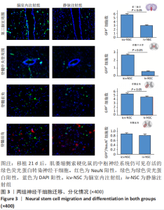
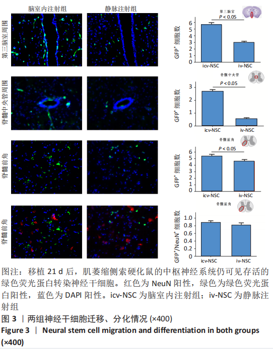
2.2 神经干细胞迁移、分化情况 免疫荧光显示,移植后21 d,2个移植组的绿色荧光蛋白转染神经干细胞均可迁移入中枢神经系统,且在3个解剖部位脑室内注射组的阳性细胞数均多于静脉注射组,即第三脑室周围[(5.67±0.13) vs. (2.99±0.07)个/mm2,P < 0.05],脊髓中央管[(2.68±0.06) vs. (0.54±0.04)个/mm2,P < 0.05],腰骶膨大处脊髓前角[(5.35±0.13) vs. (4.59±0.11)个/mm2,P < 0.05]。脊髓前角绿色荧光蛋白和NeuN的荧光双标显示,两组移植的神经干细胞均能分化为神经元,但数量均较少,无统计学差异[(0.89±0.02) vs. (0.82±0.02)个/mm2,P > 0.05]。两组移植源性神经干细胞在脊髓前角向神经元分化的比例也较小[(16±3)% vs. (17±2)%,P > 0.05],见图3。"

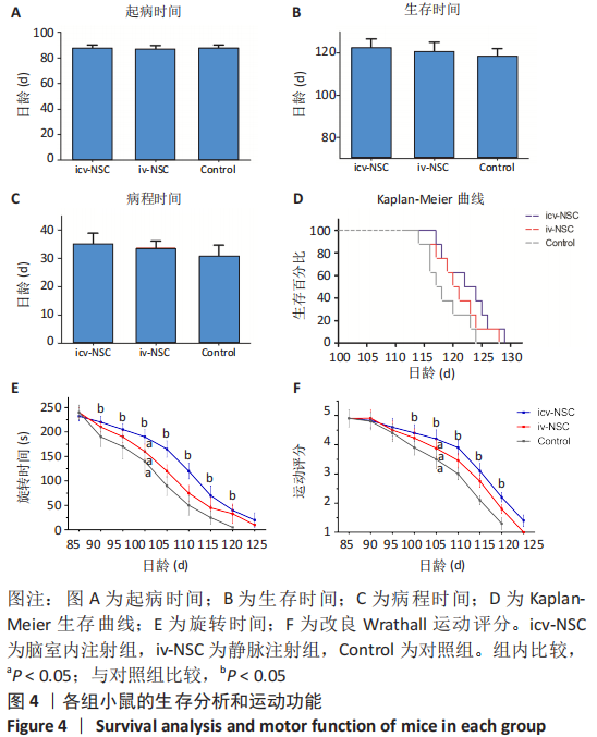
2.3 行为学表现 2.3.1 生存分析 脑室内注射组、静脉注射组、对照组的起病时间无明显差异[(87.63±4.35),(87.00±3.93),(87.86±3.62) d,P > 0.05),见图4A。生存时间方面,脑室内注射组最长,静脉注射组次之,对照组最短,但差异无显著性意义[(122.5±6.44),(120.75±5.87),(118.5± 6.88) d,P > 0.05),见图4B。KalPan-Meier生存分析也显示出相似结果(P > 0.05),见图4D。病程时间上,脑室内注射组最长,静脉注射组次之,对照组最短,但3组间也无统计学差异[(34.88±5.13),(33.75±4.85),(30.63±4.96) d,P > 0.05),见图4C。 2.3.2 运动功能检测结果 由于旋转时间和运动功能评分不满足球形检验,采用Greenhouse-Geisser统计方法。旋转实验和改良Wrathall评分均显示脑室内注射组的运动能力下降速度较对照组慢(P < 0.05),见图4E,F。与对照组相比,静脉注射组的运动能力下降速度有减慢趋势,但差异无显著性意义(P > 0.05)。与静脉注射组相比,脑室内注射组运动能力也有下降趋势,但差异无显著性意义(P > 0.05)。"
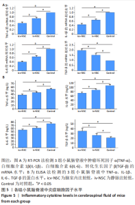
2.4.1 RT-PCR检测结果 脑室内注射组和静脉注射组促炎细胞因子肿瘤坏死因子α、白细胞介素1β、白细胞介素6的mRNA水平均低于对照组(P < 0.05),而抗炎性细胞因子转化生长因子β的mRNA水平高于对照组(P < 0.05)。与静脉注射组相比,脑室内注射组3种促炎细胞因子肿瘤坏死因子α、白细胞介素1β、白细胞介素6的mRNA水平较低,而抗炎细胞因子转化生长因子β的mRNA水平较高(P < 0.05),见图5A。 2.4.2 ELISA检测结果 脑室内注射组和静脉注射组的促炎细胞因子肿瘤坏死因子α、白细胞介素1β、白细胞介素6蛋白水平均低于对照组(P < 0.05),而抗炎细胞因子转化生长因子β水平高于对照组(P < 0.05)。与静脉注射组相比,脑室内注射组的3种促炎细胞因子肿瘤坏死因子α、白细胞介素1β、白细胞介素6的蛋白水平也较低,而抗炎细胞因子转化生长因子β的蛋白水平较高(P < 0.05),见图5B。"
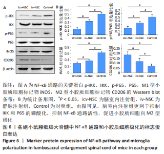
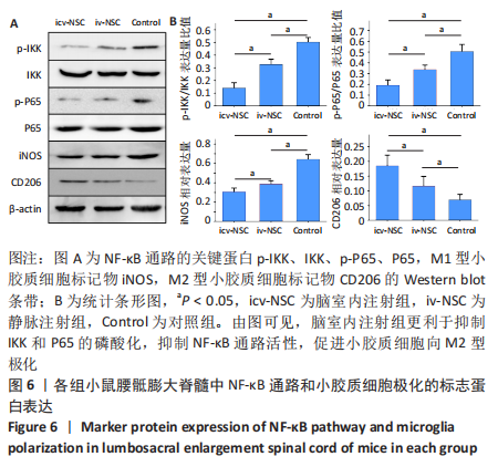
2.5 脊髓小胶质细胞极化方向及NF-κB通路活性 腰骶膨大脊髓的Western blot显示,脑室内注射组和静脉注射组M1型小胶质细胞特异性标志物iNOS的表达水平均低于对照组(P < 0.05)。与静脉注射组相比,脑室内注射组iNOS表达更低(P < 0.05)。脑室内注射组和静脉注射组M2型小胶质细胞特异性标志物CD206的表达水平均高于对照组(P < 0.05)。与静脉注射组相比,脑室内注射组CD206表达更高(P < 0.05)。脑室内注射组和静脉注射组的p-IKK/IKK和p-P65/P65比值低于对照组(P < 0.05),脑室内注射组的p-IKK/IKK和p-P65/P65比值均低于静脉注射组(P < 0.05),见图6。"
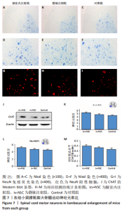
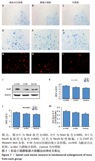
2.6 运动神经元数量 Nissl染色显示3组运动神经元数量组间无统计学差异,分别为(5.16±1.21),(4.72±1.04),(4.43±0.87)个/mm2(P > 0.05)。NeuN阳性细胞数组间无统计学差异,分别为(5.11±1.32),(4.65±0.97),(4.47±0.84)个/mm2(P > 0.05)。ChAT的相对表达量组间无统计学差异,分别为(0.39±0.07),(0.36±0.06),(0.32±0.08)个/mm2(P > 0.05),见图7。"
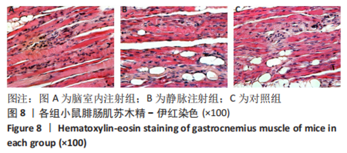
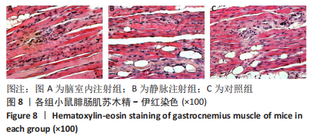
2.7 腓肠肌苏木精-伊红染色结果 3组肌肉均有不同程度的变性、坏死、萎缩等损伤,但也表现出各自的特点。脑室内注射组细胞核数量增加,肌肉细胞变薄,见图8A;静脉注射组肌肉细胞颜色变浅,肌肉条纹消失,见图8B;对照组出现大量脂肪变性和大量炎性细胞浸润,见图8C。"
| [1] GRAD LI, ROULEAU GA, RAVITS J, et al. Clinical Spectrum of Amyotrophic Lateral Sclerosis (ALS). Cold Spring Harb Perspect Med. 2017;7(8):a024117. [2] XU RS, YUAN M. Considerations on the concept, definition, and diagnosis of amyotrophic lateral sclerosis. Neural Regen Res. 2021;16(9):1723-1729. [3] BOILLÉE S, YAMANAKA K, LOBSIGER CS, et al. Onset and progression in inherited ALS determined by motor neurons and microglia. Science. 2006; 312(5778):1389-1392. [4] BEERS DR, APPEL SH. Immune dysregulation in amyotrophic lateral sclerosis: mechanisms and emerging therapies. Lancet Neurol. 2019;18(2):211-220. [5] LU Y, JIANG L, LI W, et al. Optogenetic Inhibition of Striatal Neuronal Activity Improves the Survival of Transplanted Neural Stem Cells and Neurological Outcomes after Ischemic Stroke in Mice. Stem Cells Int. 2017;2017:4364302. [6] PRAMANIK S, SULISTIO YA, HEESE K. Neurotrophin Signaling and Stem Cells-Implications for Neurodegenerative Diseases and Stem Cell Therapy. Mol Neurobiol. 2017;54(9):7401-7459. [7] CHEN T, LI Y, NI W, et al. Human Neural Stem Cell-Conditioned Medium Inhibits Inflammation in Macrophages Via Sirt-1 Signaling Pathway In Vitro and Promotes Sciatic Nerve Injury Recovery in Rats. Stem Cells Dev. 2020; 29(16):1084-1095. [8] BOESE AC, LE QE, PHAM D, et al. Neural stem cell therapy for subacute and chronic ischemic stroke. Stem Cell Res Ther. 2018;9(1):154. [9] ZHOU Q, YUAN M, QIU W, et al. Preclinical studies of mesenchymal stem cells transplantation in amyotrophic lateral sclerosis: a systemic review and metaanalysis. Neurol Sci. 2021;42(9):3637-3646. [10] DE MARCHI F, MUNITIC I, AMEDEI A, et al. Interplay between immunity and amyotrophic lateral sclerosis: Clinical impact. Neurosci Biobehav Rev. 2021;127:958-978. [11] ZHONG SJ, GONG YH, LIN YC. Combined intranasal nerve growth factor and ventricle neural stem cell grafts prolong survival and improve disease outcome in amyotrophic lateral sclerosis transgenic mice. Neurosci Lett. 2017;656:1-8. [12] 肖昆太,梁晓媚,高雅,等.hAMSCs静脉移植对ALS转基因小鼠运动功能的影响[J].中华神经医学杂志,2017,16(7):649-656. [13] LIN TJ, CHENG GC, WU LY, et al. Potential of Cellular Therapy for ALS: Current Strategies and Future Prospects. Front Cell Dev Biol. 2022;10:851613. [14] BETTE M, CORS E, KRESSE C, et al. Therapeutic Treatment of Superoxide Dismutase 1 (G93A) Amyotrophic Lateral Sclerosis Model Mice with Medical Ozone Decelerates Trigeminal Motor Neuron Degeneration, Attenuates Microglial Proliferation, and Preserves Monocyte Levels in Mesenteric Lymph Nodes. Int J Mol Sci. 2022;23(6):3403. [15] OHASHI N, TERASHIMA T, KATAGI M, et al. GLT1 gene delivery based on bone marrow-derived cells ameliorates motor function and survival in a mouse model of ALS. Sci Rep. 2021;11(1):12803. [16] TRIAS E, BEILBY PR, KOVACS M, et al. Emergence of Microglia Bearing Senescence Markers During Paralysis Progression in a Rat Model of Inherited ALS. Front Aging Neurosci. 2019;11:42. [17] MAGOTA H, SASAKI M, KATAOKA-SASAKI Y, et al. Intravenous infusion of mesenchymal stem cells delays disease progression in the SOD1G93A transgenic amyotrophic lateral sclerosis rat model. Brain Res. 2021;1757: 147296. [18] TAVAKOL-AFSHARI J, BOROUMAND AR, FARKHAD NK, et al. Safety and efficacy of bone marrow derived-mesenchymal stem cells transplantation in patients with amyotrophic lateral sclerosis. Regen Ther. 2021;18:268-274. [19] MITRECIĆ D, NICAISE C, GAJOVIĆ S, et al. Distribution, differentiation, and survival of intravenously administered neural stem cells in a rat model of amyotrophic lateral sclerosis, Cell Transplant. 2010;19(8):537-548. [20] SRIVASTAVA AK, GROSS SK, ALMAD AA, et al. Serial in vivo imaging of transplanted allogeneic neural stem cell survival in a mouse model of amyotrophic lateral sclerosis. Exp Neurol. 2017;289(4):96-102. [21] AEBISCHER J, MOUMEN A, SAZDOVITCH V, et al. Elevated levels of IFNγ and LIGHT in the spinal cord of patients with sporadic amyotrophic lateral sclerosis. Eur J Neurol. 2012;19(4):752-759,e45-46. [22] HERRANZ-PÉREZ V, NAKATANI J, ISHII M, et al. Ependymoma associated protein Zfta is expressed in immature ependymal cells but is not essential for ependymal development in mice. Sci Rep. 2022;12(1):1493. [23] BANG E, FINCHER AS, NADER S, et al. Late-Onset, Short-Term Intermittent Fasting Reverses Age-Related Changes in Calcium Buffering and Inhibitory Synaptic Transmission in Mouse Basal Forebrain Neurons. J Neurosci. 2022; 42(6):1020-1034. [24] QUARESIMA S, ISTIAQ A, JONO H, et al. Assessing the Role of Ependymal and Vascular Cells as Sources of Extracellular Cues Regulating the Mouse Ventricular-Subventricular Zone Neurogenic Niche. Front Cell Dev Biol. 2022;10:845567. [25] HENG AHS, HAN CW, ABBOTT C, et al. Chemokine-Driven Migration of Pro-Inflammatory CD4+ T Cells in CNS Autoimmune Disease. Front Immunol. 2022;13:817473. [26] MARTÍNEZ-MORENO CG, CALDERÓN-VALLEJO D, HARVEY S, et al. Growth Hormone (GH) and Gonadotropin-Releasing Hormone (GnRH) in the Central Nervous System: A Potential Neurological Combinatory Therapy? Int J Mol Sci. 2018;19(2):375. [27] LÓPEZ-MUGURUZA E, VILLAR-GÓMEZ N, MATIAS-GUIU JA, et al. The Integration of Cell Therapy and Biomaterials as Treatment Strategies for Remyelination. Life (Basel). 2022;12(4):474. [28] SIMITZI C, KARALI K, RANELLA A, et al. Controlling the Outgrowth and Functions of Neural Stem Cells: The Effect of Surface Topography. Chemphyschem. 2018;19(10):1143-1163. [29] DARVISHI M, TIRAIHI T, MESBAH-NAMIN SA, et al. Motor Neuron Transdifferentiation of Neural Stem Cell from Adipose-Derived Stem Cell Characterized by Differential Gene Expression. Cell Mol Neurobiol. 2017; 37(2):275-289. [30] DRANNIK A, MARTIN J, PETERSON R, et al. Cerebrospinal fluid from patients with amyotrophic lateral sclerosis inhibits sonic hedgehog function. PLoS One. 2017;12(2):e0171668. [31] IWATA R, CASIMIR P, VANDERHAEGHEN P. Mitochondrial dynamics in postmitotic cells regulate neurogenesis. Science. 2020;369(6505):858-862. [32] MEHTA AR, GREGORY JM, DANDO O, et al. Mitochondrial bioenergetic deficits in C9orf72 amyotrophic lateral sclerosis motor neurons cause dysfunctional axonal homeostasis. Acta Neuropathol. 2021;141(2):257-279. [33] CHEN J, JIN J, LI K, et al. Progresses and Prospects of Neuroprotective Agents-Loaded Nanoparticles and Biomimetic Material in Ischemic Stroke. Front Cell Neurosci. 2022;16:868323. [34] JE G, KEYHANIAN K, GHASEMI M. Overview of stem cells therapy in amyotrophic lateral sclerosis. Neurol Res. 2021;43(8):616-632. [35] HUANG X, FEI GQ, LIU WJ, et al. Adipose-derived mesenchymal stem cells protect against CMS-induced depression-like behaviors in mice via regulating the Nrf2/HO-1 and TLR4/NF-κB signaling pathways. Acta Pharmacol Sin. 2020;41(5):612-619. [36] LI C, PANG D, LIN J, et al. Shared genetic links between frontotemporal dementia and psychiatric disorders. BMC Med. 2022;20(1):131. [37] LIU X, MA J, DING G, et al. Microglia Polarization from M1 toward M2 Phenotype Is Promoted by Astragalus Polysaccharides Mediated through Inhibition of miR-155 in Experimental Autoimmune Encephalomyelitis. Oxid Med Cell Longev. 2021;2021:5753452. [38] KANO SI, CHOI EY, DOHI E, et al. Glutathione S-transferases promote proinflammatory astrocyte-microglia communication during brain inflammation. Sci Signal. 2019;12(569):eaar2124. [39] NI YQ, XU H, LIU YS. Roles of Long Non-coding RNAs in the Development of Aging-Related Neurodegenerative Diseases. Front Mol Neurosci. 2022;15:844193. [40] DE MENA L, LOPEZ-SCARIM J, RINCON-LIMAS DE. TDP-43 and ER Stress in Neurodegeneration: Friends or Foes?. Front Mol Neurosci. 2021;14:772226. [41] CHAILLOU T. Ribosome specialization and its potential role in the control of protein translation and skeletal muscle size. J Appl Physiol (1985). 2019; 127(2):599-607. [42] CABRAL BMI, EDDING SN, PORTOCARRERO JP, et al. Rhabdomyolysis. Dis Mon. 2020;66(8):101015. [43] BALOH RH, GLASS JD, SVENDSEN CN. Stem cell transplantation for amyotrophic lateral sclerosis. Curr Opin Neurol. 2018;31(5):655-661. [44] LYON MS, WOSISKI-KUHN M, GILLESPIE R, et al. Inflammation, Immunity, and amyotrophic lateral sclerosis: I. Etiology and pathology. Muscle Nerve. 2019;59(1):10-22. [45] LI G, XIAO L, QIN H, et al. Exosomes-carried microRNA-26b-5p regulates microglia M1 polarization after cerebral ischemia/reperfusion. Cell Cycle. 2020;19(9):1022-1035. [46] EITAN C, SIANY A, BARKAN E, et al. Whole-genome sequencing reveals that variants in the Interleukin 18 Receptor Accessory Protein 3’UTR protect against ALS. Nat Neurosci. 2022;25(4):433-445. |
| [1] | Guo Shuhui, Yang Ye, Jiang Yangyang, Xu Jianwen. Screening and validation of neurogenic bladder miRNA-mRNA regulatory network [J]. Chinese Journal of Tissue Engineering Research, 2023, 27(在线): 1-8. |
| [2] | Fang Xingyan, Tian Zhenli, Zhao Zheyi, Wen Ping, Xie Tingting. Effects of sodium arsenite on human umbilical vein endothelial cell injury and sphingosine kinases 1/sphingosine 1-phosphate signaling axis [J]. Chinese Journal of Tissue Engineering Research, 2023, 27(在线): 1-7. |
| [3] | Tang Liang, Li Xiheng, Niu Ruijuan, Li Xinyue, Zou Xinying, Mao Tianjiao, Li Jiang. Naringin regulates the function of RAW264.7 macrophages to affect the osteogenic differentiation of MC-3T3-E1 cells [J]. Chinese Journal of Tissue Engineering Research, 2023, 27(8): 1205-1210. |
| [4] | Liang Jiaqi, Liu Hengxu, Yang Jinxin, Yang Yi, Deng Xuhui, Tan Mingjian, Luo Jiong. Health benefit relationship between exercise and intestinal bacteria [J]. Chinese Journal of Tissue Engineering Research, 2023, 27(8): 1292-1299. |
| [5] | Gao Yu, Han Jiahui, Ge Xin. Immunoinflammatory microenvironment after spinal cord ischemia-reperfusion injury [J]. Chinese Journal of Tissue Engineering Research, 2023, 27(8): 1300-1305. |
| [6] | Li Wenjie, You Aijia, Zhou Junli, Fang Sujuan, Li Chun. Effects of different dressings in the treatment of burn wounds: a network meta-analysis [J]. Chinese Journal of Tissue Engineering Research, 2023, 27(7): 1141-1148. |
| [7] | Wang Min, Yin Xiushan, Wang Yingxi, Zhang Yan, Zhao Long, Xia Shuyue. Inhalation of bone marrow mesenchymal stem cells-derived exosomes alleviates inflammatory injury in chronic obstructive pulmonary disease [J]. Chinese Journal of Tissue Engineering Research, 2023, 27(6): 827-834. |
| [8] | Hao Liufang, Duan Hongmei, Wang Zijue, Hao Fei, Hao Peng, Zhao Wen, Gao Yudan, Yang Zhaoyang, Li Xiaoguang. Spatiotemporal dynamic changes of ependymal cells after spinal cord injury in transgenic mice [J]. Chinese Journal of Tissue Engineering Research, 2023, 27(6): 883-889. |
| [9] | Li Xiaoyin, Yang Xiaoqing, Chen Shulian, Li Zhengchao, Wang Ziqi, Song Zhen, Zhu Daren, Chen Xuyi. Collagen/silk fibroin scaffold combined with neural stem cells in the treatment of traumatic spinal cord injury [J]. Chinese Journal of Tissue Engineering Research, 2023, 27(6): 890-896. |
| [10] | Li Zhichao, Tan Guoqing, Su Hui, Xu Zhanwang, Xue Haipeng. Regulatory role of non-coding RNAs as potential therapeutic targets in spinal cord injury [J]. Chinese Journal of Tissue Engineering Research, 2023, 27(5): 758-764. |
| [11] | Yang Heng, Zheng Liying, Liu Zhili, Wang Qinzhang, Hao Zhiqiang, Wang Jingshen, Wang Yuan, Li Yongle, Tan Minghui, Zou Xiaofeng, Zhang Guoxi, Huang Ruohui, Jiang Bo, Qian Biao. Physiological function of the testis in rats undergoing different parts of vasectomy [J]. Chinese Journal of Tissue Engineering Research, 2023, 27(5): 720-725. |
| [12] | Zhang Lichen, Chen Liang, Gu Yong. Inorganic ion bionic periosteum regulates immune microenvironment to promote bone repair [J]. Chinese Journal of Tissue Engineering Research, 2023, 27(3): 346-353. |
| [13] | Zhao Feng, Fan Shaoqing, Cheng Xiaoyan, Li Xiaona, Li Changsheng, Ma Haojie. miR-132-3p targets and modulates Ptch1 to reduce neuropathic pain in mice with chronic constriction injury [J]. Chinese Journal of Tissue Engineering Research, 2023, 27(2): 230-236. |
| [14] | Ying Chunmiao, Ma Yucheng, Fan Feiyan, Gao Chen, Wang Shaona, Yang Xing, Zhang Yunke. Action mechanism of traditional Chinese medicine to regulate neural stem cells in treatment of Alzheimer’s disease [J]. Chinese Journal of Tissue Engineering Research, 2023, 27(15): 2385-2394. |
| [15] | Zheng Xiaohan, Wei Yanzhao, Huang Ting, Wei Xufang, Sun Shengtong, Wang Tan, Zhao Zhenqiang. Role of macrophage migration inhibitory factor for stem cells#br# [J]. Chinese Journal of Tissue Engineering Research, 2023, 27(15): 2395-2403. |
| Viewed | ||||||
|
Full text |
|
|||||
|
Abstract |
|
|||||