Chinese Journal of Tissue Engineering Research ›› 2022, Vol. 26 ›› Issue (26): 4101-4106.doi: 10.12307/2022.809
Transcriptome sequencing of the uterine in a mouse model of vitamin D deficiency
Chen Xia1, Shang Yuwei1, Wang Linxiao2, Shi Yichao1, Liu Huijun1, Sun Huiting1
- 1Department of Reproductive Center, 2Laboratory of Neurological Diseases, Changzhou Second People’s Hospital Affiliated to Nanjing Medical University, Changzhou 213003, Jiangsu Province, China
-
Received:2021-05-12Accepted:2021-07-14Online:2022-09-18Published:2022-03-08 -
Contact:Sun Huiting, Master, Attending physician, Department of Reproductive Center, Changzhou Second People’s Hospital Affiliated to Nanjing Medical University, Changzhou 213003, Jiangsu Province, China -
About author:Chen Xia, Master, Assistant researcher, Department of Reproductive Center, Changzhou Second People’s Hospital Affiliated to Nanjing Medical University, Changzhou 213003, Jiangsu Province, China -
Supported by:Changzhou Sci & Tech Program, No. CJ20200103 (to SHT); Changzhou Sci & Tech Program, No. CJ20210110 (to CX); the National Natural Science Foundation of China (Youth Fund), No. 81803498 (to WLX)
CLC Number:
Cite this article
Chen Xia, Shang Yuwei, Wang Linxiao, Shi Yichao, Liu Huijun, Sun Huiting. Transcriptome sequencing of the uterine in a mouse model of vitamin D deficiency[J]. Chinese Journal of Tissue Engineering Research, 2022, 26(26): 4101-4106.
share this article
Add to citation manager EndNote|Reference Manager|ProCite|BibTeX|RefWorks
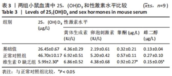
2.1 实验动物数量分析 共18只小鼠随机分组,维生素D缺乏组及正常对照组每组9只,饲养过程中无小鼠伤亡,全部进入结果分析。 2.2 维生素D缺乏小鼠表型分析 喂养8周后,ELISA法检测血清25,(OH)D3水平显著下降,说明维生素D缺乏饲料喂养8周后可以引起小鼠体内维生素D缺乏,可用于后续转录组测序分析。此外,维生素D缺乏组雌二醇水平较正常组小鼠降低(P < 0.05),睾酮水平升高(P < 0.05),卵泡刺激素、黄体生成素水平降低,但两组差异无显著性意义,见表3。"
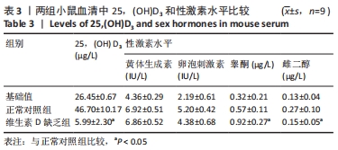
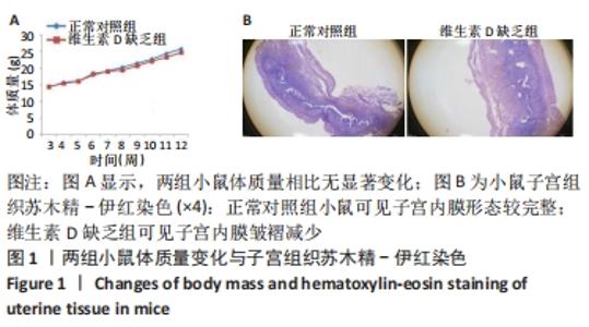
与正常对照组相比,维生素D缺乏组小鼠体质量无显著变化,见图1A。子宫组织苏木精-伊红染色结果显示,维生素D缺乏后可导致子宫内膜皱褶减少,间质中腺体数量减少并且没有腺体的乳头状增生,提示子宫着床条件变差,见图1B。"
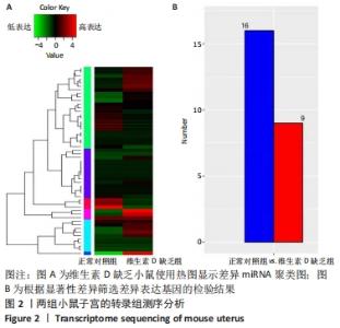
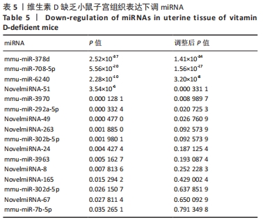
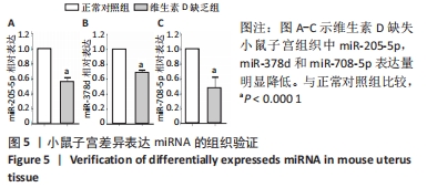
2.3 两组小鼠子宫组织差异表达基因统计及基因本体、KEGG 富集分析 对两组样本进行miRNAs表达量差异统计分析,结果筛查到561个miRNAs表达差异。对差异表达miRNAs进行聚类分析,其中显著上调的9个,显著下调的16个,见图2及表4,5。其中发现了与卵巢颗粒细胞凋亡和自噬相关的miR-378d显著下调,与子宫内膜癌密切相关的miR-205-5p显著下调,与胚胎干细胞自我更新和多能性相关的miR-541-5p明显上调,与调节妊娠早期胎盘相关miR-708-5p显著下调等,这些差异表达的miRNA都与女性生殖系统密切相关。"

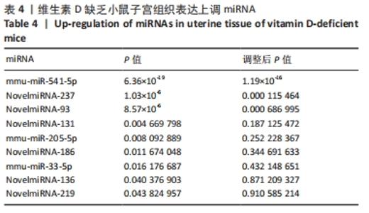
"
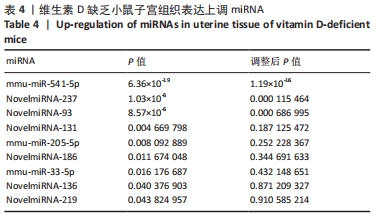
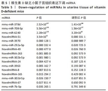
"
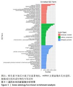
对差异表达基因进行基因本体富集分析,以探究这些差异表达基因的生物学功能。位于前 20 项显著富集中,11 项属于生物过程类(前 10 项分别是:生物调节、细胞过程、单细胞生物过程、代谢过程、定位、发育过程、细胞成分组织或生物发生、刺激反应、多细胞生物过程、免疫系统过程),7 项属于细胞组分类(细胞部分、细胞器、膜、细胞器部分、膜部分、胞外区部分、大分子复合物),2 项属于分子功能类(结合、催化活性) ,见图3。结果表明, 差异表达基因与细胞代谢和发育进程密切相关。"

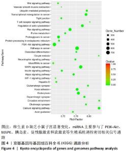
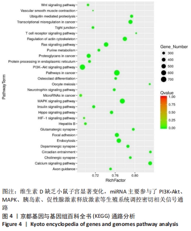
KEGG 富集分析发现,这些差异表达基因分布在139条通路中。主要参与的信号转导途径分别是PI3K-Akt信号通路、MAPK信号通路、人类Ⅰ型嗜T细胞病毒感染、RAS信号通路、胞吞作用、癌症中蛋白多糖、肌动蛋白细胞骨架的调节、胰岛素信号通路、紧密连接、促性腺激素释放激素信号通路和卵母细胞减数分裂信号通路,见图4。"
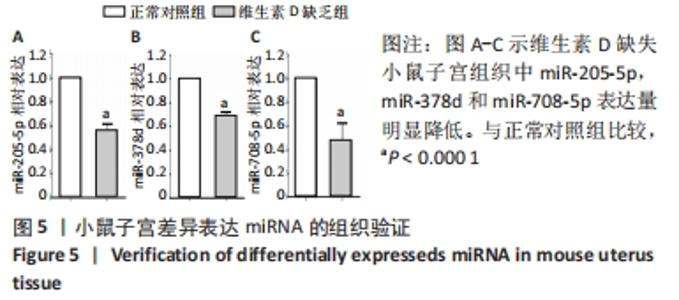
这些结果表明,维生素D缺乏后影响了女性生殖相关的多个信号通路。小鼠子宫组织实时荧光定量PCR验证结果与测序一致,miR-378d和miR-205-5p均显著下调,见图5。"
| [1] LI G, LIN L, WANG YL, et al. 1,25(OH)2D3 Protects Trophoblasts Against Insulin Resistance and Inflammation Via Suppressing mTOR Signaling. Reprod Sci. 2019; 26(2):223-232. [2] SUN J. Dietary vitamin D, vitamin D receptor, and microbiome. Curr Opin Clin Nutr Metab Care. 2018; 21(6):471-474. [3] LEI M, LIU Z, GUO J. The Emerging Role of Vitamin D and Vitamin D Receptor in Diabetic Nephropathy. Biomed Res Int. 2020;2020:4137268. [4] FOGACCI S, FOGACCI F, BANACH M, et al. Vitamin D supplementation and incident preeclampsia: A systematic review and meta-analysis of randomized clinical trials. Clin Nutr. 2020;39(6):1742-1752. [5] 汤斐, 赵云.子宫动脉介入栓塞合并氨甲喋呤注射在胎盘完全滞留中的应用[J].中华卫生应急电子杂志,2019,5(3):141-146. [6] AGARWAL S, KOVILAM O, AGRAWAL DK. Vitamin D and its impact on maternal-fetal outcomes in pregnancy: A critical review. Crit Rev Food Sci Nutr. 2018;58(5): 755-769. [7] PURSWANI JM, GALA P, DWARKANATH P, et al. The role of vitamin D in pre-eclampsia: a systematic review. BMC Pregnancy Childbirth. 2017;17(1):231. [8] BENACHI A, BAPTISTE A, TAIEB J, et al. Relationship between vitamin D status in pregnancy and the risk for preeclampsia: A nested case-control study. Clin Nutr. 2020;39(2):440-446. [9] VAN SCHOOR N, LIPS P. Global Overview of Vitamin D Status. Endocrinol Metab Clin North Am. 2017;46(4):845-870. [10] ZHANG X, JI S, CAI G, et al. H19 Increases IL-17A/IL-23 Releases via Regulating VDR by Interacting with miR675-5p/miR22-5p in Ankylosing Spondylitis. Mol Ther Nucleic Acids. 2020;19:393-404. [11] 袁长深, 容伟明, 卢智贤, 等. 基于生物信息学构建骨肉瘤miRNA-mRNA的调控网络[J]. 中国组织工程研究,2021,25(17):2740-2746. [12] KLIMAN HJ, FRANKFURTER D. Clinical approach to recurrent implantation failure: evidence-based evaluation of the endometrium. Fertil Steril. 2019;111(4):618-628. [13] MARQUARDT RM, KIM TH, SHIN JH, et al. Progesterone and Estrogen Signaling in the Endometrium: What Goes Wrong in Endometriosis? Int J Mol Sci. 2019; 20(15):3822. [14] AGIC A, XU H, ALTGASSEN C, et al. Relative expression of 1,25-dihydroxyvitamin D3 receptor, vitamin D 1 alpha-hydroxylase, vitamin D 24-hydroxylase, and vitamin D 25-hydroxylase in endometriosis and gynecologic cancers. Reprod Sci. 2007;14(5):486-497. [15] MUSCOGIURI G, ALTIERI B, DE ANGELIS C, et al. Shedding new light on female fertility: The role of vitamin D. Rev Endocr Metab Disord. 2017;18(3):273-283. [16] 王波, 王雪云. 维生素D缺乏影响受孕及胚胎发育的小鼠动物模型研究[J]. 分子诊断与治疗杂志,2016,8(5):341-345+360. [17] AUGUSTE A, BESSIÈRE L, TODESCHINI AL, et al. Molecular analyses of juvenile granulosa cell tumors bearing AKT1 mutations provide insights into tumor biology and therapeutic leads. Hum Mol Genet. 2015;24(23):6687-6698. [18] 黄冬梅, 黄光英, 陆付耳. 胚泡围着床期子宫内膜细胞的增殖分化与凋亡[J]. 生殖医学杂志,2004,13(5):313-316. [19] NEGAMI AI. Implantation model in vitro. Nihon Sanka Fujinka Gakkai Zasshi. 1992; 44(8):949-959. [20] FUNG JL, HARTMAN TJ, SCHLEICHER RL, et al. Association of vitamin D intake and serum levels with fertility: results from the Lifestyle and Fertility Study. Fertil Steril. 2017;108(2):302-311. [21] 张迪, 苏姗, 王强梅, 等. 维生素D 与多囊卵巢综合征关系的研究进展[J]. 徐州医科大学学报,2021,41(1):76-78. [22] LIANG Y, RIDZON D, WONG L, et al. Characterization of microRNA expression profiles in normal human tissues. BMC Genomics. 2007;8:166. [23] WANG B, CRUZ ITHIER M, PAROBCHAK N, et al. Vitamin D stimulates multiple microRNAs to inhibit CRH and other pro-labor genes in human placenta. Endocr Connect. 2018;7(12):1380-1388. [24] QI Y, WANG D, HUANG W, et al. CyclinD1 inhibits dicer and crucial miRNA expression by chromatin modification to promote the progression of intrahepatic cholangiocarcinoma. J Exp Clin Cancer Res. 2019;38(1):413. [25] XIN W, LIU X, DING J, et al. Long non-coding RNA derived miR-205-5p modulates human endometrial cancer by targeting PTEN. Am J Transl Res. 2015;7(11):2433-2441. [26] ABDELGHAFFAR S, SHORA H, ABDELATTY S, et al. MicroRNAs and Risk Factors for Diabetic Nephropathy in Egyptian Children and Adolescents with Type 1 Diabetes. Diabetes Metab Syndr Obes. 2020; 13: 2485-2494. [27] LAN L, LIANG Z, ZHAO Y, et al. LncRNA MCM3AP-AS1 inhibits cell proliferation in cervical squamous cell carcinoma by down-regulating miRNA-93. Biosci Rep. 2020;40(2):BSR20193794. [28] LIN L, SUN J, WU D, et al. MicroRNA-186 is associated with hypoxia-inducible factor-1α expression in chronic obstructive pulmonary disease. Mol Genet Genomic Med. 2019;7(3):e531. [29] MOTAWI T, SABRY D, MAURICE NW, et al. Role of mesenchymal stem cells exosomes derived microRNAs; miR-136, miR-494 and miR-495 in pre-eclampsia diagnosis and evaluation. Arch Biochem Biophys. 2018;659:13-21. [30] CHEN X, HUANG Z, CHEN R. Microrna-136 promotes proliferation and invasion ingastric cancer cells through Pten/Akt/P-Akt signaling pathway. Oncol Lett. 2018; 15(4):4683-4689. [31] ISHIDA M, SHIMABUKURO M, YAGI S, et al. MicroRNA-378 regulates adiponectin expression in adipose tissue: a new plausible mechanism. PLoS One. 2014;9(11): e111537. [32] MEERSON A, ELIRAZ Y, YEHUDA H, et al. Obesity impacts the regulation of miR-10b and its targets in primary breast tumors. BMC Cancer. 2019;19(1):86. [33] GUNGORMEZ C, GUMUSHAN AKTAS H, DILSIZ N, et al. Novel miRNAs as potential biomarkers in stage II colon cancer: microarray analysis. Mol Biol Rep. 2019;46(4): 4175-4183. [34] ZOU X, WANG J, QU H, et al. Comprehensive analysis of miRNAs, lncRNAs, and mRNAs reveals potential players of sexually dimorphic and left-right asymmetry in chicken gonad during gonadal differentiation. Poult Sci. 2020;99(5):2696-2707. |
| [1] | Zhou Hongqin, Wu Dandan, Yang Kun, Liu Qi. Exosomes that deliver specific miRNAs can regulate osteogenesis and promote angiogenesis [J]. Chinese Journal of Tissue Engineering Research, 2022, 26(7): 1107-1112. |
| [2] | Xu Jing, Yan Yongmin, Cai Mengjie . miR-373 inhibits hepatic stellate cell activation by downregulating transforming growth factor beta type II receptor [J]. Chinese Journal of Tissue Engineering Research, 2022, 26(5): 756-761. |
| [3] | Wei Yufan, Fan Feiyan, Li Shuangli, Zhang Yunke. Future of exosomes and mesenchymal stem cell-derived exosomes in the diagnosis and treatment of Parkinson’s disease [J]. Chinese Journal of Tissue Engineering Research, 2022, 26(25): 4076-4083. |
| [4] | Yu Chunbo, Li Dayu, Fan Fang, Li Changfu. Transcription factor-miRNA-mRNA network analysis of osteogenic differentiation of adipose-derived stem cells [J]. Chinese Journal of Tissue Engineering Research, 2022, 26(24): 3908-3913. |
| [5] | Qian Xiaofen, Zeng Ping, Liu Jinfu, Chen Cai, Fan Siqi, Nong Jiao, Deng Wei. Comprehensive analysis of competing endogenous RNA network and potential biomarkers in alcohol-induced osteonecrosis of the femoral head [J]. Chinese Journal of Tissue Engineering Research, 2022, 26(23): 3670-3675. |
| [6] | Liang Xuezhen, Xie Guoxin, Li Jiacheng, Wen Mingtao, Xu Bo, Li Gang. Identification and analysis of potential key genes in osteonecrosis of the femoral head based on miRNA-mRNA regulatory network [J]. Chinese Journal of Tissue Engineering Research, 2022, 26(11): 1720-1727. |
| [7] | Huang Yangjun, Zhou Honghai, Chen Longhao, Li Jilin, Lu Qingwang, Li Dongyang, He Xinyu. Regulatory role and mechanism of non-coding RNA on steroid-induced osteonecrosis of the femoral head [J]. Chinese Journal of Tissue Engineering Research, 2022, 26(11): 1765-1771. |
| [8] | Cui Shuaishuai, Yang Xiaohong. Effects of miRNA in self-renewal, multidirectional differentiation, fate and function regulation of bone marrow mesenchymal stem cells [J]. Chinese Journal of Tissue Engineering Research, 2022, 26(1): 101-105. |
| [9] | Mei Yunyun, Zhang Jianjun, Wang Dong. Hyperbaric oxygen combined with NgR gene silencing bone marrow mesenchymal stem cells transplantation for spinal cord injury in rats [J]. Chinese Journal of Tissue Engineering Research, 2022, 26(1): 12-19. |
| [10] | Geng Yao, Yin Zhiliang, Li Xingping, Xiao Dongqin, Hou Weiguang. Role of hsa-miRNA-223-3p in regulating osteogenic differentiation of human bone marrow mesenchymal stem cells [J]. Chinese Journal of Tissue Engineering Research, 2021, 25(7): 1008-1013. |
| [11] | Liu Cong, Liu Su. Molecular mechanism of miR-17-5p regulation of hypoxia inducible factor-1α mediated adipocyte differentiation and angiogenesis [J]. Chinese Journal of Tissue Engineering Research, 2021, 25(7): 1069-1074. |
| [12] | Li Zhao, Cai Yuqiang. Advances in exosome miRNAs in bone reconstruction and bone metabolic diseases [J]. Chinese Journal of Tissue Engineering Research, 2021, 25(29): 4697-4702. |
| [13] | Chen Ziyang, Pu Rui, Deng Shuang, Yuan Lingyan. Regulatory effect of exosomes on exercise-mediated insulin resistance diseases [J]. Chinese Journal of Tissue Engineering Research, 2021, 25(25): 4089-4094. |
| [14] | Zhao Shuangdan, Zheng Jiahua, Qi Wenbo, Huang Xianghua. Role and mechanism of exosomes derived from mesenchymal stem cells in reproductive system diseases [J]. Chinese Journal of Tissue Engineering Research, 2021, 25(19): 3097-3102. |
| [15] | Yuan Changshen, Rong Weiming, Lu Zhixian, Duan Kan, Guo Jinrong, Mei Qijie. Construction of osteosarcoma miRNA-mRNA regulatory network based on bioinformatics [J]. Chinese Journal of Tissue Engineering Research, 2021, 25(17): 2740-2746. |
| Viewed | ||||||
|
Full text |
|
|||||
|
Abstract |
|
|||||