Chinese Journal of Tissue Engineering Research ›› 2021, Vol. 25 ›› Issue (19): 3071-3076.doi: 10.3969/j.issn.2095-4344.3520
Previous Articles Next Articles
Application prospects and problems of peripheral blood derived mesenchymal stem cells in cartilage repair of osteoarthritis
Yang Tengyun, Li Yanlin, Liu Dejian, Wang Guoliang
- Department of Sports Medicine, the First Affiliated Hospital of Kunming Medical University, Kunming 650032, Yunnan Province, China
-
Received:2020-06-04Revised:2020-06-12Accepted:2020-07-20Online:2021-07-09Published:2021-01-14 -
Contact:Li Yanlin, MD, Professor, Department of Sports Medicine, the First Affiliated Hospital of Kunming Medical University, Kunming 650032, Yunnan Province, China -
About author:Yang Tengyun, Department of Sports Medicine, the First Affiliated Hospital of Kunming Medical University, Kunming 650032, Yunnan Province, China -
Supported by:the National Natural Science Foundation of China, No. 81960409 (to LYL), No. 81760403 (to LYL); the Key Project of Joint Fund between Yunnan Provincial Department of Science and Technology and Kunming Medical University, No. [2017FE467(-007)] (to LYL); the Yunnan Expert Workstation Project, No.2018IC102 (to LYL)
CLC Number:
Cite this article
Yang Tengyun, Li Yanlin, Liu Dejian, Wang Guoliang. Application prospects and problems of peripheral blood derived mesenchymal stem cells in cartilage repair of osteoarthritis[J]. Chinese Journal of Tissue Engineering Research, 2021, 25(19): 3071-3076.
share this article
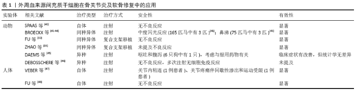
2.1 外周血来源间充质干细胞提取、分离、培养与鉴定 研究初期,外周血来源间充质干细胞存在与否一直颇受争议,随着研究的不断深入,学者们成功从猫、猪、绵羊、马等动物和人外周血中分离出间充质干细胞[25-30],并通过实验证实其与骨髓、脂肪等来源间充质干细胞具有相似生物学特性,但分离效率较低。研究初期,KASSIS等[30]通过使用集落刺激因子激活健康成年人外周血,从外周血祖细胞中分离出大量的间充质干细胞,这一实验使得外周血来源间充质干细胞分离效率得到了很大的提高。目前,许多研究者联合集落刺激因子和AMD3100对大鼠、兔等动物外周血进行动员,成功提取外周血来源间充质干细胞,并认为2种动员剂具有协同作用[31-32]。ZHAO等[33]研究也证实集落刺激因子和AMD3100是外周血来源间充质干细胞的有效动员剂,相比于单一因子动员组,动员效率提高近4倍。 目前分离提取外周血来源间充质干细胞的方法暂无统一标准,但文献报道的分离方法主要有4种,早期KUZNETSOV等[34]提出的全血细胞法、FERNANDEZ等[35]实验采用的血细胞分离机法和READING等[36]采用的免疫磁珠分离法,以及目前国内外实验采用最多的密度梯度离心法[25-33]。免疫磁珠分选法和流式细胞仪分离法虽然获得的外周血来源间充质干细胞较纯,但因其费用高、操作复杂且分选后的细胞活性低等原因现在已较少采用;密度梯度离心法因其符合正常生理性渗透压而不易破坏生物膜,对细胞无损害,分离效果好且简便易行,成为目前国内外常用的分选方案。 HOPPER等[37]从健康人体中分离出外周血来源间充质干细胞,在低氧条件下培养2周后呈成纤维细胞样形态,94%的细胞表达间充质干细胞标记物(CD90、CD105、CD146),而在正常氧条件下只有41%的细胞表达这些标记物,这为后续培养条件提供了有用的参考价值。赵岚等[38]在实验中发现即使纯度较高的外周血来源间充质干细胞,体外培养40 d即发生衰老和凋亡,但β-联蛋白可延长细胞存活时间至90 d 以上,增加其体外增殖率1. 08倍,明显提高了细胞增殖能力,为进一步提高外周血来源间充质干细胞的存活和增殖提供了新的突破点。大量实验皆证实外周血来源间充质干细胞具有和骨髓间充质干细胞相似的增殖及多向分化能力,流式细胞学分析CD44、CD54、CD105、CD166均为阳性,CD14、CD34、CD45、CD31均为阴性,Ⅰ型胶原和Ⅲ型胶原免疫组化法染色均为阳性[25-33],这与国际干细胞治疗协会(ISCT)[39]提出的间充质干细胞的3个定义标准相符:①贴壁生长;②表达特定表面抗原;③多向分化能力。外周血获取具有更简便、更微创的特点,操作所带来并发症也显著降低,具有良好的临床应用前景。不仅如此,因为来源于自体,可以更有效降低疾病传播和移植物抗宿主病等风险。 2.2 外周血来源间充质干细胞在骨关节炎及软骨修复中的应用 近年来,不同来源的间充质干细胞在软骨修复中的应用取得了不错的成就,如来自骨髓、脂肪和滑膜组织的间充质干细胞,通过注射或体外培养后移植等方式促进软骨修复[17-19]。 随着研究的进行,外周血来源间充质干细胞也进行了体内外的动物实验与人体相关试验,取得了积极的研究成果,见表1[32-33,40-48]。 2.2.1 动物实验 相关实验主要包括3方面的应用:同种同体移植、同种异体移植与异种移植。SPAAS等[40]从1匹5岁龄的患有骨关节炎的马外周血中分离提取出外周血间充质干细胞,经过体外培养后注入其骹关节,通过视觉步态评估和客观的压力板分析,结果证实了外周血来源间充质干细胞具有促进马骨关节炎恢复的作用。随后,BROECKX及其团队在2014年与2019年的实验中,分别对自发骨关节炎或者诱发导致骨关节炎的马,使用同种异体外周血来源间充质干细胞移植联合富含血小板血浆处理其关节软骨面的轻中度受损,治疗后定期行视觉步态、跛行评分和客观压力板分析,以及影像学和实验结束尸检等一系列评估,结果显示马的临床疼痛与跛行表现均有明显改善[41-44]。FU等[32]在实验中探讨了外周血来源间充质干细胞用于兔软骨修复的可行性,利用集落刺激因子和CXCR4拮抗剂AMD3100动员兔外周血后分离和扩增出间充质干细胞,然后与脱钙骨基质复合植入膝关节修复缺损软骨,并与骨髓间充质干细胞相对比,显示出相同的体内修复软骨缺损的能力。ZHAO等[33]从滇南小耳猪外周血中提取培养外周血间充质干细胞,而后制备载细胞因子壳聚糖缓释微球与脱钙骨基质复合工程骨,同种异体移植修复膝关节软骨缺损,结果显示此工程骨能较好地修复滇南小耳猪关节软骨缺损,并且接近于周围正常软骨组织。基于同种异体治疗的成功,异种移植的实验也被实施,所采用的间充质干细胞均来源于马外周血。DAEMS等[45]应用马外周血来源间充质干细胞治疗犬自发骨关节炎,治疗后行走时跛足的程度明显低于安慰剂治疗,此外,狗在行走时突然转向时的疼痛明显减轻,但体检、影像学、生化检查等其他各方面均无明显差异,其原因可能是样本量不足与无法进行组织学检查的局限性,但该实验表明了异种应用外周血来源间充质干细胞的安全性。DEBOSSCHERE等[46]在最近的研究中也得出相同结论,他们认为与同种异体间充质干细胞相比,异种马外周血来源间充质干细胞对猫免疫调节特性更优,并且多次注射无明显细胞免疫反应,这将是一种更有效和潜在有效的间充质干细胞替代治疗方法。 2.2.2 人体应用 外周血来源间充质干细胞在人体的试验研究远远少于动物实验,但试验结果有力支持其临床应用。VEBER等[47]对52例晚期骨关节炎软骨缺损ICRS Ⅲ或Ⅳ级病变患者进行治疗,这些患者采用微骨折联合自体外周血来源间充质干细胞移植,然后覆盖胶原膜,约90%的病例取得了良好的效果。FU等[48]病例报告显示了使用外周血来源间充质干细胞修复关节软骨的积极的治疗结果。外周血来源间充质干细胞来源于1例19岁软骨损伤程度为ICRS Ⅳ级男性拳击运动员,通过在移植骨膜瓣下注射外周血来源间充质干细胞并结合髌股关节重建来修复软骨缺损,术后8个月复查关节镜检查表面光滑,经过7.5年的随访,患者恢复了竞技跆拳道运动;各类关节评分以及CT和MRI评估显示与术前相比有显著改善。"

| [1] KIM SJ, SHETTY AA, KURIAN NM, et al. Articular cartilage repair using autologous collagen-induced chondrogenesis (ACIC): a pragmatic and cost-effective enhancement of a traditional technique. Knee Surg Sports Traumatol Arthrosc. 2020;28(8):2598-2603. [2] LIU Y, MA Y, ZHANG J, et al. Exosomes: A Novel Therapeutic Agent for Cartilage and Bone Tissue Regeneration. Dose Response. 2019; 17(4):1559325819892702. [3] RIFF AJ, HUDDLESTON HP, COLE BJ, et al. Autologous Chondrocyte Implantation and Osteochondral Allograft Transplantation Render Comparable Outcomes in the Setting of Failed Marrow Stimulation. Am J Sports Med. 2020;48(4):861-870. [4] GUGJOO MB, AMARPAL A, SHARMA GT, et al. Cartilage tissue engineering: Role of mesenchymal stem cells along with growth factors & scaffolds. Indian J Med Res. 2016;144(3):339-347. [5] PUNWAR S, KHAN WS. Mesenchymal stem cells and articular cartilage repair: clinical studies and future direction. Open Orthop J. 2011;5 Suppl 2:296-301. [6] PELTTARI K, WINTER A, STECK E, et al. Premature induction of hypertrophy during in vitro chondrogenesis of human mesenchymal stem cells correlates with calcification and vascular invasion after ectopic transplantation in SCID mice. Arthritis Rheum. 2006;54(10): 3254-3266. [7] LOLLI A, PENOLAZZI L, NARCISI R, et al. Emerging potential of gene silencing approaches targeting anti-chondrogenic factors for cell-based cartilage repair. Cell Mol Life Sci. 2017;74(19):3451-3465. [8] RAGNI E, PERUCCA ORFEI C, DE LUCA P, et al. Secreted Factors and EV-miRNAs Orchestrate the Healing Capacity of Adipose Mesenchymal Stem Cells for the Treatment of Knee Osteoarthritis. Int J Mol Sci. 2020;21(5):1582. [9] LIN W, XU L, LIN S, et al. Characterisation of multipotent stem cells from human peripheral blood using an improved protocol. J Orthop Translat. 2019;19:18-28. [10] PIÑEIRO-RAMIL M, SANJURJO-RODRÍGUEZ C, CASTRO-VIÑUELAS R, et al. Usefulness of Mesenchymal Cell Lines for Bone and Cartilage Regeneration Research. Int J Mol Sci. 2019;20(24):6286. [11] JAIN A, KHADWAL A, SACHDEVA MUS, et al. Variables affecting the presence of mesenchymal stromal cells in peripheral blood and their relationship with apheresis products. Br J Haematol. 2020;189(4):772-776. [12] LOTFY A, EL-SHERBINY YM, CUTHBERT R, et al. Comparative study of biological characteristics of mesenchymal stem cells isolated from mouse bone marrow and peripheral blood. Biomed Rep. 2019;11(4): 165-170. [13] KIM J, SHAPIRO L, FLYNN A. The clinical application of mesenchymal stem cells and cardiac stem cells as a therapy for cardiovascular disease. Pharmacol Ther. 2015;151:8-15. [14] DABROWSKA S, ANDRZEJEWSKA A, LUKOMSKA B, et al. Neuroinflammation as a target for treatment of stroke using mesenchymal stem cells and extracellular vesicles. J Neuroinflammation. 2019;16(1):178. [15] GU J, HUANG L, ZHANG C, et al. Therapeutic evidence of umbilical cord-derived mesenchymal stem cell transplantation for cerebral palsy: a randomized, controlled trial. Stem Cell Res Ther. 2020;11(1):43. [16] GUGJOO MB, HUSSAIN S, AMARPAL A, et al. Mesenchymal Stem Cell-Mediated Immuno-Modulatory and Anti- Inflammatory Mechanisms in Immune and Allergic Disorders. Recent Pat Inflamm Allergy Drug Discov. 2020;14(1):3-14. [17] IIJIMA H, ISHO T, KUROKI H, et al. Effectiveness of mesenchymal stem cells for treating patients with knee osteoarthritis: a meta-analysis toward the establishment of effective regenerative rehabilitation. NPJ Regen Med. 2018;3:15. [18] GARZA JR, CAMPBELL RE, TJOUMAKARIS FP, et al. Clinical Efficacy of Intra-articular Mesenchymal Stromal Cells for the Treatment of Knee Osteoarthritis: A Double-Blinded Prospective Randomized Controlled Clinical Trial. Am J Sports Med. 2020;48(3):588-598. [19] BASTOS R, MATHIAS M, ANDRADE R, et al. Intra-articular injections of expanded mesenchymal stem cells with and without addition of platelet-rich plasma are safe and effective for knee osteoarthritis. Knee Surg Sports Traumatol Arthrosc. 2018;26(11):3342-3350. [20] FREITAG J, BATES D, WICKHAM J, et al. Adipose-derived mesenchymal stem cell therapy in the treatment of knee osteoarthritis: a randomized controlled trial. Regen Med. 2019;14(3):213-230. [21] RANMUTHU CDS, RANMUTHU CKI, KHAN WS. Evaluating the Current Literature on Treatments Containing Adipose-Derived Stem Cells for Osteoarthritis: a Progress Update. Curr Rheumatol Rep. 2018;20(11):67. [22] DRAGANSKI E, DEASON T, CRAIG FE. Bone Marrow Aspiration and Biopsy Performed by RNs: A Review of Clinical Practice. Am J Nurs. 2019;119(9):47-53. [23] 杨春水,杨志刚,李建英,等.成人外周血间充质干细胞诱导为神经元样细胞的体外研究[J].交通医学,2010,24(6):602-604. [24] WANG SJ, JIANG D, ZHANG ZZ, et al. Chondrogenic Potential of Peripheral Blood Derived Mesenchymal Stem Cells Seeded on Demineralized Cancellous Bone Scaffolds. Sci Rep. 2016;6:36400. [25] SATO K, YAMAWAKI-OGATA A, KANEMOTO I, et al. Isolation and characterisation of peripheral blood-derived feline mesenchymal stem cells. Vet J. 2016;216:183-188. [26] CASADO JG, GOMEZ-MAURICIO G, ALVAREZ V, et al. Comparative phenotypic and molecular characterization of porcine mesenchymal stem cells from different sources for translational studies in a large animal model. Vet Immunol Immunopathol. 2012;147(1-2):104-112. [27] LYAHYAI J, MEDIANO DR, RANERA B, et al. Isolation and characterization of ovine mesenchymal stem cells derived from peripheral blood. BMC Vet Res. 2012;8:169. [28] SPAAS JH, DE SCHAUWER C, CORNILLIE P, et al. Culture and characterisation of equine peripheral blood mesenchymal stromal cells. Vet J. 2013;195(1):107-113. [29] CHONG PP, SELVARATNAM L, ABBAS AA, et al. Human peripheral blood derived mesenchymal stem cells demonstrate similar characteristics and chondrogenic differentiation potential to bone marrow derived mesenchymal stem cells. J Orthop Res. 2012;30(4):634-642. [30] KASSIS I, ZANGI L, RIVKIN R, et al. Isolation of mesenchymal stem cells from G-CSF-mobilized human peripheral blood using fibrin microbeads. Bone Marrow Transplant. 2006;37(10):967-976. [31] FU Q, ZHANG Q, JIA LY, et al. Isolation and Characterization of Rat Mesenchymal Stem Cells Derived from Granulocyte Colony-Stimulating Factor-Mobilized Peripheral Blood. Cells Tissues Organs. 2015 -2016; 201(6):412-422. [32] FU WL, ZHOU CY, YU JK. A new source of mesenchymal stem cells for articular cartilage repair: MSCs derived from mobilized peripheral blood share similar biological characteristics in vitro and chondrogenesis in vivo as MSCs from bone marrow in a rabbit model. Am J Sports Med. 2014;42(3):592-601. [33] ZHAO D, LI Y, ZHOU X, et al. Peripheral Blood Mesenchymal Stem Cells Combined with Modified Demineralized Bone Matrix Promote Pig Cartilage Defect Repair. Cells Tissues Organs. 2018;206(1-2):26-34. [34] KUZNETSOV SA, MANKANI MH, GRONTHOS S, et al. Circulating skeletal stem cells. J Cell Biol. 2001;153(5):1133-1140. [35] FERNÁNDEZ M, SIMON V, HERRERA G, et al. Detection of stromal cells in peripheral blood progenitor cell collections from breast cancer patients. Bone Marrow Transplant. 1997;20(4):265-271. [36] READING L, STILL K, BISHOP N, et al. Peripheral blood as an alternative source of mesenchymal stem cells. J Bone Mineral Res. 2000;15(6): 1239-1239. [37] HOPPER N, WARDALE J, BROOKS R, et al. Peripheral Blood Mononuclear Cells Enhance Cartilage Repair in in vivo Osteochondral Defect Model. PLoS One. 2015;10(8):e0133937. [38] 赵岚,季洲,张锐,等.β-联蛋白促进大鼠外周血间充质干细胞存活及扩增[J].中国生物化学与分子生物学报,2019,35(8):880-887. [39] LI Z, CHEN S, MA K, et al. Comparison of different methods for the isolation and purification of rat nucleus pulposus-derived mesenchymal stem cells. Connect Tissue Res. 2019:1-9. [40] SPAAS JH, OOSTERLINCK M, BROECKX S, et al. Treatment of equine degenerative joint disease with autologous peripheral blood-derived mesenchymal stem cells: A case report. Vlaams Diergeneeskundig Tijdschrift. 2012;81(1):11-15. [41] BROECKX S, SULS M, BEERTS C, et al. Allogenic mesenchymal stem cells as a treatment for equine degenerative joint disease: a pilot study. Curr Stem Cell Res Ther. 2014;9(6):497-503. [42] BROECKX S, BORENA BM, ZIMMERMAN M, et al. Intravenous application of allogenic peripheral blood-derived mesenchymal stem cells: a safety assessment in 291 equine recipients. Curr Stem Cell Res Ther. 2014;9(6):452-457. [43] BROECKX SY, SEYS B, SULS M, et al. Equine Allogeneic Chondrogenic Induced Mesenchymal Stem Cells Are an Effective Treatment for Degenerative Joint Disease in Horses. Stem Cells Dev. 2019;28(6): 410-422. [44] BROECKX SY, MARTENS AM, BERTONE AL, et al. The use of equine chondrogenic-induced mesenchymal stem cells as a treatment for osteoarthritis: A randomised, double-blinded, placebo-controlled proof-of-concept study. Equine Vet J. 2019;51(6):787-794. [45] DAEMS R, VAN HECKE L, SCHWARZKOPF I, et al. A Feasibility Study on the Use of Equine Chondrogenic Induced Mesenchymal Stem Cells as a Treatment for Natural Occurring Osteoarthritis in Dogs. Stem Cells Int. 2019;2019:4587594. [46] DEBOSSCHERE Y, DEPUYDT E, PAUWELYN G, et al. Safety and immunomodulatory properties of equine peripheral blood-derived mesenchymal stem cells in healthy cats. Vet Immunol Immunopathol. 2020;227:110083. [47] VEBER M, VOGLER J, KNEŽEVIĆ M, et al. Combination of Filtered Bone Marrow Aspirate and Biomimetic Scaffold for the Treatment of Knee Osteochondral Lesions: Cellular and Early Clinical Results of a Single Centre Case Series. Tissue Eng Regen Med. 2020;17(3):375-386. [48] FU WL, AO YF, KE XY, et al. Repair of large full-thickness cartilage defect by activating endogenous peripheral blood stem cells and autologous periosteum flap transplantation combined with patellofemoral realignment. Knee. 2014;21(2):609-612. [49] ARSHI A, PETRIGLIANO FA, WILLIAMS RJ, et al. Stem Cell Treatment for Knee Articular Cartilage Defects and Osteoarthritis. Curr Rev Musculoskelet Med. 2020;13(1):20-27. [50] JAFRI MA, KALAMEGAM G, ABBAS M, et al. Deciphering the Association of Cytokines, Chemokines, and Growth Factors in Chondrogenic Differentiation of Human Bone Marrow Mesenchymal Stem Cells Using an ex vivo Osteochondral Culture System. Front Cell Dev Biol. 2020;7:380. [51] CHEN S, FU P, CONG R, et al. Strategies to minimize hypertrophy in cartilage engineering and regeneration. Genes Dis. 2015;2(1):76-95. [52] BOS PK, VAN OSCH GJ, FRENZ DA, et al. Growth factor expression in cartilage wound healing: temporal and spatial immunolocalization in a rabbit auricular cartilage wound model. Osteoarthritis Cartilage. 2001;9(4):382-389. [53] BRANLY T, BERTONI L, CONTENTIN R, et al. Characterization and use of Equine Bone Marrow Mesenchymal Stem Cells in Equine Cartilage Engineering. Study of their Hyaline Cartilage Forming Potential when Cultured under Hypoxia within a Biomaterial in the Presence of BMP-2 and TGF-ß1. Stem Cell Rev Rep. 2017;13(5):611-630. [54] FAN L, CHEN J, TAO Y, et al. Enhancement of the chondrogenic differentiation of mesenchymal stem cells and cartilage repair by ghrelin. J Orthop Res. 2019;37(6):1387-1397. [55] 陈松,符培亮,丛锐军,等.TGF-β3、BMP-2及地塞米松诱导兔滑膜MSCs成软骨分化的研究[J].中国修复重建外科杂志,2014,28(1): 92-99. [56] CHIJIMATSU R, KOBAYASHI M, EBINA K, et al. Impact of dexamethasone concentration on cartilage tissue formation from human synovial derived stem cells in vitro. Cytotechnology. 2018;70(2):819-829. [57] LONGHINI ALF, SALAZAR TE, VIEIRA C, et al. Peripheral blood-derived mesenchymal stem cells demonstrate immunomodulatory potential for therapeutic use in horses. PLoS One. 2019;14(3):e0212642. |
| [1] | Lin Qingfan, Xie Yixin, Chen Wanqing, Ye Zhenzhong, Chen Youfang. Human placenta-derived mesenchymal stem cell conditioned medium can upregulate BeWo cell viability and zonula occludens expression under hypoxia [J]. Chinese Journal of Tissue Engineering Research, 2021, 25(在线): 4970-4975. |
| [2] | Pu Rui, Chen Ziyang, Yuan Lingyan. Characteristics and effects of exosomes from different cell sources in cardioprotection [J]. Chinese Journal of Tissue Engineering Research, 2021, 25(在线): 1-. |
| [3] | Huang Dengcheng, Wang Zhike, Cao Xuewei. Comparison of the short-term efficacy of extracorporeal shock wave therapy for middle-aged and elderly knee osteoarthritis: a meta-analysis [J]. Chinese Journal of Tissue Engineering Research, 2021, 25(9): 1471-1476. |
| [4] | Peng Zhihao, Feng Zongquan, Zou Yonggen, Niu Guoqing, Wu Feng. Relationship of lower limb force line and the progression of lateral compartment arthritis after unicompartmental knee arthroplasty with mobile bearing [J]. Chinese Journal of Tissue Engineering Research, 2021, 25(9): 1368-1374. |
| [5] | Liu Xiangxiang, Huang Yunmei, Chen Wenlie, Lin Ruhui, Lu Xiaodong, Li Zuanfang, Xu Yaye, Huang Meiya, Li Xihai. Ultrastructural changes of the white zone cells of the meniscus in a rat model of early osteoarthritis [J]. Chinese Journal of Tissue Engineering Research, 2021, 25(8): 1237-1242. |
| [6] | Zhang Xiumei, Zhai Yunkai, Zhao Jie, Zhao Meng. Research hotspots of organoid models in recent 10 years: a search in domestic and foreign databases [J]. Chinese Journal of Tissue Engineering Research, 2021, 25(8): 1249-1255. |
| [7] | Wang Zhengdong, Huang Na, Chen Jingxian, Zheng Zuobing, Hu Xinyu, Li Mei, Su Xiao, Su Xuesen, Yan Nan. Inhibitory effects of sodium butyrate on microglial activation and expression of inflammatory factors induced by fluorosis [J]. Chinese Journal of Tissue Engineering Research, 2021, 25(7): 1075-1080. |
| [8] | Wang Xianyao, Guan Yalin, Liu Zhongshan. Strategies for improving the therapeutic efficacy of mesenchymal stem cells in the treatment of nonhealing wounds [J]. Chinese Journal of Tissue Engineering Research, 2021, 25(7): 1081-1087. |
| [9] | Liao Chengcheng, An Jiaxing, Tan Zhangxue, Wang Qian, Liu Jianguo. Therapeutic target and application prospects of oral squamous cell carcinoma stem cells [J]. Chinese Journal of Tissue Engineering Research, 2021, 25(7): 1096-1103. |
| [10] | Xie Wenjia, Xia Tianjiao, Zhou Qingyun, Liu Yujia, Gu Xiaoping. Role of microglia-mediated neuronal injury in neurodegenerative diseases [J]. Chinese Journal of Tissue Engineering Research, 2021, 25(7): 1109-1115. |
| [11] | Li Shanshan, Guo Xiaoxiao, You Ran, Yang Xiufen, Zhao Lu, Chen Xi, Wang Yanling. Photoreceptor cell replacement therapy for retinal degeneration diseases [J]. Chinese Journal of Tissue Engineering Research, 2021, 25(7): 1116-1121. |
| [12] | Jiao Hui, Zhang Yining, Song Yuqing, Lin Yu, Wang Xiuli. Advances in research and application of breast cancer organoids [J]. Chinese Journal of Tissue Engineering Research, 2021, 25(7): 1122-1128. |
| [13] | Wang Shiqi, Zhang Jinsheng. Effects of Chinese medicine on proliferation, differentiation and aging of bone marrow mesenchymal stem cells regulating ischemia-hypoxia microenvironment [J]. Chinese Journal of Tissue Engineering Research, 2021, 25(7): 1129-1134. |
| [14] | Zeng Yanhua, Hao Yanlei. In vitro culture and purification of Schwann cells: a systematic review [J]. Chinese Journal of Tissue Engineering Research, 2021, 25(7): 1135-1141. |
| [15] | Kong Desheng, He Jingjing, Feng Baofeng, Guo Ruiyun, Asiamah Ernest Amponsah, Lü Fei, Zhang Shuhan, Zhang Xiaolin, Ma Jun, Cui Huixian. Efficacy of mesenchymal stem cells in the spinal cord injury of large animal models: a meta-analysis [J]. Chinese Journal of Tissue Engineering Research, 2021, 25(7): 1142-1148. |
| Viewed | ||||||
|
Full text |
|
|||||
|
Abstract |
|
|||||