Chinese Journal of Tissue Engineering Research ›› 2020, Vol. 24 ›› Issue (32): 5179-5185.doi: 10.3969/j.issn.2095-4344.2868
Previous Articles Next Articles
Hippocampal astrocytes in juvenile rats with persistent epilepsy: the role of cannabinoid receptor type 2 in regulating MAPK pathway
Cao Qingjun, Yang Fenghua, Wang Hua
Department of Pediatric Neurology in Shengjing Hospital of China Medical University, Shenyang 110004, Liaoning Province, China
-
Received:2018-12-14Revised:2018-12-19Accepted:2019-09-07Online:2020-11-18Published:2020-09-25 -
Contact:Wang Hua, MD, Professor, Doctoral supervisor, Chief physician, Department of Pediatric Neurology in Shengjing Hospital of China Medical University, Shenyang 110004, Liaoning Province, China -
About author:Cao Qingjun, MD, Department of Pediatric Neurology in Shengjing Hospital of China Medical University, Shenyang 110004, Liaoning Province, China -
Supported by:the Science and Technology Plan of Liaoning Province, No. 2014225007
CLC Number:
Cite this article
Cao Qingjun, Yang Fenghua, Wang Hua. Hippocampal astrocytes in juvenile rats with persistent epilepsy: the role of cannabinoid receptor type 2 in regulating MAPK pathway[J]. Chinese Journal of Tissue Engineering Research, 2020, 24(32): 5179-5185.
share this article
1.4.5 Real-time PCR检测大鼠脑海马组织中GFAP mRNA的表达 取出-80 ℃冷冻保存的幼年大鼠海马组织,在EP管中加入50 mg组织与1 mL Trizol,剪碎脑组织并用5 mL注射器抽打,提取细胞沉淀中的总RNA,紫外分光光度计测定总RNA的浓度和纯度,按Takara(RR047A)反转录试剂盒条件配20 μL体系进行反转录,取出标本,按Takara(RR047A)扩增试剂盒说明加入引物(Takara公司设计合成,见表1)及试剂配20 μL体系进行PCR扩增,反转录得到cDNA;新鲜稀释阳性模板,取不同梯度上机,得出RT-PCR标准曲线;上样,每个样本2个指标,每个指标3个重复孔。 "
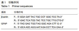
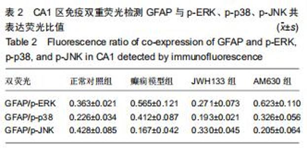
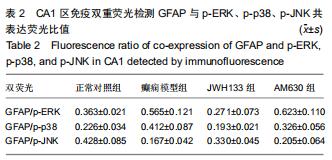
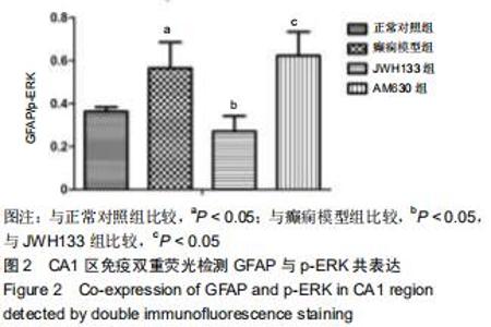
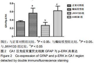
2.3.1 GFAP+p-ERK荧光显示结果 见图1。癫痫模型组匹鲁卡品致幼年大鼠癫痫持续状态后,其GFAP+p-ERK共表达较正常对照组海马区CA1、CA3和DG区有明显的增加,以CA1区较明显(P < 0.05),而JWH133组较癫痫模型组有所减少(P < 0.05),JWH133组与正常对照组相差不多;AM630组在经过激动剂JWH133和拮抗剂AM630处理后,GFAP+p-ERK共表达较JWH133组有明显增多(P < 0.05),并且较癫痫模型组也有所增加,双重荧光比值分析见表2和图2。 "

2.3.1 GFAP+p-ERK荧光显示结果 见图1。癫痫模型组匹鲁卡品致幼年大鼠癫痫持续状态后,其GFAP+p-ERK共表达较正常对照组海马区CA1、CA3和DG区有明显的增加,以CA1区较明显(P < 0.05),而JWH133组较癫痫模型组有所减少(P < 0.05),JWH133组与正常对照组相差不多;AM630组在经过激动剂JWH133和拮抗剂AM630处理后,GFAP+p-ERK共表达较JWH133组有明显增多(P < 0.05),并且较癫痫模型组也有所增加,双重荧光比值分析见表2和图2。 "
2.3.1 GFAP+p-ERK荧光显示结果 见图1。癫痫模型组匹鲁卡品致幼年大鼠癫痫持续状态后,其GFAP+p-ERK共表达较正常对照组海马区CA1、CA3和DG区有明显的增加,以CA1区较明显(P < 0.05),而JWH133组较癫痫模型组有所减少(P < 0.05),JWH133组与正常对照组相差不多;AM630组在经过激动剂JWH133和拮抗剂AM630处理后,GFAP+p-ERK共表达较JWH133组有明显增多(P < 0.05),并且较癫痫模型组也有所增加,双重荧光比值分析见表2和图2。 "
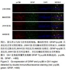
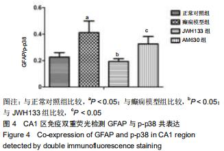
2.3.2 GFAP+p-p38荧光显示结果 见图3。癫痫模型组匹鲁卡品致幼年大鼠癫痫持续状态后,其GFAP+p-p38共表达较正常对照组海马区CA1、CA3和DG区有明显增加,以CA1区较明显,而JWH133组较癫痫模型组有所减少,JWH133组与正常对照组相差不多;AM630组在经过激动剂JWH133和拮抗剂AM630处理后,GFAP+p-p38共表达较JWH133组有明显增多,并且较癫痫模型组也有所增加,双重荧光比值分析见表2和图4。 "
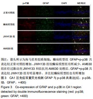
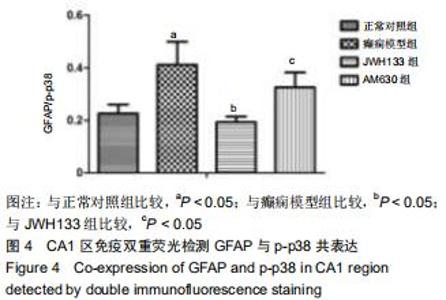
2.3.2 GFAP+p-p38荧光显示结果 见图3。癫痫模型组匹鲁卡品致幼年大鼠癫痫持续状态后,其GFAP+p-p38共表达较正常对照组海马区CA1、CA3和DG区有明显增加,以CA1区较明显,而JWH133组较癫痫模型组有所减少,JWH133组与正常对照组相差不多;AM630组在经过激动剂JWH133和拮抗剂AM630处理后,GFAP+p-p38共表达较JWH133组有明显增多,并且较癫痫模型组也有所增加,双重荧光比值分析见表2和图4。 "
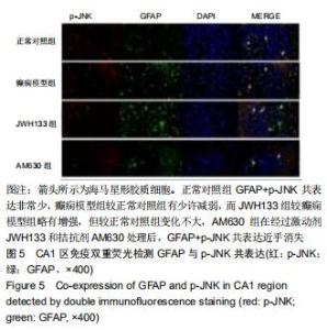
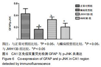
2.3.3 GFAP+p-JNK荧光显示结果 见图5。正常对照组GFAP+p-JNK存在共表达,癫痫模型组匹鲁卡品致幼年大鼠癫痫持续状态后,其GFAP+p-JNK共表达较正常对照组海马区CA1、CA3和DG区有减弱,而JWH133组较癫痫模型组有增强,AM630组在经过激动剂JWH133和拮抗剂AM630处理后,GFAP+p-JNK共表达减弱,与单纯癫痫模型组无明显差异,可见通过调节CB2R会影响JNK在匹鲁卡品致幼年大鼠癫痫持续状态后的海马区表达,见表2和图6。 "
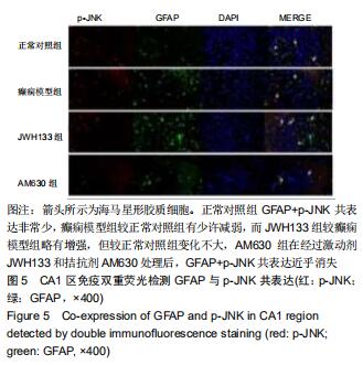
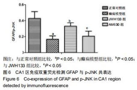
2.3.3 GFAP+p-JNK荧光显示结果 见图5。正常对照组GFAP+p-JNK存在共表达,癫痫模型组匹鲁卡品致幼年大鼠癫痫持续状态后,其GFAP+p-JNK共表达较正常对照组海马区CA1、CA3和DG区有减弱,而JWH133组较癫痫模型组有增强,AM630组在经过激动剂JWH133和拮抗剂AM630处理后,GFAP+p-JNK共表达减弱,与单纯癫痫模型组无明显差异,可见通过调节CB2R会影响JNK在匹鲁卡品致幼年大鼠癫痫持续状态后的海马区表达,见表2和图6。 "
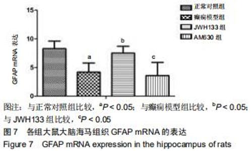
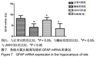
2.4 癫痫幼年大鼠海马GFAP mRNA的动态表达 癫痫模型组GFAP的mRNA表达较正常对照组减少,JWH133组较癫痫模型组有明显增多,而AM630组GFAP的表达降低,见图7。 "
|
[1] DANZER SC. Contributions of Adult-Generated Granule Cells to Hippocampal Pathology in Temporal Lobe Epilepsy: A Neuronal Bestiary. Brain Plast. 2018;3(2):169-181.
[2] MOSHÉ SL, PERUCCA E, RYVLIN P, et al. Epilepsy: new advances. Lancet. 2015;385(9971):884-898.
[3] CHO CH. New mechanism for glutamate hypothesis in epilepsy. Front Cell Neurosci. 2013;7:127.
[4] LIANG J, CHAO D, SANDHU HK, et al. δ-Opioid receptors up-regulate excitatory amino acid transporters in mouse astrocytes. Br J Pharmacol. 2014;171(23):5417-5430.
[5] BIZIÈRE K, CHAMBON JP. Animal models of epilepsy and experimental seizures. Rev Neurol(Paris). 1987;143(5): 329-340.
[6] JANSZKY J, TÉNYI D, BÓNÉ B. Valproate in the treatment of epilepsy and status epilepticus. Ideggyogy Sz. 2017;70(7-8): 258-264.
[7] MASLIAH-PLANCHON J, GARINET S, PASMANT E. RAS-MAPK pathway epigenetic activation in cancer: miRNAs in action. Oncotarget. 2016;7(25):38892-38907.
[8] LAKE D, CORRÊA SA, MÜLLER J. Negative feedback regulation of the ERK1/2 MAPK pathway. Cell Mol Life Sci. 2016;73(23):4397-4413.
[9] RAUCH N, RUKHLENKO OS, KOLCH W, et al. MAPK kinase signalling dynamics regulate cell fate decisions and drug resistance. Curr Opin Struct Biol. 2016;41:151-158.
[10] FISHER RS. Animal models of the epilepsies. Brain Res Brain Res Rev. 1989;14(3):245-278.
[11] BRODIE MJ, ELDER AT, KWAN P. Epilepsy in later life. Lancet Neurol. 2009;8(11):1019-1030.
[12] LAZAROWSKI A, CZORNYJ L. Potential role of multidrug resistant proteins in refractory epilepsy and antiepileptic drugs interactions. Drug Metabol Drug Interact. 2011;26(1):21-26.
[13] MINJAREZ B, CAMARENA HO, HARAMATI J, et al. Behavioral changes in models of chemoconvulsant-induced epilepsy: A review. Neurosci Biobehav Rev. 2017;83:373-380.
[14] LEE M, CHOI BY, SUH SW. Unexpected Effects of Acetylcholine Precursors on Pilocarpine Seizure- Induced Neuronal Death. Curr Neuropharmacol. 2018;16(1):51-58.
[15] KANDRATAVICIUS L, BALISTA PA, LOPES-AGUIAR C, et al. Animal models of epilepsy: use and limitations. Neuropsychiatr Dis Treat. 2014;10:1693-1705.
[16] SAUVANT C, NOWAK M, WIRTH C, et al. Acidosis induces multi-drug resistance in rat prostate cancer cells (AT1) in vitro and in vivo by increasing the activity of the p-glycoprotein via activation of p38. Int J Cancer. 2008;123(11):2532-2542.
[17] COPPOLA G, AURICCHIO G, FEDERICO R, et al. Lamotrigine versus valproic acid as first-line monotherapy in newly diagnosed typical absence seizures: an open-label, randomized, parallel-group study. Epilepsia. 2004;45(9): 1049-1053.
[18] REGESTA G, TANGANELLI P. Clinical aspects and biological bases of drug-resistant epilepsies. Epilepsy Res.1999;34(2-3): 109-122.
[19] BORST P, EVERS R, KOOL M, et al. A family of drug transporters: the multidrug resistance-associated proteins. J Natl Cancer Inst. 2000;92(16):1295-1302.
[20] CÁRCEL-TRULLOLS J, KOVÁCS AD, PEARCE DA. Cell biology of the NCL proteins: What they do and don't do. Biochim Biophys Acta. 2015;1852(10 Pt B):2242-2255.
[21] EID T, LEE TW, PATRYLO P, et al. Astrocytes and Glutamine Synthetase in Epileptogenesis. J Neurosci Res. 2019;97(11): 1345-1362.
[22] KOZELA E, JUKNAT A, VOGEL Z. Modulation of Astrocyte Activity by Cannabidiol, a Nonpsychoactive Cannabinoid. Int J Mol Sci. 2017;18(8): E1669.
[23] WIXEY JA, CHAND KK, COLDITZ PB, et al. Review: Neuroinflammation in intrauterine growth restriction. Placenta. 2017;54:117-124.
[24] NAVARRO G, BORROTO-ESCUELA DO, FUXE K, et al. Purinergic signaling in Parkinson's disease. Relevance for treatment. Neuropharmacology. 2016;104:161-168.
[25] MILOSO M, SCUTERI A, FOUDAH D, et al. MAPKs as mediators of cell fate determination: an approach to neurodegenerative diseases. Curr Med Chem. 2008;15(6): 538-548.
[26] ZIEMBA BP, BURKE JE, MASSON G, et al. Regulation of PI3K by PKC and MARCKS: Single-Molecule Analysis of a Reconstituted Signaling Pathway. Biophys J. 2016;110(8): 1811-1825.
[27] KANNER AM, MEADOR KJ. Remember…there is more to epilepsy than seizures! Neurology. 2015;85(13):1094-1095.
[28] MAC TL, TRAN DS, QUET F, et al. Epidemiology, aetiology, and clinical management of epilepsy in Asia: a systematic review. Lancet Neurol. 2007;6(6):533-543.
[29] HUANG Y, LIU X, LIAO Y, et al. MiR-181a influences the cognitive function of epileptic rats induced by pentylenetetrazol. Int J Clin Exp Pathol. 2015;8(10):12861-12868.
[30] ZHANG C, HOU D, FENG X. Mir-181b Functions as Anti-Apoptotic Gene in Post-Status Epilepticus via Modulation of Nrarp and Notch Signaling Pathway. Ann Clin Lab Sci. 2015;45(5):550-555.
[31] YU PM, DING D, ZHU GX, et al. International Bureau for Epilepsy survey of children, teenagers, and young people with epilepsy: data in China. Epilepsy Behav. 2009;16(1):99-104.
[32] SRIVASTAVA A, DIXIT AB, BANERJEE J, et al. Role of inflammation and its miRNA based regulation in epilepsy: Implications for therapy. Clin Chim Acta. 2016;452:1-9.
[33] ZHENG H, TANG R, YAO Y, et al. MiR-219 Protects Against Seizure in the Kainic Acid Model of Epilepsy. Mol Neurobiol. 2016;53(1):1-7.
[34] CUI L, TAO H, WANG Y, et al. A functional polymorphism of the microRNA-146a gene is associated with susceptibility to drug-resistant epilepsy and seizures frequency. Seizure. 2015; 27:60-65.
[35] LORIGADOS PEDRE L, MORALES CHACÓN LM, OROZCO SUÁREZ S, et al. Inflammatory mediators in epilepsy. Curr Pharm Des. 2013;19(38):6766-6772.
[36] CHEN Y, LIU T, LANGFORD P, et al. Haemophilus parasuis induces activation of NF-κB and MAP kinase signaling pathways mediated by toll-like receptors. Mol Immunol. 2015; 65(2):360-366.
[37] OHNO Y. Astrocytic Kir4.1 potassium channels as a novel therapeutic target for epilepsy and mood disorders. Neural Regen Res. 2018;13(4):651-652.
[38] XU JJ, DIAZ P, BIE B, et al. Spinal gene expression profiling and pathways analysis of a CB2 agonist (MDA7)-targeted prevention of paclitaxel-induced neuropathy. Neuroscience. 2014;260:185-194.
[39] BALENGA NA, MARTÍNEZ-PINILLA E, KARGL J, et al. Heteromerization of GPR55 and cannabinoid CB2 receptors modulates signalling. Br J Pharmacol.2014;171(23): 5387-5406.
[40] SU SH, WU YF, LIN Q, et al. Cannabinoid receptor agonist WIN55,212-2 and fatty acid amide hydrolase inhibitor URB597 suppress chronic cerebral hypoperfusion-induced neuronal apoptosis by inhibiting c-Jun N-terminal kinase signaling. Neuroscience. 2015;301:563-575. [41] WANG ZY, WANG P, BJORLING DE. Activation of cannabinoid receptor 2 inhibits experimental cystitis. Am J Physiol Regul Integr Comp Physiol. 2013;304(10):R846-853. |
| [1] | Zhang Chao, Lü Xin. Heterotopic ossification after acetabular fracture fixation: risk factors, prevention and treatment progress [J]. Chinese Journal of Tissue Engineering Research, 2021, 25(9): 1434-1439. |
| [2] | Wu Xun, Meng Juanhong, Zhang Jianyun, Wang Liang. Concentrated growth factors in the repair of a full-thickness condylar cartilage defect in a rabbit [J]. Chinese Journal of Tissue Engineering Research, 2021, 25(8): 1166-1171. |
| [3] | Li Jing, Xie Jianshan, Cui Huilin, Cao Ximei, Yang Yanping, Li Hairong. Expression and localization of diacylglycerol kinase zeta and protein kinase C beta II in mouse back skin with different coat colors [J]. Chinese Journal of Tissue Engineering Research, 2021, 25(8): 1196-1200. |
| [4] | Tan Jingyu, Liu Haiwen. Genome-wide identification, classification and phylogenetic analysis of Fasciclin gene family for osteoblast specific factor 2 [J]. Chinese Journal of Tissue Engineering Research, 2021, 25(8): 1243-1248. |
| [5] | Wang Zhengdong, Huang Na, Chen Jingxian, Zheng Zuobing, Hu Xinyu, Li Mei, Su Xiao, Su Xuesen, Yan Nan. Inhibitory effects of sodium butyrate on microglial activation and expression of inflammatory factors induced by fluorosis [J]. Chinese Journal of Tissue Engineering Research, 2021, 25(7): 1075-1080. |
| [6] | Kong Desheng, He Jingjing, Feng Baofeng, Guo Ruiyun, Asiamah Ernest Amponsah, Lü Fei, Zhang Shuhan, Zhang Xiaolin, Ma Jun, Cui Huixian. Efficacy of mesenchymal stem cells in the spinal cord injury of large animal models: a meta-analysis [J]. Chinese Journal of Tissue Engineering Research, 2021, 25(7): 1142-1148. |
| [7] | Shi Yangyang, Qin Yingfei, Wu Fuling, He Xiao, Zhang Xuejing. Pretreatment of placental mesenchymal stem cells to prevent bronchiolitis in mice [J]. Chinese Journal of Tissue Engineering Research, 2021, 25(7): 991-995. |
| [8] | Fan Quanbao, Luo Huina, Wang Bingyun, Chen Shengfeng, Cui Lianxu, Jiang Wenkang, Zhao Mingming, Wang Jingjing, Luo Dongzhang, Chen Zhisheng, Bai Yinshan, Liu Canying, Zhang Hui. Biological characteristics of canine adipose-derived mesenchymal stem cells cultured in hypoxia [J]. Chinese Journal of Tissue Engineering Research, 2021, 25(7): 1002-1007. |
| [9] | Geng Yao, Yin Zhiliang, Li Xingping, Xiao Dongqin, Hou Weiguang. Role of hsa-miRNA-223-3p in regulating osteogenic differentiation of human bone marrow mesenchymal stem cells [J]. Chinese Journal of Tissue Engineering Research, 2021, 25(7): 1008-1013. |
| [10] | Lun Zhigang, Jin Jing, Wang Tianyan, Li Aimin. Effect of peroxiredoxin 6 on proliferation and differentiation of bone marrow mesenchymal stem cells into neural lineage in vitro [J]. Chinese Journal of Tissue Engineering Research, 2021, 25(7): 1014-1018. |
| [11] | Duan Liyun, Cao Xiaocang. Human placenta mesenchymal stem cells-derived extracellular vesicles regulate collagen deposition in intestinal mucosa of mice with colitis [J]. Chinese Journal of Tissue Engineering Research, 2021, 25(7): 1026-1031. |
| [12] | Guan Qian, Luan Zuo, Ye Dou, Yang Yinxiang, Wang Zhaoyan, Wang Qian, Yao Ruiqin. Morphological changes in human oligodendrocyte progenitor cells during passage [J]. Chinese Journal of Tissue Engineering Research, 2021, 25(7): 1045-1049. |
| [13] | Song Chengjie, Chang Hengrui, Shi Mingxin, Meng Xianzhong. Research progress in biomechanical stability of lateral lumbar interbody fusion [J]. Chinese Journal of Tissue Engineering Research, 2021, 25(6): 923-928. |
| [14] | Xie Yang, Zhang Shujiang, Liu Menglan, Luo Ying, Yang Yang, Li Zuoxiao. Mechanism by which rapamycin protects spinal cord neurons in experimental autoimmune encephalomyelitis mice [J]. Chinese Journal of Tissue Engineering Research, 2021, 25(5): 695-700. |
| [15] | Ma Binxiang, He Wanqing, Zhou Guangchao, Guan Yonglin. Triptolide improves motor dysfunction in rats following spinal cord injury [J]. Chinese Journal of Tissue Engineering Research, 2021, 25(5): 701-706. |
| Viewed | ||||||
|
Full text |
|
|||||
|
Abstract |
|
|||||