Chinese Journal of Tissue Engineering Research ›› 2020, Vol. 24 ›› Issue (31): 4954-4960.doi: 10.3969/j.issn.2095-4344.2147
Previous Articles Next Articles
Relationship between mitochondrial autophagy and chondrogenesis of bone marrow mesenchymal stem cells
Li Yuanqi1, 2, Lin Hai2, Luo Hongrong2, Zhang Xingdong1, 2
1Guangxi Collaborative Innovation Center for Biomedicine, Guangxi Medical University, Nanning 530021, Guangxi Zhuang Autonomous Region, China; 2Engineering Research Center in Biomaterials, Sichuan University, Chengdu 610064, Sichuan Province, China
-
Received:2020-04-16Revised:2020-04-20Accepted:2020-05-09Online:2020-11-08Published:2020-09-03 -
Contact:Zhang Xingdong, MD, Professor, Guangxi Collaborative Innovation Center for Biomedicine, Guangxi Medical University, Nanning 530021, Guangxi Zhuang Autonomous Region, China; Engineering Research Center in Biomaterials, Sichuan University, Chengdu 610064, Sichuan Province, China Lin Hai, MD, Associate researcher, Engineering Research Center in Biomaterials, Sichuan University, Chengdu 610064, Sichuan Province, China -
About author:Li Yuanqi, Master candidate, Guangxi Collaborative Innovation Center for Biomedicine, Guangxi Medical University, Nanning 530021, Guangxi Zhuang Autonomous Region, China; Engineering Research Center in Biomaterials, Sichuan University, Chengdu 610064, Sichuan Province, China -
Supported by:the National Key Research and Development Program of China during the 13th Five-Year Plan Period, No. 2018YFC1106203 ; the Key Science and Technology Program of Guangxi Zhuang Autonomous Region, No. Guike AA17204085-2
CLC Number:
Cite this article
Li Yuanqi, Lin Hai, Luo Hongrong, Zhang Xingdong. Relationship between mitochondrial autophagy and chondrogenesis of bone marrow mesenchymal stem cells[J]. Chinese Journal of Tissue Engineering Research, 2020, 24(31): 4954-4960.
share this article
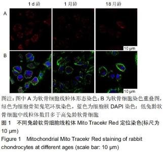
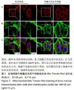
2.1 不同兔龄软骨细胞的线粒体染色结果 不同兔龄软骨细胞的线粒体染色结果见图1。出生1 d的兔软骨细胞中线粒体呈点状分布,胞膜胞核完整,核膜清晰,细胞骨架的微丝结构比较明显。出生1个月的兔软骨细胞骨架的微丝贯穿整个细胞,胞核胞膜也完整,但线粒体数目较刚出生的少。出生18个月的兔软骨细胞胞膜断断续续,界限不清,细胞骨架的微丝失去正常结构,细胞核变形严重,胞内线粒体数目在3组中最少。 "
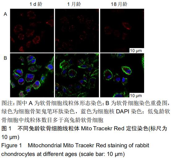
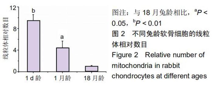
由图2实验结果得出,幼年关节软骨细胞的线粒体数目最多,随着兔龄的增长,软骨细胞线粒体数目减少,软骨细胞中线粒体数量与细胞发育衰老有关。 "
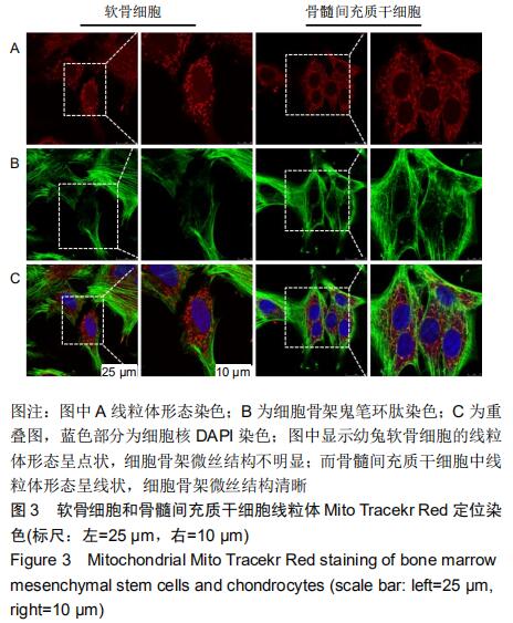
2.2 幼兔骨髓间充质干细胞和幼兔软骨细胞的线粒体染色结果 幼兔骨髓间充质干细胞和幼兔软骨细胞的线粒体染色结果见图3。从图中可以看出,软骨细胞线粒体网络结构被分裂,大部分呈碎片状,成堆聚集,形成自噬小体,细胞形态呈圆形或椭圆形,骨架边界模糊。骨髓间充质干细胞形态呈梭形,细胞骨架边界清晰,线粒体网络结构显著。软骨细胞中线粒体的形态与骨髓间充质干细胞的线粒体形态存在明显差异。 "
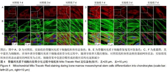
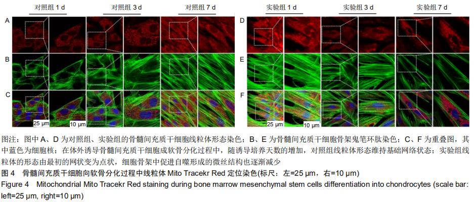
2.3 骨髓间充质干细胞分化过程中的线粒体染色结果 在观察正常骨髓间充质干细胞和软骨细胞线粒体形态的基础上,探究骨髓间充质干细胞成软骨诱导分化过程中线粒体形态变化。骨髓间充质干细胞分化过程中的线粒体染色结果见图4。第1天时,与对照组线粒体形态相比,实验组线粒体网络结构开始破坏,线粒体开始分裂,但大部分线粒体仍通过管状连接;第3天时,对照组线粒体仍保持基础状态的网络管状结构,而实验组线粒体网络结构大面积破坏;第7天时,对照组线粒体仍处于基础状态,而实验组线粒体状态发生显著变化。线粒体网状结构明显破坏,碎片化程度加深,点状线粒体增多,受损的线粒体已被包裹形成自噬小体,并进一步降解其所包裹的内容物,完成受损线粒体的清除。骨架的微丝结构也较诱导前期减少。细胞肌动蛋白丝是自噬发生发展的动力基础[13],促进自噬体与溶酶体融合,完成自噬过程。因此,对照组、实验组的线粒体标记染色结果说明线粒体自噬存在于骨髓间充质干细胞成软骨诱导分化过程中。 "
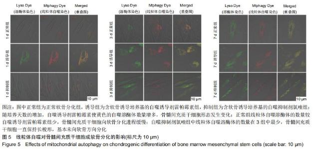
2.4 线粒体自噬对骨髓间充质干细胞成软骨分化的影响 前期线粒体染色结果表明线粒体自噬发生在骨髓间充质干细胞成软骨分化过程中,但线粒体自噬的变化是否直接影响到骨髓间充质干细胞的成软骨分化尚不清楚。故该实验通过加入自噬抑制剂氯喹和自噬诱导剂雷帕霉素,考察抑制或增强线粒体自噬对骨髓间充质干细胞成软骨分化的影响,结果见图5,从图中可以看出,随诱导天数的增加,自噬诱导组的自噬溶酶体数量显著增加,骨髓间充质干细胞形态发生明显变化,由最初的长梭形变为圆形或椭圆形;正常组在培养过程中仅有部分自噬溶酶体出现,骨髓间充质干细胞形态变化不明显;应用氯喹抑制自噬体与溶酶体的融合后,随培养天数的增加,自噬溶酶体较雷帕霉素组少,骨髓间充质干细胞形态一直保持长梭形,未向软骨方向分化。该结果初步说明雷帕霉素促进了线粒体自噬,进而影响骨髓间充质干细胞向软骨分化的过程。 "
|
[1] BLANCO FJ, REGO-PÉREZ I. Mitochondria and mitophagy: biosensors for cartilage degradation and osteoarthritis. Osteoarthritis Cartilage. 2018;26(8):989-991.
[2] JUNG YH, LEE HJ, KIM JS, et al. EphB2 signaling-mediated Sirt3 expression reduces MSC senescence by maintaining mitochondrial ROS homeostasis. Free Radic Biol Med. 2017;110: 368-380.
[3] WOLFF P, HEIMANN L, LIEBSCH G, et al. Oxygen-distribution within 3-D collagen I hydrogels for bone tissue engineering. Mater Sci Eng C Mater Biol Appl. 2019;95:422-427.
[4] XIANG G, YANG L, LONG Q, et al. BNIP3L-dependent mitophagy accounts for mitochondrial clearance during 3 factors-induced somatic cell reprogramming. Autophagy. 2017;13(9):1543-1555.
[5] SINHA KM, TSENG C, GUO P, et al. Hypoxia-inducible factor 1α (HIF-1α) is a major determinant in the enhanced function of muscle-derived progenitors from MRL/MpJ mice. FASEB J. 2019; 33(7):8321-8334.
[6] PEI DD, SUN JL, ZHU CH, et al. Contribution of Mitophagy to Cell-Mediated Mineralization: Revisiting a 50-Year-Old Conundrum. Adv Sci (Weinh). 2018;5(10):1800873.
[7] LAMPERT MA, OROGO AM, NAJOR RH, et al. BNIP3L/NIX and FUNDC1-mediated mitophagy is required for mitochondrial network remodeling during cardiac progenitor cell differentiation. Autophagy. 2019;15(7):1182-1198.
[8] FU W, LIU Y, YIN H. Mitochondrial Dynamics: Biogenesis, Fission, Fusion, and Mitophagy in the Regulation of Stem Cell Behaviors. Stem Cells Int. 2019;2019:9757201.
[9] ZENG R, FANG Y, ZHANG Y, et al. p62 is linked to mitophagy in oleic acid-induced adipogenesis in human adipose-derived stromal cells. Lipids Health Dis. 2018;17(1):133.
[10] LUO P, GAO F, NIU D, et al. The Role of Autophagy in Chondrocyte Metabolism and Osteoarthritis: A Comprehensive Research Review. Biomed Res Int. 2019;2019:5171602.
[11] MARYCZ K, KORNICKA K, GRZESIAK J, et al. Macroautophagy and Selective Mitophagy Ameliorate Chondrogenic Differentiation Potential in Adipose Stem Cells of Equine Metabolic Syndrome: New Findings in the Field of Progenitor Cells Differentiation. Oxid Med Cell Longev. 2016;2016:3718468.
[12] 杨俊丽,韩霞,孙明启,等.兔骨髓间充质干细胞的生物学特征及原代培养[J].中国组织工程研究,2015,19(50):8043-8047.
[13] ZHENG J, CROTEAU DL, BOHR VA, et al. Diminished OPA1 expression and impaired mitochondrial morphology and homeostasis in Aprataxin-deficient cells. Nucleic Acids Res. 2019;47(8):4086-4110.
[14] LI Q, GAO S, KANG Z, et al. Rapamycin Enhances Mitophagy and Attenuates Apoptosis After Spinal Ischemia-Reperfusion Injury. Front Neurosci. 2018;12:865.
[15] RODENAS-ROCHINA J, KELLY DJ, GÓMEZ RIBELLES JL, et al. Influence of oxygen levels on chondrogenesis of porcine mesenchymal stem cells cultured in polycaprolactone scaffolds. J Biomed Mater Res A. 2017;105(6):1684-1691.
[16] 王玉全,李程程,孟爱民,等.线粒体在细胞衰老中的作用[J].医学综述, 2019,25(8):1457-1462.
[17] LIN H, SOHN J, SHEN H, et al. Bone marrow mesenchymal stem cells: Aging and tissue engineering applications to enhance bone healing. Biomaterials. 2019;203:96-110.
[18] 刘恒,刘超,曹永平.软骨细胞线粒体功能异常在关节软骨早期退变中的作用[J].中国矫形外科杂志,2011,19(8):652-654.
[19] WAI T, LANGER T. Mitochondrial Dynamics and Metabolic Regulation. Trends Endocrinol Metab. 2016;27(2):105-117.
[20] MURATA D, ARAI K, IIJIMA M, et al. Mitochondrial division, fusion and degradation. J Biochem. 2020;167(3):233-241.
[21] PODESTÀ MA, REMUZZI G, CASIRAGHI F. Mesenchymal Stromal Cells for Transplant Tolerance. Front Immunol. 2019;10: 1287.
[22] LI H, LI X, JING X, et al. Hypoxia promotes maintenance of the chondrogenic phenotype in rat growth plate chondrocytes through the HIF-1α/YAP signaling pathway. Int J Mol Med. 2018;42(6): 3181-3192.
[23] VINCENT G, NOVAK EA, SIOW VS, et al. Nix-Mediated Mitophagy Modulates Mitochondrial Damage During Intestinal Inflammation. Antioxid Redox Signal. 2020 Mar 31. doi: 10.1089/ars.2018.7702. [Epub ahead of print]
[24] ESTEBAN-MARTÍNEZ L, BOYA P. BNIP3L/NIX-dependent mitophagy regulates cell differentiation via metabolic reprogramming. Autophagy. 2018;14(5):915-917.
[25] HO TT, WARR MR, ADELMAN ER, et al. Autophagy maintains the metabolism and function of young and old stem cells. Nature. 2017;543(7644):205-210.
[26] HARPER JW, ORDUREAU A, HEO JM. Building and decoding ubiquitin chains for mitophagy. Nat Rev Mol Cell Biol. 2018;19(2): 93-108.
[27] RAMBOLD AS, KOSTELECKY B, ELIA N, et al. Tubular network formation protects mitochondria from autophagosomal degradation during nutrient starvation. Proc Natl Acad Sci U S A. 2011;108(25):10190-10195.
[28] GOMES LC, DI BENEDETTO G, SCORRANO L. During autophagy mitochondria elongate, are spared from degradation and sustain cell viability. Nat Cell Biol. 2011;13(5):589-598.
[29] MAUTHE M, ORHON I, ROCCHI C, et al. Chloroquine inhibits autophagic flux by decreasing autophagosome-lysosome fusion. Autophagy. 2018;14(8):1435-1455.
[30] IWAI-KANAI E, YUAN H, HUANG C, et al. A method to measure cardiac autophagic flux in vivo. Autophagy. 2008;4(3):322-329.
[31] XU LX, TANG XJ, YANG YY, et al. Neuroprotective effects of autophagy inhibition on hippocampal glutamate receptor subunits after hypoxiaischemia-induced brain damage in newborn rats. Neural Regen Res. 2017;12(3):417-424.
[32] SINGH AK, SINGH S, TRIPATHI VK, et al. Rapamycin Confers Neuroprotection Against Aging-Induced Oxidative Stress, Mitochondrial Dysfunction, and Neurodegeneration in Old Rats Through Activation of Autophagy. Rejuvenation Res. 2019;22(1): 60-70.
[33] 陈哲,白海,潘耀柱,等.辐射诱导人骨髓间充质干细胞自噬反应的研究[J].中华血液学杂志,2011,32(9):602-605.
[34] YUAN T, LUO H, GUO L, et al. In vivo immunological properties research on mesenchymal stem cells based engineering cartilage by a dialyzer pocket model. J Mater Sci Mater Med. 2017;28(10):150.
[35] NI Y, TANG Z, YANG J, et al. Collagen structure regulates MSCs behavior by MMPs involved cell-matrix interactions. J Mater Chem B. 2018;6(2):312-326. [36] DHANABALAN K, HUISAMEN B, LOCHNER A. Mitochondrial oxidative phosphorylation and mitophagy in myocardial ischaemia/reperfusion: effects of chloroquine. Cardiovasc J Afr. 2019;30:1-11. |
| [1] | Fan Haixia, Tan Qingkun, Wang Hong, Cheng Huanzhi, Liu Xue, Ching-chang Ko, Geng Haixia. Rabbit skull defects repaired by the hydroxyapatite/geltin scaffold combined with bone marrow mesenchymal stem cells and umbilical vein endothelial cells [J]. Chinese Journal of Tissue Engineering Research, 2021, 25(10): 1495-1499. |
| [2] | Wei Yashu, Liu Hongjing, Wang Huifeng, Wang Junduo, Zhao Wenjing, Chen Weiping. Cardiomyocyte-like differentiation of bone marrow mesenchymal stem cells induced by myocardial tissue lysates from different parts of the myocardium [J]. Chinese Journal of Tissue Engineering Research, 2021, 25(1): 32-37. |
| [3] | Gui Xianwei, Jiang Huijiao, Wu Jie, Liang Xueqi, Xu Xiaodan, Wang Erqiang, Zou Hailiang, Chen Hejie, Chen Xueling, Wu Xiangwei. Mesenchymal stem cell calcification induced by protoscolex of two species of Echinococcus: a differential analysis [J]. Chinese Journal of Tissue Engineering Research, 2021, 25(1): 38-43. |
| [4] | Ye Dou, , Ma Xuexia , Guan Qian, , Luan Zuo , Yang Yinxiang , Wang Zhaoyan , Wang Qian , He Ying , Yao Ruiqin. Proportion and morphological characteristics of human oligodendrocyte precursor cells in different cell culture vessels [J]. Chinese Journal of Tissue Engineering Research, 2021, 25(1): 44-49. |
| [5] | Gu Jingjing, Zhou Rui, Yang Tingting, Yang Xiaoping, Xu Fei, Zheng Bo. Supporting effect of human skeletal muscle-derived myoendothelial cells on hematopoietic stem/progenitor cells in vitro [J]. Chinese Journal of Tissue Engineering Research, 2021, 25(1): 50-55. |
| [6] | Tao Guilu, Chu Tongbin, Zhang Lei. Proliferation and migration of endothelial progenitor cells promoted by bone marrow mesenchymal stem cells conditioned medium with rosiglitazone [J]. Chinese Journal of Tissue Engineering Research, 2021, 25(1): 56-60. |
| [7] | Zhu Bingbing, He Haibin, Deng Jianghua, Wang Wenqiang, Mu Xiaoling. Human umbilical cord mesenchymal stem cells overexpressing interleukin 8 receptor inhibit inflammation and promote vascular repair [J]. Chinese Journal of Tissue Engineering Research, 2021, 25(1): 61-66. |
| [8] | Jing Yucheng, Wang Le, Wang Xianyun, Wei Mei, Li Min, Ji Lishuang, Ma Fangfang, Liu Gang , Zheng Mingqi. Umbilical cord mesenchymal stem cell transplantation in the treatment of ischemic heart disease: a 3-year follow-up [J]. Chinese Journal of Tissue Engineering Research, 2021, 25(1): 6-12. |
| [9] | Zhou Pengfei, Lin Jing, Chen Yuying, Lin Minkui. Canine dental pulp stem cells-polyglycolic acid scaffold complex for canine periodontal tissue defect [J]. Chinese Journal of Tissue Engineering Research, 2020, 24(34): 5526-5531. |
| [10] | Liu Xin, Du Bin, Sun Guangquan, Cao Jinxing, Jiang Xiaohong. Porous beta-tricalcium phosphate-polypyrrole-biotin-icariin composite scaffold promotes recruitment of bone marrow mesenchymal stem cells [J]. Chinese Journal of Tissue Engineering Research, 2020, 24(34): 5532-5537. |
| [11] | Yu Xingge, Lin Kaili. Application of nanocomposite hydrogels in bone tissue engineering [J]. Chinese Journal of Tissue Engineering Research, 2020, 24(34): 5441-5446. |
| [12] | Zhang Huxiong, Li Wei, Yang Wupeng, New Suyaratu. 3D-printed icariin/decalcified bone matrix material promotes the repair of femoral condyle defects in rabbits [J]. Chinese Journal of Tissue Engineering Research, 2020, 24(34): 5461-5466. |
| [13] | Zhang Shuang, Xu Xiaomei, Zeng Yang, Yuan Xiaoping, Lin Fuwei. Rev-erbα’s effect on osteoblastogenesis of mouse bone marrow mesenchymal stem cells [J]. Chinese Journal of Tissue Engineering Research, 2020, 24(31): 4921-4926. |
| [14] | Xun Chong, Wang Qiang, Li Changzhou, Liu Xiaofeng. Potential molecular targets and therapeutic mechanisms underlying transplantation of autologous bone marrow stem cells for the treatment of spinal cord injury based on bioinformatics [J]. Chinese Journal of Tissue Engineering Research, 2020, 24(31): 4927-4933. |
| [15] | Chen Jia, Yang Yiqiang, Hu Chen, Chen Qi, Zhao Tian, Yong Min, Ma Dongyang, Ren Liling. Fabrication of prevascularized osteogenic differentiated cell sheet based on human bone marrow mesenchymal stem cells and human umbilical vein endothelial cells [J]. Chinese Journal of Tissue Engineering Research, 2020, 24(31): 4934-4940. |
| Viewed | ||||||
|
Full text |
|
|||||
|
Abstract |
|
|||||