Chinese Journal of Tissue Engineering Research ›› 2021, Vol. 25 ›› Issue (1): 61-66.doi: 10.3969/j.issn.2095-4344.2124

Previous Articles     Next Articles

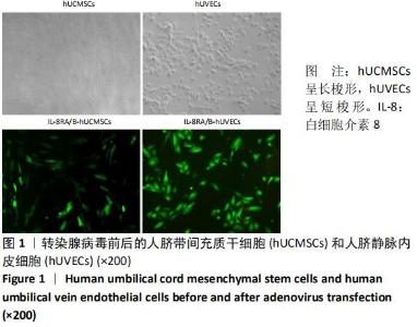
Human umbilical cord mesenchymal stem cells overexpressing interleukin 8 receptor inhibit inflammation and promote vascular repair

Zhu Bingbing, He Haibin, Deng Jianghua, Wang Wenqiang, Mu Xiaoling   

  1. School of Medicine, Shihezi University, Shihezi 832000, Xinjiang Uygur Autonomous Region, China
  • Received:2019-11-01 Revised:2019-11-12 Accepted:2020-01-07 Online:2021-01-08 Published:2020-10-29
  • Contact: Mu Xiaoling, MD, Professor, School of Medicine, Shihezi University, Shihezi 832000, Xinjiang Uygur Autonomous Region, China
  • About author:Zhu Bingbing, Master candidate, School of Medicine, Shihezi University, Shihezi 832000, Xinjiang Uygur Autonomous Region, China
  • Supported by:
    新疆兵团社会发展科技攻关与成果转化计划(2016AD013),项目负责人:慕晓玲;自治区研究生教育创新计划科研创新项目(XJGRI201604)

Abstract: Abstract
BACKGROUND: Vascular injury is a common complication after balloon dilatation. The development of umbilical cord mesenchymal stem cells (UC-MSCs) provides a new method for treating vascular injury.
OBJECTIVE: To investigate the mechanism underlying the repair of damaged blood vessels by human UC-MSCs (hUC-MSCs) transfected with interleukin-8RA/B (IL-8RA/B) adenovirus. 
METHODS: hUC-MSCs and human umbilical vein endothelial cells (hUVECs) were collected and transfected with adenovirus vectors containing human IL-8RA and/or IL-8RB cDNAs and green fluorescent protein. A rat model of carotid artery injury was established. Sprague-Dawley rats were randomly divided into four groups: IL-8RA/B-hUCMSCs group, Il-8ra/B-hUVECs group, Null-hUCMSCs group, and control group, followed by injection of 0.5×10⁶ corresponding cells 
(500 μL) and same volume of normal saline via the tail vein respectively at 1, 3, and 5 hours post-surgery. After 30 minutes of injection, the carotid artery was taken and the expression of green fluorescent protein was observed. After 24 hours, the serum levels of inflammatory and anti-inflammatory factors were measured by ELISA; and the infiltration of neutrophil cells and mononuclear macrophages was observed by immunohistochemistry. After 14 days, Evans blue staining was used to observe vascular endothelialization and fibrosis. After 28 days, the neointimal hyperplasia was observed by hematoxylin-eosin staining. 
RESULTS AND CONCLUSION: (1) After 30 minutes of IL-8RA/B-hUC-MSCs infusion, the expression of green fluorescent protein was observed in the injured vascular intima, and the fluorescence expression was higher than that of the other three groups. (2) After 24 hours of IL-8RA/B-hUC-MSCs infusion, the expression of inflammatory factors in the serum was significantly lower than that of the other three groups, while the expression of anti-inflammatory factor interleukin-10 was higher than that of the other three groups (P < 0.05). In addition, inflammatory cell infiltration in the IL-8RA/B-hUC-MSCs group decreased significantly. (3) hUC-MSCs overexpressing interleukin-8 receptor promoted re-endothelialization of injured vessels and reduced vascular fibrosis after 14 days of infusion. (4) IL-8RA/B-hUC-MSCs reduced vascular neointimal hyperplasia after 28 days of infusion. (5) Interleukin-8 receptor enhances the targeted homing ability of hUC-MSCs, allowing MSCs to migrate to the site of vascular injury, inhibit inflammation, reduce neointimal hyperplasia, and promote vascular repair.

Key words: human umbilical cord mesenchymal stem cells,  interleukin-8RA/B,  vascular injury,  inflammatory factor,  vascular endothelium

CLC Number: