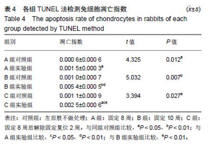
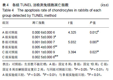
| [1]尹志强,潘惟丽,金昊,等.骨性关节炎病因的研究进展[J].现代生物医学进展,2016,16(7):1369-1371,89.[2]韦宜山,刘万林,丁良甲,等.发育性髋脱位髋臼软骨细胞病理学改变的实验研究[J].内蒙古医学院学报,2012,34(6):447-452.[3]王炳海,韦宜山,丁良甲,等.发育性髋关节脱位早期髋臼软骨细胞凋亡与Bcl-2、Bax表达的相关性研究[J].实用医学杂志,2013,29(20): 3301-3303.[4]边红飞,韦宜山.发育性髋关节脱位的病理与细胞凋亡的研究进展[J].内蒙古医学院学报, 2012, (S1): 211-215.[5]Eskes R,Desagher S,Antonsson B,et al.Bid induces the oligomerization and insertion of Bax into the outer mitochondrial membrane.Mol Cell Biol.2000;20(3):929-935.[6]Vervloessem T,Kerkhofs M,La Rovere RM,et al.Bcl-2 inhibitors as anti-cancer therapeutics: The impact of and on calcium signaling.Cell Calcium.2017 Jun 4. pii: S0143-4160(17)30103-3.[7]Birkinshaw RW,Czabotar PE.The BCL-2 family of proteins and mitochondrial outer membrane permeabilisation.Semin Cell Dev Biol. 2017 Apr 8. pii: S1084-9521(17)30189-1. [8]Peña-Blanco A,García-Sáez AJ.Bax,Bak and beyond: mitochondrial performance in apoptosis.FEBS J. 2017 Jul 29.[9]Mosadegh M,Hasanzadeh S,Razi M.Nicotine-induced damages in testicular tissue of rats; evidences for bcl-2, p53 and caspase-3 expression.Iran J Basic Med Sci.2017;20(2):199-208.[10]Xia Z,Dickens M,Raingeaud J,et al.Opposing effects of ERK and JNK-p38 MAP kinases on apoptosis.Science (New York, NY). 1995;270(5240):1326-1331.[11]Kluck RM,Bossy-Wetzel E,Green DR,et al.The release of cytochrome c from mitochondria: a primary site for Bcl-2 regulation of apoptosis.Science (New York, NY). 1997;275(5303): 1132-1136.[12]Oltvai ZN, Milliman CL,Korsmeyer SJ.Bcl-2 heterodimerizes in vivo with a conserved homolog, Bax, that accelerates programmed cell death.Cell.1993;74(4): 609-619.[13]Elmore S.Apoptosis: A review of programmed cell death.Toxicol Pathol.2007;35(4):495-516.[14]Dang AC, Kim HT. Chondrocyte apoptosis after simulated intraarticular fracture: a comparison of histologic detection methods.Clin Orthop Relat Res. 2009;467(7):1877-1884.[15]Pelletier JP,Jovanovic DV,Lascau-Coman V,et al.Selective inhibition of inducible nitric oxide synthase reduces progression of experimental osteoarthritis in vivo: possible link with the reduction in chondrocyte apoptosis and caspase 3 level.Arthritis Rheum. 2000;43(6):1290-1299.[16]Gustafsson AB,Gottlieb RA.Bcl-2 family members and apoptosis, taken to heart.Am J Physiol-Cell Ph.2007;292(1): C45-C51.[17]Brunelle JK, Letai A. Control of mitochondrial apoptosis by the Bcl-2 family. J Cell Sci.2009;122(4): 437-441.[18]Zhang H,Li Q,Li Z,et al.The protection of Bcl-2 overexpression on rat cortical neuronal injury caused by analogous ischemia/ reperfusion in vitro.Neurosci Res.2008;62(2):140-146.[19]朱玉山,卢铁元,王蕊,等.Bcl-2家族蛋白调控线粒体膜通透性和细胞色素C释放的新机制[J].生命科学,2011,23(11):1076-1080.[20]Pu X,Storr SJ,Zhang Y,et al.Caspase-3 and caspase-8 expression in breast cancer: caspase-3 is associated with survival.Apoptosis. 2017;22(3):357-368. [21]Cory S,Roberts AW,Colman PM,et al.Targeting BCL-2-like Proteins to Kill Cancer Cells.Trends Cancer.2016;2(8):443-460.[22]Eichhorn L,Nietzel C,Schröder S,et al.A single air dive induces apoptotic gene regulation but no increase in nucleosomes. Undersea Hyperb Med.2016;43(7):813-819.[23]Garner TP,Lopez A,Reyna DE,et al.Progress in targeting the BCL-2 family of proteins.Curr Opin Chem Biol.2017;39:133-142.[24]Edlich F. BCL-2 proteins and apoptosis: Recent insights and unknowns. Biochem Biophys Res Commun. 2017 Jul 1. pii: S0006-291X(17)31321-31329. |