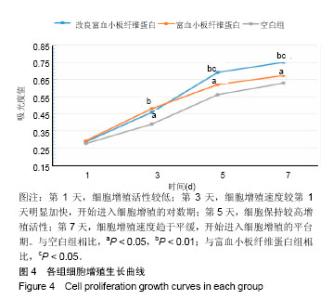
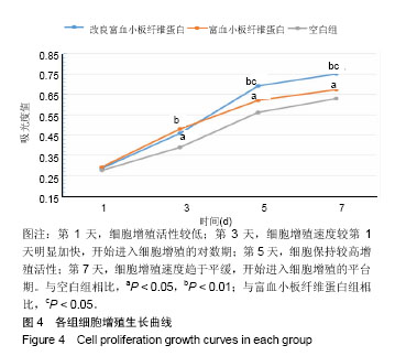
| [1] Puišys A, ?ukauskas S, Kubilius R, et al. Bone augmentation and simultaneous soft tissue thickening with collagen tissue matrix derivate membrane in an aesthetic area A case report. Stomatologija. 2017; 19(2): 64-68.[2] Miron RJ, Zucchelli G, Pikos MA, et al. Use of platelet-rich fibrin in regenerative dentistry: a systematic review. Clin Oral Investig. 2017; 21(6): 1913-1927.[3] Vahabi S, Vaziri S, Torshabi M. Effects of plasma rich in growth factors and platelet-rich fibrin on proliferation and viability of human gingival fibroblasts. J Dent (Tehran). 2015; 12(7): 504-512.[4] Polimeni G, Xiropaidis AV, Wikesjö UME. Biology and principles of periodontal wound healing/regeneration. Periodontology. 2006; 41(1): 30.[5] Garg AK. The use of platelet-rich plasma to enhance the success of bone grafts around dental implants. Dental Implantology Update. 2000;11(3): 17.[6] Landesberg R, Moses M. Risks of using platelet rich plasma gel. J Oral Maxillofac Surg. 1998; 56(9): 1116-1117.[7] Choukroun J, Adda F, Schoefer C, et al. An opportunity in perio-implantology: The PRF. Implantodontie. 2000; 42: 55-62.[8] Rodella LF, Favero G, Boninsegna R, et al. Growth factors, CD34 positive cells, and fibrin network analysis in concentrated growth factors fraction. Microsc Res Tech. 2011; 74(8): 772-777.[9] Gupta S, Banthia R, Singh P, et al. Clinical evaluation and comparison of the efficacy of coronally advanced flap alone and in combination with platelet rich fibrin membrane in the treatment of Miller Class I and II gingival recessions. Contemp Clin Dent. 2015; 6(2): 153-160.[10] Bishara M, Kurtzman GM, Khan W, et al. Soft-tissue grafting techniques associated with immediate implant placement. Compend Contin Educ Dent. 2018; 39(2): e1-4.[11] Ocak H, Kutuk N, Demetoglu U, et al. Comparison of bovine bone-autogenic bone mixture versus platelet-rich fibrin for maxillary sinus grafting: histologic and histomorphologic study. J Oral Implantol. 2017; 43(3): 194-201.[12] Ghanaati S, Booms P, Orlowska A, et al. Advanced platelet-rich fibrin: a new concept for cell-based tissue engineering by means of inflammatory cells. J Oral Implantol. 2014; 40(6): 679-689.[13] Taoufik K, Mavrogonatou E, Eliades T, et al. Effect of blue light on the proliferation of human gingival fibroblasts. Dental Materials. 2008; 24(7): 895-900.[14] Xu QC, Wang ZG, Ji QX, et al. Systemically transplanted human gingiva-derived mesenchymal stem cells contributing to bone tissue regeneration. Int J Clin Exp Pathol. 2014; 7(8): 4922-4929.[15] Soundara Rajan T, Scionti D, Diomede F, et al. Prolonged expansion induces spontaneous neural progenitor differentiation from human gingiva-derived mesenchymal stem cells. Cell Reprogram. 2017; 19(6): 389-401.[16] 杨琴秋.富血小板纤维蛋白诱导口腔软组织缺损修复再生的实验研究[D].成都:四川医科大学,2015.[17] Eren G. Platelet-rich fibrin in the treatment of localized gingival recessions: a split-mouth randomized clinical trial. Clin Oral Investig. 2014; 18(8): 1941-1948.[18] Dohan Ehrenfest DM, Rasmusson L. Classification of platelet concentrates: from pure platelet-rich plasma (P-PRP) to leucocyte- and platelet-rich fibrin (L-PRF). Trends Biotechnol. 2009; 27(3): 158-167.[19] Dohan Ehrenfest D M. How to optimize the preparation of leukocyte- and platelet-rich fibrin (L-PRF, Choukroun's technique) clots and membranes: introducing the PRF Box. Oral Surg Oral Med Oral Pathol Oral Radiol Endod. 2010; 110(3): 278-280.[20] 练轶,毛俊丽,孙嵩,等.改良型富血小板纤维蛋白与富血小板纤维蛋白的成分及超微结构观察[J].西南国防医药. 2017; 27(6): 548-551.[21] Dohan Ehrenfest DM, Diss A, Odin G, et al. In vitro effects of Choukroun's platelet-rich fibrin on human gingival fibroblasts, dermal prekeratinocytes, preadipocytes, and maxillofacial osteoblasts in primary cultures. Oral Surg Oral Med Oral Pathol Oral Radiol Endod. 2009; 108(3): 341-352.[22] Fujioka-Kobayashi M, Miron RJ, Hernandez M, et al. Optimized platelet rich fibrin with the low speed concept: growth factor release, biocompatibility and cellular response. J Periodontol. 2016;88(1): 112-121.[23] Dohan Ehrenfest DM, Pinto NR, Pereda A, et al. The impact of the centrifuge characteristics and centrifugation protocols on the cells, growth factors, and fibrin architecture of a leukocyte- and platelet-rich fibrin (L- PRF) clot and membrane. Platelets. 2018; 29(2): 171-184.[24] Kobayashi E, Flückiger L, Fujioka-Kobayashi M, et al. Comparative release of growth factors from PRP, PRF, and advanced-PRF. Clin Oral Investig. 2016; 20(9): 2353-2360.[25] Liu Y, Kalén A, Risto O, et al. Fibroblast proliferation due to exposure to a platelet concentrate in vitro is pH dependent. Wound Repair Regen. 2002; 10(5): 336-340.[26] Graziani F, Ivanovski S, Cei S, et al. The in vitro effect of different PRP concentrations on osteoblasts and fibroblasts. Clin Oral Implants Res. 2006; 17(2): 212-219. [27] Goto H, Matsuyama T, Miyamoto M, et al. Platelet-rich plasma/osteoblasts complex induces bone formation via osteoblastic differentiation following subcutaneous transplantation. J Periodontal Res. 2006;41(5): 455-62.[28] Slapnicka J, Fassmann A, Strasak L, et al. Effects of activated and nonactivated platelet-rich plasma on proliferation of human osteoblasts in vitro. J Oral Maxillofac Surg. 2008; 66(2): 297-301.[29] 何婷婷.两种富血小板纤维蛋白的降解特性研究[D].泸州:西南医科大学, 2017. |