Chinese Journal of Tissue Engineering Research ›› 2019, Vol. 23 ›› Issue (10): 1520-1525.doi: 10.3969/j.issn.2095-4344.1574
Previous Articles Next Articles
Feasibility of repairing articular cartilage defects with particulated juvenile cartilage allograft
You Qi1, Duan Xiaojun2, Zhang Jun1, Jin Ying1, Peng Xu1, Ge Zhen1, Liu Yi1
- 1Department of Joint Surgery, the Affiliated Hospital of Zunyi Medical University, Zunyi 563000, Guizhou Province, China; 2Department of Joint Surgery, Southwest Hospital, Army Medical University, Chongqing 400038, China
-
Received:2018-10-15Online:2019-04-08Published:2019-04-08 -
Contact:Liu Yi, Professor, Department of Joint Surgery, the Affiliated Hospital of Zunyi Medical University, Zunyi 563000, Guizhou Province, China. -
About author:You Qi, Master candidate, Department of Joint Surgery, the Affiliated Hospital of Zunyi Medical University, Zunyi 563000, Guizhou Province, China -
Supported by:the National Natural Science Foundation of China, No. 81071484 (to DXJ); the Combined Foundation of Science & Technology Department of Guizhou Province, No. LH[2017]7015
CLC Number:
Cite this article
You Qi, Duan Xiaojun, Zhang Jun, Jin Ying, Peng Xu, Ge Zhen, Liu Yi. Feasibility of repairing articular cartilage defects with particulated juvenile cartilage allograft[J]. Chinese Journal of Tissue Engineering Research, 2019, 23(10): 1520-1525.
share this article
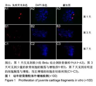
2.1 幼年软骨微粒体外增殖情况检测 倒置荧光显微镜观察显示,幼年软骨微粒体外培养第1天仅见到极少的Brdu结合到软骨微粒中,第3天可见到少量的软骨细胞的脱落与增殖,第7天见到有明显的细胞脱落与增殖,而且增殖的细胞在切缘周围,见图1。"
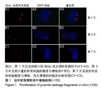
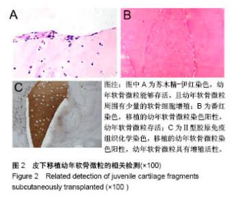
2.2 皮下移植幼年软骨微粒的相关检测 石蜡切片苏木精-伊红染色结果示,幼年软骨微粒能够存活,且幼年软骨微粒周围有少量的软骨细胞增殖,见图2A。番红染色结果示,移植的幼年软骨微粒染色阳性,幼年软骨微粒仍存活,见图2B。Ⅱ型胶原免疫组织化学结果示,移植的幼年软骨微粒染色阳性,幼年软骨微粒具有增殖活性,见图2C。"
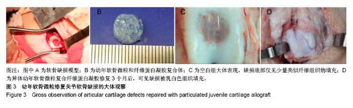
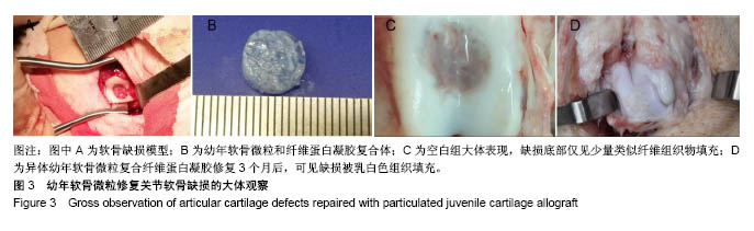
2.3 膝关节软骨缺损修复效果 2.3.1 大体观察 所有动物伤口愈合良好,膝关节活动良好,关节液清亮,关节腔未见粘连。空白组缺损区域可见明显凹陷,无明显填充物,或仅有少量纤维组织样物填充;实验组缺损区域被乳白色组织填充,表面光滑平整,缺损边缘整合良好,见图3。"
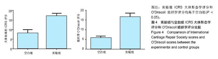
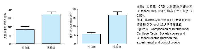
2.3.2 大体评分及组织学评分 空白组、实验组大体形态评分分别为6.2±1.2,17.4±0.9,两组评分比较差异有显著性意义(P < 0.05)。空白组、实验组组织学评分分别为4.8±0.8,15.8±0.8,两组评分比较差异有显著性意义(P < 0.05),见图4。"
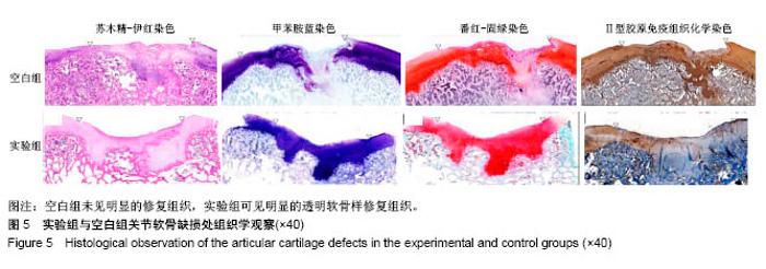
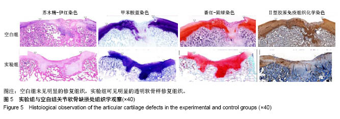
2.3.3 组织学观察 苏木精-伊红染色结果显示,空白组无明显填充物,仅有少量纤维组织样物填充;实验组缺损被软骨样组织填充,与周围正常软骨整合良好。甲苯胺蓝染色结果显示,空白组缺损未见明显着色的修复组织;实验组缺损可见蓝染的修复组织填充,着色强度与周围正常软骨组织类似,界面整合良好。番红-固绿染色结果显示,空白组缺损中修复组织着色不明显;实验组缺损中可见红染的修复组织填充,且着色强度与周围正常软骨类似,潮线结构完整,软骨与骨界面整合良好。Ⅱ型胶原免疫组织化学染色结果显示,空白组缺损中未见明显着色的Ⅱ型胶原;实验组缺损中可见着色阳性的Ⅱ型胶原,且着色强度与周围正常软骨组织类似。实验组与对照组关节软骨缺损组织学观察结果,见图5。"
| [1] Schindler OS.Current concepts of articular cartilage repair. Acta orthop Belg. 2011;77(6):709-726.[2] Min BH,Choi WH,Lee YS,et al.Effect of different bone marrow stimulation techniques (BSTs) on MSCs mobilization.J Orthop Res. 2013;31(11):1814-1819.[3] Bedi A,Feeley BT,Williams RJ.Management of Articular Cartilage Defects of the Knee. J Bone Joint Surg Am. 2010; 92(4):994-1009.[4] Perterson L,Minas T,Brittberg M,et al.Two-to-9-year outcome after autologous chondrocyte transplantation of the knee.Clin Orthop Relat Res.2000;374:212-214.[5] Haddo O,Mahroof S,Higgs D,et al.The use of chondrogide membrane in autologous chondrocyte implantation.The knee. 2004;(11)1:51-55.[6] Filardo G,Kon E,Di Martino A,et al.Second-generation arthroscopic autologous chondrocyte implantation for the treatment of degenerative cartilage lesions.Knee Surg Sports Traumatol Arthrosc. 2012;20(9):1704-1713.[7] Kon E,Delcogliano M,Filardo G,et al.A novel nano-composite multi-layered biomaterial for treatment of osteochondral lesion:technique note and an early stability pilot clinical trial. Injury. 2010;41(7):693-701.[8] Gille J,Behrens P,Schulz AP,et al.Matrix-Associated Autologous Chondrocyte Implantation: A Clinical Follow-Up at 15 Years. Cartilage.2016;7(4):309-315.[9] Karoubi G, Ormiston ML,Stewart DJ,et al. Single-cell hydrogel encapsulation for enhanced survival of human marrow stromal cells. Biomaterials.2009;30(29):5445-5455.[10] Shimizu H,Ohashi K,Utoh R,et al.Bioengineering of a functional sheet of islet cells for the treatment of diabetes mellitus. Biomaterials. 2009;30(30):5943-5949.[11] Hayashi S,Kamei N,Ikuta Y,et al.Chondrocyte cell-sheet transplantation for treating monoiodoacetate-induced arthritis in rats. Tissue Eng Part C Methods.2017;23(6):346-356.[12] Xu X,Shi D,Liu Y,et al.Synovium-derived mesenchymal stem cell sheet enhance autologous osteochondral transplantation in a rabbit momdel. Int J Clin Exp Med. 2016;9(6):10322-10332.[13] Shaari CM,Farber D,Brandwein MS,et al.Chracterizing the antigenic profile of the human trachea:Implications for tracheal transplantation.Head Neck.1998;20(6):522-527 [14] Liu K,Zhou GD,Liu W,et al.The dependence of in vivo stable ectopic chondrogenesis by human mesenchymal stem cells on chondrogenic differentiation in vitro. Biomaterials. 2008;29(14): 2183-2192.[15] Ge Y, Gong YY,Xu Z,et al.The application of sheet technology in cartilage tissue engineering.Tissue Eng Part B Rev. 2016; 22(2):114-124.[16] Mouthuy PA,Elsherbini Y,Cui Z,et al.Layering PLGA-based electrospum membranes and cell sheets for engineering cartilage-bone transition.Tissue Eng Regen Med. 2016;10(4): 263-274.[17] Verdonk P,Dhollander A,Almqvist KF,et al.Treatment of osteochondral lesions in the knee using a cell-free scaffold. Bone Joint J. 2015;97-B(3):318-323.[18] Brix M,Kaipel M,Kellner R,et al. Successful osteoconduction but limited cartilage tissue quality following osteochondral repair by a cell-free multilayered nano-composite scaffold at the knee. Int Orthop. 2016;40(3):625-632.[19] Filardo G,Kon E,Di MA,et al.Treatment of knee osteochondritis dissecans with a cell-free biomimetic osteochondral scaffold: clinical and imaging evaluation at 2-year follow up.am J Sports Med. 2013;41(8):1786-1793.[20] Albrecht F,Roessner A,Zimmermann E.Closure of osteochondral lesions using chondral fragments and fibrin adhesive. Arch Orthop Trauma Surg.1983;101(3):213-217.[21] Riboh JC,Cole BJ,Farr J.Particulated articular cartilage for symptomatic chondral defects of the knee.Curr Rev Musculoskelet Med.2015;11(1):21-34.[22] Bonasia DE,Marmotti A,Rosso F,et al.Use of chondral fragments for one stage caitilage repair:a systematic review. World J Orthop. 2015;6(11):1006.[23] 宁志刚,杨柳,王富友,等.保留钙化层结构的猪股骨滑车全厚软骨缺损模型建立[J].中国修复重建外科杂志,2012,26(5):527-531.[24] Brittberg M,Aglietti P,Gambardella R,et al.ICRS Cartilage Injury Evaluation Package. 2002:16-18. https://www.secot.es/ uploads/descargas/formacion/escalas_valoracion/ICRS._TRAUMA_CARTaILAGO.pdf [25] Mainil-Varlet P,Aigner T,Brittberg M,et al.Histological assessment of cartilage repair: a report by the Histology Endpoint Committee of the International Cartilage Repair Society (ICRS). J Bone Joint Surg Am. 2003;85-A 2(1):45-57.[26] Kontturi LS,Jarvinen E,Muhonen V,et al.An injectable,insitu forming type IIcollagen/hyaluronic acid hydrogel vehicle for chondrocyte delivery in cartilage tissue engineering.Drug Deliv Transl Res. 2014;4(2):149-158.[27] Cole BJ,Farr J,Winalski CS,et al.Outcomes after a single-stage procedure for cell-based cartilage repair: a prospective clinical safety trial with 2-year follow-up.Am J Sports Med. 2011;39(6): 1170-1179.[28] Kon E,Filardo G,Zaffagnini S,et al.Biodegradable polyurethane meniscal scaffold for isolated partial lesions or as combined procedure for knees with multiple comorbidities: clinical results at 2 years. Knee Surg Sports Traumatol Arthrosc. 2014;22(1): 128-134.[29] Farr J,Yao JQ.Chondral Defect Repair with Particulated Juvenile Cartilage Allograft. Cartilage. 2011;2(4):346-353.[30] Farr J,Tabet SK,Margerrison E,et al.Clinical,Radiographic,and histological outcomes after cartilage repair with particulated juvenile articular cartilage:a 2-year prospective study.Am J Sports Med. 2014:42(6):1417.[31] Mcmillan S,Light GT, Brtz C.All-arthroscopic implantation of minced juvenile chondral allograft for an isolated, full-thickness chondral lesion in the trochlea of an adult knee. Arthrosc Tech. 2016;5(2):397-401.[32] Arshi A,Wang D,Jones KJ.Combined particulated juvenile cartilage allograft transplantation and medial patellofemoral ligament reconstruction for symptomatic chondral defects in the setting of recurrent patellar instability.Arthrosc Tech. 2016;5(5): 1149-1154.[33] Buckwalter JA,Bowman GN,Albright JP,et al.Clinical outcomes of patellar chondral lesions treated with juvenile particulated cartilage allografts.lowa Orthop J. 2013;34(1)44-49.[34] Kruse DL,Ng A,Paden M,et al.Athroscopic De Novo NT(R) juvenile allograft cartilage implantation in the talus: a case presentation.J Foot Ankle Surg.2012;51(2):218.[35] Dunn JC,kusnezov N,Orr J,et al.Ostochondral defects of the upper extremity treated with particulated juvenile cartilage transfer. HAND. 2015;10(4):683-687.[36] Pascual-Garrido C,Hao J,Schrock J,et al.Arthroscopic juvenile allograft cartilage implantation for cartilage lesions of the hip. Arthrosc Tech. 2016;5(4)929-933.[37] Adkisson HD,Martin JA,Amendola RL,et al.The potential of human allogeneic juvenile chondrocytes for restoration of articular cartilage. Am J Sports Med.2010;38(7):1324-1333.[38] Smeriglio P,Lai JH,Dhulipala L,et al.Comparative potential of juvenile and adult human articular chondrocytes for cartilage tissue formation in three-dimensional biomimetic hydrogels. Tissue Eng Part A. 2015;21(1-2)147-155.[39] Skaalure SC,Milligan IL,Bryant SJ.Age impacts extracellular matrix metabolism in chondrocytes encapsulated in degradable hydrogels. Biomed Mater.2012;7(2):024111.[40] Bonasia DE,Martin JA,Marmotti A,et al.Cocultures of adult and juvenile chondrocytes compared with adult and juvenile chondral fragments: in vitro matrix production.Am J Sports Med. 2011;39(11):2355-2361.[41] Marmotti A,Bonasia DE,Bruzzone M,et al.Human cartilage fragments in a composite scaffold for single-stage cartilage repair:an in vitro study of the chondrocyte migration and the influence of TGF-beta1 and G-CSF.Knee Surg Sports Traumatol Arthrosc.2013;21(8):1819-1833.[42] Adkisson HD,Milliman C,Zhang X,et al.Immune evasion by neocartilage-derived chondrocytes: Implications for biologic repair of joint articular cartilage.Stem Cell Res. 2010;4(1): 57-68.[43] Tompkins M,Adkisson HD, Bonner KF. DeNovo NT Allograft. Oper Tech Sports Med. 2013;21(2):82-89. |
| [1] | Zhang Tongtong, Wang Zhonghua, Wen Jie, Song Yuxin, Liu Lin. Application of three-dimensional printing model in surgical resection and reconstruction of cervical tumor [J]. Chinese Journal of Tissue Engineering Research, 2021, 25(9): 1335-1339. |
| [2] | Zeng Yanhua, Hao Yanlei. In vitro culture and purification of Schwann cells: a systematic review [J]. Chinese Journal of Tissue Engineering Research, 2021, 25(7): 1135-1141. |
| [3] | Xu Dongzi, Zhang Ting, Ouyang Zhaolian. The global competitive situation of cardiac tissue engineering based on patent analysis [J]. Chinese Journal of Tissue Engineering Research, 2021, 25(5): 807-812. |
| [4] | Wu Zijian, Hu Zhaoduan, Xie Youqiong, Wang Feng, Li Jia, Li Bocun, Cai Guowei, Peng Rui. Three-dimensional printing technology and bone tissue engineering research: literature metrology and visual analysis of research hotspots [J]. Chinese Journal of Tissue Engineering Research, 2021, 25(4): 564-569. |
| [5] | Cheng Jun, Tan Jun, Zhao Yun, Cheng Fangdong, Shi Guojia. Effect of thrombin concentration on the prevention of postoperative cerebrospinal leakage by fibrin glue [J]. Chinese Journal of Tissue Engineering Research, 2021, 25(4): 570-575. |
| [6] | Chang Wenliao, Zhao Jie, Sun Xiaoliang, Wang Kun, Wu Guofeng, Zhou Jian, Li Shuxiang, Sun Han. Material selection, theoretical design and biomimetic function of artificial periosteum [J]. Chinese Journal of Tissue Engineering Research, 2021, 25(4): 600-606. |
| [7] | Liu Fei, Cui Yutao, Liu He. Advantages and problems of local antibiotic delivery system in the treatment of osteomyelitis [J]. Chinese Journal of Tissue Engineering Research, 2021, 25(4): 614-620. |
| [8] | Li Xiaozhuang, Duan Hao, Wang Weizhou, Tang Zhihong, Wang Yanghao, He Fei. Application of bone tissue engineering materials in the treatment of bone defect diseases in vivo [J]. Chinese Journal of Tissue Engineering Research, 2021, 25(4): 626-631. |
| [9] | Zhang Zhenkun, Li Zhe, Li Ya, Wang Yingying, Wang Yaping, Zhou Xinkui, Ma Shanshan, Guan Fangxia. Application of alginate based hydrogels/dressings in wound healing: sustained, dynamic and sequential release [J]. Chinese Journal of Tissue Engineering Research, 2021, 25(4): 638-643. |
| [10] | Chen Jiana, Qiu Yanling, Nie Minhai, Liu Xuqian. Tissue engineering scaffolds in repairing oral and maxillofacial soft tissue defects [J]. Chinese Journal of Tissue Engineering Research, 2021, 25(4): 644-650. |
| [11] | Xing Hao, Zhang Yonghong, Wang Dong. Advantages and disadvantages of repairing large-segment bone defect [J]. Chinese Journal of Tissue Engineering Research, 2021, 25(3): 426-430. |
| [12] | Chen Siqi, Xian Debin, Xu Rongsheng, Qin Zhongjie, Zhang Lei, Xia Delin. Effects of bone marrow mesenchymal stem cells and human umbilical vein endothelial cells combined with hydroxyapatite-tricalcium phosphate scaffolds on early angiogenesis in skull defect repair in rats [J]. Chinese Journal of Tissue Engineering Research, 2021, 25(22): 3458-3465. |
| [13] | Wang Hao, Chen Mingxue, Li Junkang, Luo Xujiang, Peng Liqing, Li Huo, Huang Bo, Tian Guangzhao, Liu Shuyun, Sui Xiang, Huang Jingxiang, Guo Quanyi, Lu Xiaobo. Decellularized porcine skin matrix for tissue-engineered meniscus scaffold [J]. Chinese Journal of Tissue Engineering Research, 2021, 25(22): 3473-3478. |
| [14] | Mo Jianling, He Shaoru, Feng Bowen, Jian Minqiao, Zhang Xiaohui, Liu Caisheng, Liang Yijing, Liu Yumei, Chen Liang, Zhou Haiyu, Liu Yanhui. Forming prevascularized cell sheets and the expression of angiogenesis-related factors [J]. Chinese Journal of Tissue Engineering Research, 2021, 25(22): 3479-3486. |
| [15] | Liu Chang, Li Datong, Liu Yuan, Kong Lingbo, Guo Rui, Yang Lixue, Hao Dingjun, He Baorong. Poor efficacy after vertebral augmentation surgery of acute symptomatic thoracolumbar osteoporotic compression fracture: relationship with bone cement, bone mineral density, and adjacent fractures [J]. Chinese Journal of Tissue Engineering Research, 2021, 25(22): 3510-3516. |
| Viewed | ||||||
|
Full text |
|
|||||
|
Abstract |
|
|||||