Chinese Journal of Tissue Engineering Research ›› 2018, Vol. 22 ›› Issue (29): 4637-4642.doi: 10.3969/j.issn.2095-4344.0620
Previous Articles Next Articles
Cryopreserved human umbilical cord mesenchymal stem cells: isolation and culture
Liu Ting, Jiang Hui, Liu Zhi-hua, Sun Ning, Li Xiao-mei, Weng Jin-sheng
- Department of Central Laboratory, the Fifth Affiliated Hospital of Guangzhou Medical University, Guangzhou 510700, Guangdong Province, China
-
Revised:2018-07-11Online:2018-10-18Published:2018-10-18 -
Contact:Weng Jin-sheng, MD, Researcher, Department of Central Laboratory, the Fifth Affiliated Hospital of Guangzhou Medical University, Guangzhou 510700, Guangdong Province, China -
About author:Liu Ting, MD, Assistant researcher, Department of Central Laboratory, the Fifth Affiliated Hospital of Guangzhou Medical University, Guangzhou 510700, Guangdong Province, China -
Supported by:the National Natural Science Foundation of China, No. 81570189, 8177110782; the Project of Guangdong Provincial Science and Technology Department, No. 2016ZC0145
CLC Number:
Cite this article
Liu Ting, Jiang Hui, Liu Zhi-hua, Sun Ning, Li Xiao-mei, Weng Jin-sheng. Cryopreserved human umbilical cord mesenchymal stem cells: isolation and culture[J]. Chinese Journal of Tissue Engineering Research, 2018, 22(29): 4637-4642.
share this article
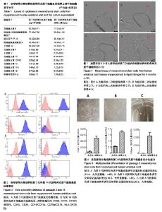
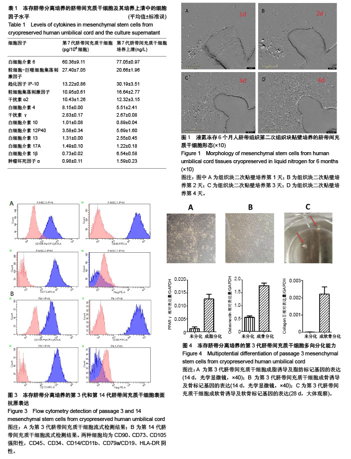
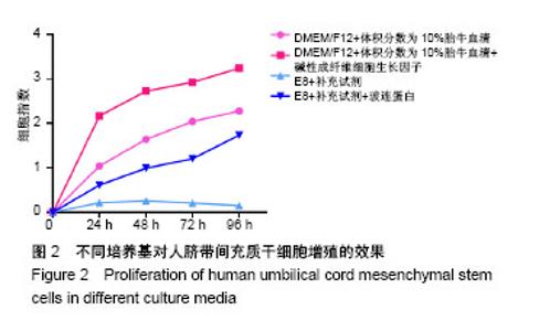
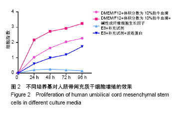
2.1 从冻存人脐带中原代培养间充质干细胞的生物学特性 组织块接种7 d后,在光镜下可见较少的细胞从组织块中移行出来,形态多为梭形,少数为三角形或多角形,向四周呈单层贴壁生长;14 d时组织块转移至新的培养皿,加培养基二次贴壁培养,1 d后显微镜下可以观察到细胞从组织块边缘爬出,随后快速生长增殖,4 d后便可长满培养皿,大部分细胞贴壁,形态为典型成纤维细胞样,以长梭形为主,大量细胞呈漩涡状生长(图1)。三四天传代1次,可长期稳定传15代以上。传代后大部分细胞五六个小时贴壁,12-24 h开始生长,3 d左右达到80%-90%融合。初步判断这些贴壁生长的成纤维样细胞为间充质干细胞。 2.2 不同培养基传代细胞的增殖能力 在最常用血清培养基(DMEM/F12+体积分数为10%胎牛血清)的基础上添加碱性成纤维细胞生长因子,脐带间充质干细胞增殖速率显著提高,基础培养基E8添加补充试剂(E8+S)不能启动脐带间充质干细胞的增殖,而在添加玻连蛋白后(E8+S+V),脐带间充质干细胞是可以实现无血清培养基继代培养,但增殖速率还远低于有血清培养基(图2),有待进一步优化。 2.3 细胞表面抗原 根据国际细胞疗法协会(International society for cellular therapy,ISCT)制定的鉴定标准,多能间充质干细胞应该是贴壁细胞,表面抗原CD90、CD73、CD105(多能干细胞标记)应呈强阳性表达,CD45(白细胞标记)、CD34(原始造血祖细胞和内皮细胞标记)、CD14/CD11b(单核细胞和巨噬细胞标记)、CD79a/CD19(B淋巴细胞标记)、HLA-DR(干细胞非激活不表达标记)应呈阴性表达,能向成脂、成骨、成软骨分化[28]。BD Human MSC Analysis Kit包含有CD73-APC,CD90-FITC,CD105-per-CP以及CD45-PE,CD34-PE,CD11b-PE,CD19-PE,HLA-DR-PE cocktail。由图3所示,冻存脐带分离培养的第3代和第14代脐带间充质干细胞表面抗原符合ISCT的标准。 2.4 细胞分化能力 图4A显示第3代脐带间充质干细胞加入成脂诱导培养基14 d后细胞形态由长梭形逐渐变为多角形、不规则形,细胞内出现大量的脂滴。酶解收集细胞,检测脂肪细胞标记基因PPARγ的表达,结果表明其与未诱导的细胞相比,表达量显著升高。图4B显示第3代脐带间充质干细胞加入成骨诱导培养基14 d后细胞形态由长梭形逐渐变为多角形、不规则形,细胞堆积,呈簇状生长。酶解收集细胞,检测骨细胞标记基因Osteonectin的表达,结果表明其与未诱导的细胞相比,表达量显著升高。图4C显示第3代脐带间充质干细胞加入成软骨诱导培养基28 d后细胞在培养皿底形成一层白膜,之后逐渐聚集成一团白色的软骨状组织。收集软骨状组织,检测软骨细胞标记基因CollagenⅡ的表达,结果表明其与未诱导的细胞相比,表达量显著升高。 2.5 分泌细胞因子水平 在第7代脐带间充质干细胞及其无血清培养上清中可检测到较多粒细胞集落刺激因子、粒细胞-巨噬细胞集落刺激因子、干扰素α2,趋化因子IP-10,白细胞介素4和白细胞介素6,而干扰素γ、白细胞介素10、白细胞介素12P40、白细胞介素13、白细胞介素17A、白细胞介素1β、白细胞介素1β等分泌量较低,见表1。"
"
| [1] Friedenstein AJ, Piatetzky-Shapiro II, Petrakova KV.Osteogenesis in transplants of bone marrow cells.J EmbryolExpMorphol. 1966; 16(3):381-390. [2] Bianco P, Robey PG, Simmons PJ.Mesenchymal stem cells: revisiting history, concepts, and assays.Cell Stem Cell. 2008;2(4):313-319. [3] Bongso A, Fong CY.The therapeutic potential, challenges and future clinical directions of stem cells from the Wharton's jelly of the human umbilical cord.Stem Cell Rev. 2013;9(2):226-240. [4] Zhang L, Tan X, Dong C,et al. In vitro differentiation of human umbilical cord mesenchymal stem cells (hUCMSCs), derived from Wharton's jelly, into choline acetyltransferase (ChAT)-positive cells.Int J Dev Neurosci. 2012;30(6):471-477. [5] Peng J, Wang Y, Zhang L, et al. Human umbilical cord Wharton's jelly-derived mesenchymal stem cells differentiate into a Schwann-cell phenotype and promote neurite outgrowth in vitro. Brain Res Bull. 2011;84(3):235-243. [6] Banas A.Purification of adipose tissue mesenchymal stem cells and differentiation toward hepatic-like cells.Methods Mol Biol. 2012;826: 61-72. [7] Shevde N.Stem Cells: Flexible friends.Nature. 2012;483(7387): S22-26. [8] Le Blanc K, Frassoni F, Ball L, et al. Mesenchymal stem cells for treatment of steroid-resistant, severe, acute graft-versus-host disease: a phase II study.Lancet. 2008;371(9624):1579-1586. [9] Lee RH, Pulin AA, Seo MJ, et al. Intravenous hMSCs improve myocardial infarction in mice because cells embolized in lung are activated to secrete the anti-inflammatory protein TSG-6.Cell Stem Cell. 2009;5(1):54-63. [10] Johnson K, Zhu S, Tremblay MS, et al. A stem cell-based approach to cartilage repair.Science. 2012;336(6082):717-721. [11] Liu R, Zhang Z, Lu Z, et al. Human umbilical cord stem cells ameliorate experimental autoimmune encephalomyelitis by regulating immunoinflammation and remyelination.Stem Cells Dev. 2013;22(7):1053-1062. [12] Lee HJ, Lee JK, Lee H, et al. Human umbilical cord blood-derived mesenchymal stem cells improve neuropathology and cognitive impairment in an Alzheimer's disease mouse model through modulation of neuroinflammation.Neurobiol Aging. 2012;33(3): 588-602. [13] Mora-Lee S, Sirerol-Piquer MS, Gutiérrez-Pérez M, et al. Therapeutic effects of hMAPC and hMSC transplantation after stroke in mice.PLoS One. 2012;7(8):e43683. [14] 毛开云,范月蕾,王跃,等.间充质干细胞治疗产品开发现状与趋势[J].中国生物工程杂志,2017,37(10):126-135.[15] Ramanathan R, Rupert S, Selvaraj S, et al. Role of Human Wharton's Jelly Derived Mesenchymal Stem Cells (WJ-MSCs) for Rescue of d-Galactosamine Induced Acute Liver Injury in Mice.J ClinExpHepatol. 2017;7(3):205-214. [16] Thomas KE, Moon LD.Will stem cell therapies be safe and effective for treating spinal cord injuries.Br Med Bull. 2011;98:127-142. [17] Nadri S, Soleimani M.Comparative analysis of mesenchymal stromal cells from murine bone marrow and amniotic fluid. Cytotherapy. 2007;9(8):729-737. [18] In 't Anker PS, Scherjon SA, Kleijburg-van der Keur C, et al. Isolation of mesenchymal stem cells of fetal or maternal origin from human placenta.Stem Cells. 2004;22(7):1338-1345. [19] 穆晓红,徐林,赵子义,等.人胎盘与骨髓源性间充质干细胞体外培养及生物学特性对比[J].中国组织工程研究与临床康复, 2009,13(19): 3708-3712.[20] Secunda R, Vennila R, Mohanashankar AM, et al. Isolation, expansion and characterisation of mesenchymal stem cells from human bone marrow, adipose tissue, umbilical cord blood and matrix: a comparative study.Cytotechnology. 2015;67(5):793-807. [21] Bojani? I, Golubi?Cepuli? B.Umbilical cord blood as a source of stem cells. Acta Med Croatica. 2006;60(3):215-225. [22] Li G, Zhang XA, Wang H, et al. Comparative proteomic analysis of mesenchymal stem cells derived from human bone marrow, umbilical cord, and placenta: implication in the migration. Proteomics. 2009;9(1):20-30. [23] Bieback K, Kern S, Kocaömer A, et al. Comparing mesenchymal stromal cells from different human tissues: bone marrow, adipose tissue and umbilical cord blood.Biomed Mater Eng. 2008;18 (1 Suppl): S71-76. [24] Kikuchi-Taura A, Taguchi A, Kanda T, et al. Human umbilical cord provides a significant source of unexpanded mesenchymal stromal cells.Cytotherapy. 2012;14(4):441-450. [25] Fong CY, Subramanian A, Biswas A, et al. Freezing of Fresh Wharton's Jelly From Human Umbilical Cords Yields High Post-Thaw Mesenchymal Stem Cell Numbers for Cell-Based Therapies.J Cell Biochem. 2016;117(4):815-827. [26]Shivakumar SB, Bharti D, Subbarao RB, et al. DMSO and Serum-Free Cryopreservation of Wharton's Jelly Tissue Isolated From Human Umbilical Cord.J Cell Biochem. 2016;117(10):2397-2412. [27] Chen G, Gulbranson DR, Hou Z, et al. Chemically defined conditions for human iPSC derivation and culture.Nat Methods. 2011;8(5):424-429. [28] Dominici M, Le Blanc K, Mueller I, et al. Minimal criteria for defining multipotent mesenchymal stromal cells. The International Society for Cellular Therapy position statement.Cytotherapy. 2006;8(4): 315-317. [29] Balci D, Can A.The assessment of cryopreservation conditions for human umbilical cord stroma-derived mesenchymal stem cells towards a potential use for stem cell banking.Curr Stem Cell Res Ther. 2013;8(1):60-72. [30] Capelli C, Pedrini O, Valgardsdottir R, et al. Clinical grade expansion of MSCs.Immunol Lett. 2015;168(2):222-227. [31] Priya N, Sarcar S, Majumdar AS, et al. Explant culture: a simple, reproducible, efficient and economic technique for isolation of mesenchymal stromal cells from human adipose tissue and lipoaspirate.J Tissue Eng Regen Med. 2014;8(9):706-716. [32] Chen PM, Yen ML, Liu KJ, et al. Immunomodulatory properties of human adult and fetal multipotent mesenchymal stem cells.J Biomed Sci. 2011;18:49. [33] Horwitz EM, Dominici M.How do mesenchymal stromal cells exert their therapeutic benefit.Cytotherapy.2008;10(8):771-774. [34] Liu S, Yuan M, Hou K, et al. Immune characterization of mesenchymal stem cells in human umbilical cord Wharton's jelly and derived cartilage cells.Cell Immunol. 2012;278(1-2):35-44. |
| [1] | Pu Rui, Chen Ziyang, Yuan Lingyan. Characteristics and effects of exosomes from different cell sources in cardioprotection [J]. Chinese Journal of Tissue Engineering Research, 2021, 25(在线): 1-. |
| [2] | Lin Qingfan, Xie Yixin, Chen Wanqing, Ye Zhenzhong, Chen Youfang. Human placenta-derived mesenchymal stem cell conditioned medium can upregulate BeWo cell viability and zonula occludens expression under hypoxia [J]. Chinese Journal of Tissue Engineering Research, 2021, 25(在线): 4970-4975. |
| [3] | Zhang Tongtong, Wang Zhonghua, Wen Jie, Song Yuxin, Liu Lin. Application of three-dimensional printing model in surgical resection and reconstruction of cervical tumor [J]. Chinese Journal of Tissue Engineering Research, 2021, 25(9): 1335-1339. |
| [4] | Hou Jingying, Yu Menglei, Guo Tianzhu, Long Huibao, Wu Hao. Hypoxia preconditioning promotes bone marrow mesenchymal stem cells survival and vascularization through the activation of HIF-1α/MALAT1/VEGFA pathway [J]. Chinese Journal of Tissue Engineering Research, 2021, 25(7): 985-990. |
| [5] | Shi Yangyang, Qin Yingfei, Wu Fuling, He Xiao, Zhang Xuejing. Pretreatment of placental mesenchymal stem cells to prevent bronchiolitis in mice [J]. Chinese Journal of Tissue Engineering Research, 2021, 25(7): 991-995. |
| [6] | Liang Xueqi, Guo Lijiao, Chen Hejie, Wu Jie, Sun Yaqi, Xing Zhikun, Zou Hailiang, Chen Xueling, Wu Xiangwei. Alveolar echinococcosis protoscolices inhibits the differentiation of bone marrow mesenchymal stem cells into fibroblasts [J]. Chinese Journal of Tissue Engineering Research, 2021, 25(7): 996-1001. |
| [7] | Fan Quanbao, Luo Huina, Wang Bingyun, Chen Shengfeng, Cui Lianxu, Jiang Wenkang, Zhao Mingming, Wang Jingjing, Luo Dongzhang, Chen Zhisheng, Bai Yinshan, Liu Canying, Zhang Hui. Biological characteristics of canine adipose-derived mesenchymal stem cells cultured in hypoxia [J]. Chinese Journal of Tissue Engineering Research, 2021, 25(7): 1002-1007. |
| [8] | Geng Yao, Yin Zhiliang, Li Xingping, Xiao Dongqin, Hou Weiguang. Role of hsa-miRNA-223-3p in regulating osteogenic differentiation of human bone marrow mesenchymal stem cells [J]. Chinese Journal of Tissue Engineering Research, 2021, 25(7): 1008-1013. |
| [9] | Lun Zhigang, Jin Jing, Wang Tianyan, Li Aimin. Effect of peroxiredoxin 6 on proliferation and differentiation of bone marrow mesenchymal stem cells into neural lineage in vitro [J]. Chinese Journal of Tissue Engineering Research, 2021, 25(7): 1014-1018. |
| [10] | Zhu Xuefen, Huang Cheng, Ding Jian, Dai Yongping, Liu Yuanbing, Le Lixiang, Wang Liangliang, Yang Jiandong. Mechanism of bone marrow mesenchymal stem cells differentiation into functional neurons induced by glial cell line derived neurotrophic factor [J]. Chinese Journal of Tissue Engineering Research, 2021, 25(7): 1019-1025. |
| [11] | Duan Liyun, Cao Xiaocang. Human placenta mesenchymal stem cells-derived extracellular vesicles regulate collagen deposition in intestinal mucosa of mice with colitis [J]. Chinese Journal of Tissue Engineering Research, 2021, 25(7): 1026-1031. |
| [12] | Pei Lili, Sun Guicai, Wang Di. Salvianolic acid B inhibits oxidative damage of bone marrow mesenchymal stem cells and promotes differentiation into cardiomyocytes [J]. Chinese Journal of Tissue Engineering Research, 2021, 25(7): 1032-1036. |
| [13] | Wang Xianyao, Guan Yalin, Liu Zhongshan. Strategies for improving the therapeutic efficacy of mesenchymal stem cells in the treatment of nonhealing wounds [J]. Chinese Journal of Tissue Engineering Research, 2021, 25(7): 1081-1087. |
| [14] | Wang Shiqi, Zhang Jinsheng. Effects of Chinese medicine on proliferation, differentiation and aging of bone marrow mesenchymal stem cells regulating ischemia-hypoxia microenvironment [J]. Chinese Journal of Tissue Engineering Research, 2021, 25(7): 1129-1134. |
| [15] | Zeng Yanhua, Hao Yanlei. In vitro culture and purification of Schwann cells: a systematic review [J]. Chinese Journal of Tissue Engineering Research, 2021, 25(7): 1135-1141. |
| Viewed | ||||||
|
Full text |
|
|||||
|
Abstract |
|
|||||