Chinese Journal of Tissue Engineering Research ›› 2018, Vol. 22 ›› Issue (27): 4364-4368.doi: 10.3969/j.issn.2095-4344.0354
Previous Articles Next Articles
Osteonecrosis of the femoral head after femoral neck fractures induced by biomechanical factors: testified by dog models of internal fixation for unilateral femoral neck fracture
Wang Hai-yang, Lin Yan-bin, Yu Guang-shu
- Department of Orthopedics, Fuzhou Second Hospital Affiliated to Xiamen University, Fuzhou 350007, Fujian Province, China
-
Online:2018-09-28Published:2018-09-28 -
Contact:Lin Yan-bin, Chief physician, Professor, Master’s supervisor, Department of Orthopedics, Fuzhou Second Hospital Affiliated to Xiamen University, Fuzhou 350007, Fujian Province, China -
About author:Wang Hai-yang, Master candidate, Department of Orthopedics, Fuzhou Second Hospital Affiliated to Xiamen University, Fuzhou 350007, Fujian Province, China -
Supported by:the Young Talents Training Project of Health System of Fujian Province, No. 2014-ZQN-JC-34
CLC Number:
Cite this article
Wang Hai-yang, Lin Yan-bin, Yu Guang-shu. Osteonecrosis of the femoral head after femoral neck fractures induced by biomechanical factors: testified by dog models of internal fixation for unilateral femoral neck fracture [J]. Chinese Journal of Tissue Engineering Research, 2018, 22(27): 4364-4368.
share this article
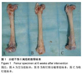
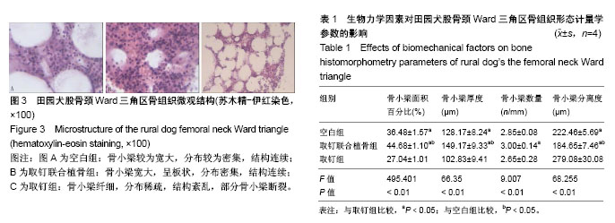
2.1 实验动物数量分析 纳入健康成年田园犬12只,随机分为空白组、取钉联合植骨组和取钉组,每组4只。全部进入结果分析,无脱失。 2.2 大体观察结果 所有田园犬股骨颈骨折造模成功及实验干预后的切口愈合情况良好,未见明显炎症渗出和切口裂开,均于术后2周拆线。 所有田园犬股骨颈骨折造模成功后第3天均能够站立并缓慢行走,2周后能正常行走,活动自如。手术侧髋关节被动屈伸、外展活动正常或近似正常。实验干预6周后各组田园犬股骨颈均骨性连接,未见明显成角及移位,股骨头均未见明显塌陷、坏死(图1)。"
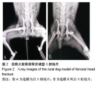
2.3 X射线片观察结果 所有田园犬股骨颈骨折造模成功当日,X射线片显示螺钉位置固定正确,骨折对位对线良好(图2A);6周后X射线片显示,有连续骨痂通过骨折线,骨折线变模糊(图2B)。"

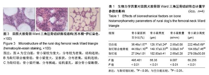
2.4 组织学观察结果 镜下组织观察见图3。生物力学因素对田园犬股骨颈Ward三角区骨组织形态计量学参数的影响见表1。取钉联合植骨组和空白组的骨小梁面积百分比、骨小梁厚度显著高于取钉组(P < 0.05);取钉联合植骨组和空白组的骨小梁分离度显著低于取钉组(P < 0.05);取钉联合植骨组的骨小梁面积百分数、骨小梁厚度显著高于空白组(P < 0.05);取钉联合植骨组的骨小梁分离度显著低于空白组(P < 0.05);取钉联合植骨组的骨小梁数量显著高于取钉组(P < 0.05);空白组的骨小梁数量与取钉组比较,差异无显著性意义(P > 0.05);取钉联合植骨组的骨小梁数量与空白组比较,差异无显著性意义(P > 0.05)。"
| [1] Purcell RL, Cody JP, Gordon W, et al. Outcomes of war related femoral neck fractures. Injury. 2015;46(12):2399-2403.[2] Della RG. Gaps and opportunities in the management of the young femoral neck fracture. Injury. 2015;46(3):515-518.[3] Slobogean GP, Sprague SA, Scott T, et al. Complications following young femoral neck fractures. Injury. 2015;46(3):484-491.[4] Zeng X, Zhan K, Zhang L, et al. The impact of high total cholesterol and high low-density lipoprotein on avascular necrosis of the femoral head in low-energy femoral neck fractures. J Orthop Surg Res. 2017; 12(1):30.[5] Takigawa N, Yasui K, Eshiro H, et al. Clinical results of surgical treatment for femoral neck fractures with the Targon((R)) FN. Injury. 2016;47 Suppl 7:S44-S48.[6] Boss JH, Misselevich I. Osteonecrosis of the femoral head of laboratory animals: the lessons learned from a comparative study of osteonecrosis in man and experimental animals. Vet Pathol. 2003; 40(4):345-354.[7] Kregor PJ. The effect of femoral neck fractures on femoral head blood flow. Orthopedics. 1996;19(12):1031-1036, 1037-1038.[8] Ehlinger M, Moser T, Adam P, et al. Early prediction of femoral head avascular necrosis following neck fracture. Orthop Traumatol Surg Res. 2011;97(1):79-88.[9] Bonnaire F, Schaefer DJ, Kuner E H. Hemarthrosis and hip joint pressure in femoral neck fractures. Clin Orthop Relat Res. 1998;(353): 148-155.[10] Han S, Oh M, Yoon S, et al. Risk stratification for avascular necrosis of the femoral head after internal fixation of femoral neck fractures by Post-Operative Bone SPECT/CT. Nucl Med Mol Imaging. 2017;51(1): 49-57.[11] Ma JX, He WW, Zhao J, et al. Bone microarchitecture and biomechanics of the necrotic femoral head. Sci Rep. 2017;7(1):13345.[12] Ueo T, Tsutsumi S, Yamamuro T, et al. Biomechanical aspects of the development of aseptic necrosis of the femoral head. Arch Orthop Trauma Surg. 1985;104(3):145-149.[13] 懋王,张长青. 股骨头坏死实验动物模型:分类与实验应用[J]. 中国组织工程研究,2014,18(36):5879-5884.[14] Jee WS. D.Sc. (hon) -- one man's association. J Musculoskelet Neuronal Interact. 2006;6(2):113-121.[15] 陈珺,张豪,杨国柱,等. 骨形态计量学目前应用专家共识[J]. 中国骨质疏松杂志,2014,20(9):1031-1038.[16] Min BW, Kim SJ. Avascular necrosis of the femoral head after osteosynthesis of femoral neck fracture. Orthopedics. 2011;34(5):349.[17] Wang T, Sun JY, Zha GC, et al. Analysis of risk factors for femoral head necrosis after internal fixation in femoral neck fractures. Orthopedics. 2014;37(12):e1117-e1123.[18] Ai ZS, Gao YS, Sun Y, et al. Logistic regression analysis of factors associated with avascular necrosis of the femoral head following femoral neck fractures in middle-aged and elderly patients. J Orthop Sci. 2013;18(2):271-276.[19] Zhang YL, Chen S, Ai ZS, et al. Osteonecrosis of the femoral head, nonunion and potential risk factors in Pauwels grade-3 femoral neck fractures: A retrospective cohort study. Medicine (Baltimore). 2016; 95(24):e3706.[20] Wang C, Xu GJ, Han Z, et al. Correlation between residual displacement and osteonecrosis of the femoral head following cannulated screw fixation of femoral neck fractures. Medicine (Baltimore). 2015;94(47):e2139.[21] Lambers FM, Bouman AR, Rimnac CM, et al. Microdamage caused by fatigue loading in human cancellous bone: relationship to reductions in bone biomechanical performance. PLoS One. 2013;8(12):e83662.[22] Ahn AC, Grodzinsky AJ. Relevance of collagen piezoelectricity to "Wolff's Law": a critical review. Med Eng Phys. 2009;31(7):733-741.[23] Gou WL, Lu Q, Wang X, et al. Key pathway to prevent the collapse of femoral head in osteonecrosis. Eur Rev Med Pharmacol Sci. 2015; 19(15):2766-2774.[24] Wang C, Wang Y, Meng H, et al. Microstructure and nanomechanical properties of singletrabecular bone in different Regions of osteonecrosis of the femoral head. J Nanosci Nanotechnol. 2016; 16(3):2264-2269.[25] 孙蕴,贺丽英,马兆坤,等. Ward三角区再研究[J]. 中国骨质疏松杂志, 2016,22(6):706-710.[26] Xiao D, Ye M, Li X, et al. Development of femoral head interior supporting device and 3D finite element analysis of its application in the treatment of femoral head avascular necrosis. Med Sci Monit. 2015;21:1520-1526.[27] Chen Z, Xu Y, Qi Z, et al. The formation and function of the sclerosis rim in the femoral head: A biomechanical point of view. Med Eng Phys. 2015;37(12):1125-1132.[28] Kim JW, Ryu JS, Baek S, et al. The timing of bone SPECT to predict osteonecrosis after internal fixation of femur neck fractures. J Orthop Sci. 2017;22(3):457-462.[29] 张洋,王楠,杨立枫,等. 骨髓间充质干细胞移植联合髓芯减压植骨修复股骨头坏死(英文)[J]. 中国组织工程研究, 2015,19(6):883-890.[30] Yu T, Xie L, Chu F. A sclerotic rim provides mechanical support for the femoral head in osteonecrosis. Orthopedics. 2015;38(5):e374-e379.[31] Escudier JC, Ollivier M, Donnez M, et al. Superimposition of maximal stress and necrosis areas at the top of the femoral head in hip aseptic osteonecrosis. Orthop Traumatol Surg Res. 2018.[32] Yi W, Tian Q, Dai Z, et al. Mechanical behaviour of umbrella-shaped, Ni-Ti memory alloy femoral head support device during implant operation: a finite element analysis study. PLoS One. 2014;9(6):e100765.[33] Ma J, Sun W, Gao F, et al. Porous tantalum implant in treating osteonecrosis of the femoral head: still a viable option? Sci Rep. 2016; 6:28227.[34] Zeng YR, He S, Feng WJ, et al. Vascularised greater trochanter bone graft, combined free iliac flap and impaction bone grafting for osteonecrosis of the femoral head. Int Orthop. 2013;37(3):391-398.[35] Yao C, Yi N, Shen J, et al. Clinical reports of surgical dislocation of the hip with sequestrum clearance and impacting bone graft for grade IIIA-IIIB aseptic necrosis of femoral head (ANFH) patients. Oncotarget. 2017;8(30):50084-50090. |
| [1] | Xu Feng, Kang Hui, Wei Tanjun, Xi Jintao. Biomechanical analysis of different fixation methods of pedicle screws for thoracolumbar fracture [J]. Chinese Journal of Tissue Engineering Research, 2021, 25(9): 1313-1317. |
| [2] | Zhang Tongtong, Wang Zhonghua, Wen Jie, Song Yuxin, Liu Lin. Application of three-dimensional printing model in surgical resection and reconstruction of cervical tumor [J]. Chinese Journal of Tissue Engineering Research, 2021, 25(9): 1335-1339. |
| [3] | Chen Xinmin, Li Wenbiao, Xiong Kaikai, Xiong Xiaoyan, Zheng Liqin, Li Musheng, Zheng Yongze, Lin Ziling. Type A3.3 femoral intertrochanteric fracture with augmented proximal femoral nail anti-rotation in the elderly: finite element analysis of the optimal amount of bone cement [J]. Chinese Journal of Tissue Engineering Research, 2021, 25(9): 1404-1409. |
| [4] | Zhou Jihui, Li Xinzhi, Zhou You, Huang Wei, Chen Wenyao. Multiple problems in the selection of implants for patellar fracture [J]. Chinese Journal of Tissue Engineering Research, 2021, 25(9): 1440-1445. |
| [5] | Li Zhongfeng, Chen Minghai, Fan Yinuo, Wei Qiushi, He Wei, Chen Zhenqiu. Mechanism of Yougui Yin for steroid-induced femoral head necrosis based on network pharmacology [J]. Chinese Journal of Tissue Engineering Research, 2021, 25(8): 1256-1263. |
| [6] | Zeng Yanhua, Hao Yanlei. In vitro culture and purification of Schwann cells: a systematic review [J]. Chinese Journal of Tissue Engineering Research, 2021, 25(7): 1135-1141. |
| [7] | Xu Yulin, Shen Shi, Zhuo Naiqiang, Yang Huilin, Yang Chao, Li Yang, Zhao Heng, Zhao Lu. Biomechanical comparison of three different plate fixation methods for acetabular posterior column fractures in standing and sitting positions [J]. Chinese Journal of Tissue Engineering Research, 2021, 25(6): 826-830. |
| [8] | Cai Qunbin, Zou Xia, Hu Jiantao, Chen Xinmin, Zheng Liqin, Huang Peizhen, Lin Ziling, Jiang Ziwei. Relationship between tip-apex distance and stability of intertrochanteric femoral fractures with proximal femoral anti-rotation nail: a finite element analysis [J]. Chinese Journal of Tissue Engineering Research, 2021, 25(6): 831-836. |
| [9] | Song Chengjie, Chang Hengrui, Shi Mingxin, Meng Xianzhong. Research progress in biomechanical stability of lateral lumbar interbody fusion [J]. Chinese Journal of Tissue Engineering Research, 2021, 25(6): 923-928. |
| [10] | Xie Chongxin, Zhang Lei. Comparison of knee degeneration after anterior cruciate ligament reconstruction with or without remnant preservation [J]. Chinese Journal of Tissue Engineering Research, 2021, 25(5): 735-740. |
| [11] | Xu Dongzi, Zhang Ting, Ouyang Zhaolian. The global competitive situation of cardiac tissue engineering based on patent analysis [J]. Chinese Journal of Tissue Engineering Research, 2021, 25(5): 807-812. |
| [12] | Wu Zijian, Hu Zhaoduan, Xie Youqiong, Wang Feng, Li Jia, Li Bocun, Cai Guowei, Peng Rui. Three-dimensional printing technology and bone tissue engineering research: literature metrology and visual analysis of research hotspots [J]. Chinese Journal of Tissue Engineering Research, 2021, 25(4): 564-569. |
| [13] | Chang Wenliao, Zhao Jie, Sun Xiaoliang, Wang Kun, Wu Guofeng, Zhou Jian, Li Shuxiang, Sun Han. Material selection, theoretical design and biomimetic function of artificial periosteum [J]. Chinese Journal of Tissue Engineering Research, 2021, 25(4): 600-606. |
| [14] | Liu Fei, Cui Yutao, Liu He. Advantages and problems of local antibiotic delivery system in the treatment of osteomyelitis [J]. Chinese Journal of Tissue Engineering Research, 2021, 25(4): 614-620. |
| [15] | Li Xiaozhuang, Duan Hao, Wang Weizhou, Tang Zhihong, Wang Yanghao, He Fei. Application of bone tissue engineering materials in the treatment of bone defect diseases in vivo [J]. Chinese Journal of Tissue Engineering Research, 2021, 25(4): 626-631. |
| Viewed | ||||||
|
Full text |
|
|||||
|
Abstract |
|
|||||