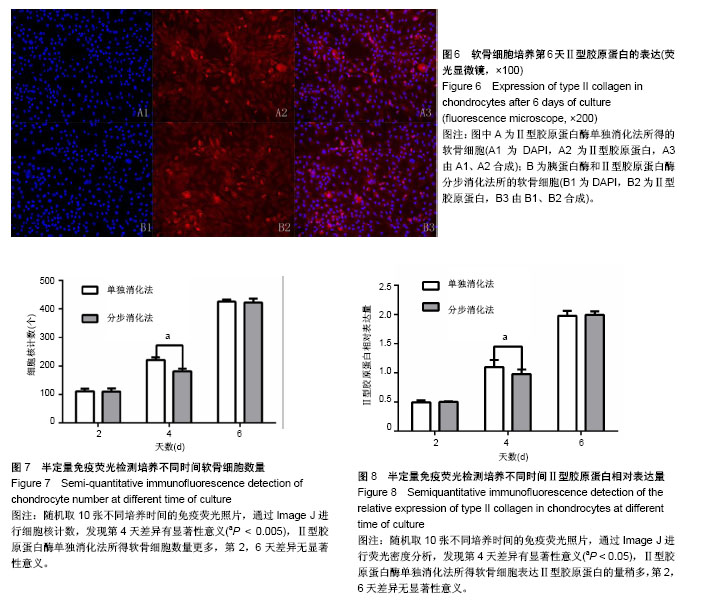
| [1] Rosenthal AK, Gohr CM, Mitton-Fitzgerald E, et al. Autophagy modulates articular cartilage vesicle formation in primary articular chondrocytes. J Biol Chem. 2015;290(21): 13028-13038.[2] Yan B, Zhang Z, Jin D, et al. mTORC1 regulates PTHrP to coordinate chondrocyte growth, proliferation and differentiation. Nat Commun. 2016;7:11151.[3] Choi H, Choi Y, Kim J, et al. Longitudinal bone growth is impaired by direct involvement of caffeine with chondrocyte differentiation in the growth plate. J Anat. 2017;230(1): 117-127.[4] Chang LH, Wu SC, Chen CH, et al. Parathyroid hormone 1-34 reduces dexamethasone-induced terminal differentiation in human articular chondrocytes. Toxicology. 2016;368-369: 116-128.[5] Li H, Li X, Liu G, et al. Bauhinia championi (Benth.) Benth. polysaccharides upregulate Wnt/β-catenin signaling in chondrocytes. Int J Mol Med. 2013;32(6):1329-1336.[6] 刘振峰,方锐,孟庆才. Ⅱ型胶原酶消化法可短时间获得大量纯化大鼠关节软骨细胞[J]. 中国组织工程研究与临床康复, 2011, 15(50): 9323-9326. [7] 韦宋谱,张晓钢,丁道芳,等. 一种获取具有旺盛增殖力的大鼠软骨细胞培养方法[J]. 上海中医药杂志, 2012, 46(7):8-12.[8] 于莹,艾虹,刘天悦,等. 周期性单轴压力对大鼠髁突软骨细胞结缔组织生长因子表达的影响[J]. 口腔材料器械杂志, 2015, 24(4):181-185.[9] Zhang Q, Deng S, Sun K, et al. MMP-2 and Notch signal pathway regulate migration of adipose-derived stem cells and chondrocytes in co-culture systems. Cell Prolif. 2017;50(6): e12385.[10] Xu XX, Zhang XH, Diao Y, et al. Achyranthes bidentate saponins protect rat articular chondrocytes against interleukin-1β-induced inflammation and apoptosis in vitro. Kaohsiung J Med Sci. 2017;33(2):62-68.[11] Kobayashi T, Kronenberg H. Minireview: transcriptional regulation in development of bone. Endocrinology. 2005; 146(3):1012-1017.[12] Beier F. Cell-cycle control and the cartilage growth plate. J Cell Physiol. 2005;202(1):1-8.[13] Kozhemyakina E, Lassar AB, Zelzer E. A pathway to bone: signaling molecules and transcription factors involved in chondrocyte development and maturation. Development. 2015;142(5):817-831.[14] Tsang KY, Chan D, Cheah KS. Fate of growth plate hypertrophic chondrocytes: death or lineage extension. Dev Growth Differ. 2015;57(2):179-192.[15] Ogawa H, Akiyama H. Analysis of Musculoskeletal Systems and Their Diseases. Regulation of chondrogenesis and cartilage regeneration by molecular and developmental biology. Clin Calcium. 2015;25(8):1116-1124.[16] Sun MM, Beier F. Chondrocyte hypertrophy in skeletal development, growth, and disease. Birth Defects Res C Embryo Today. 2014;102(1):74-82.[17] Wang M, Man XF, Liu YQ, et al. Dysfunction of collagen synthesis and secretion in chondrocytes induced by wisp3 mutation. Int J Endocrinol. 2013;2013:679763.[18] Usmani SE, Pest MA, Kim G, et al. Transforming growth factor alpha controls the transition from hypertrophic cartilage to bone during endochondral bone growth. Bone. 2012;51(1): 131-141.[19] Tian Y, Guo R, Shi B, et al. MicroRNA-30a promotes chondrogenic differentiation of mesenchymal stem cells through inhibiting Delta-like 4 expression. Life Sci. 2016; 148:220-228.[20] Liu Q, Zhang X, Hu X, et al. Circular RNA Related to the Chondrocyte ECM Regulates MMP13 Expression by Functioning as a MiR-136 'Sponge' in Human Cartilage Degradation. Sci Rep. 2016;6:22572.[21] Allen JL, Cooke ME, Alliston T. ECM stiffness primes the TGFβ pathway to promote chondrocyte differentiation. Mol Biol Cell. 2012;23(18):3731-3742.[22] Sheng MH, Zhou XD, Bonewald LF, et al. Disruption of the insulin-like growth factor-1 gene in osteocytes impairs developmental bone growth in mice. Bone. 2013;52(1): 133-144.[23] Yeung Tsang K, Wa Tsang S, Chan D, et al. The chondrocytic journey in endochondral bone growth and skeletal dysplasia. Birth Defects Res C Embryo Today. 2014;102(1):52-73.[24] Li J, Dong S. The Signaling Pathways Involved in Chondrocyte Differentiation and Hypertrophic Differentiation. Stem Cells Int. 2016;2016:2470351.[25] Choi H, Choi Y, Kim J, et al. Longitudinal bone growth is impaired by direct involvement of caffeine with chondrocyte differentiation in the growth plate. J Anat. 2017;230(1): 117-127.[26] Heinegård D, Saxne T. The role of the cartilage matrix in osteoarthritis. Nat Rev Rheumatol. 2011;7(1):50-56.[27] 潘思年,杜敏联,马华梅,等. 大鼠胫骨生长板软骨细胞的体外培养及鉴定[J]. 中山大学学报(医学科学版), 2009, 30(z2): 1-5,10.[28] Mellor LF, Baker TL, Brown RJ, et al. Optimal 3D culture of primary articular chondrocytes for use in the rotating wall vessel bioreactor. Aviat Space Environ Med. 2014;85(8): 798-804.[29] Xu XX, Zhang XH, Diao Y, et al. Achyranthes bidentate saponins protect rat articular chondrocytes against interleukin-1β-induced inflammation and apoptosis in vitro. Kaohsiung J Med Sci. 2017;33(2):62-68.[30] Avila-Rodríguez D, Paisano-Cerón K, Valdovinos-Ramírez I, et al. Three-dimensional Alginate-bead Culture of Human Pituitary Adenoma Cells. J Vis Exp. 2016;(108):53637.[31] Bhuyan MK, Rodriguez-Devora J, Tseng TL, et al. Photovoltaic surfaces enable clonal myoblastic cell release using visible light as external stimulation. Biotechnol J. 2016;11(3):393-398.[32] Huang HL, Hsing HW, Lai TC, et al. Trypsin-induced proteome alteration during cell subculture in mammalian cells. J Biomed Sci. 2010;17:36.[33] 秦苏萍,李慧,李小翠,等. CIA大鼠滑膜成纤维细胞的原代培养及鉴定[J]. 免疫学杂志, 2014, 30(12): 1096-1099. |