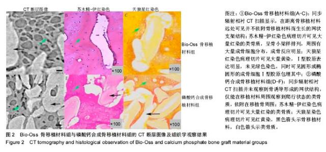
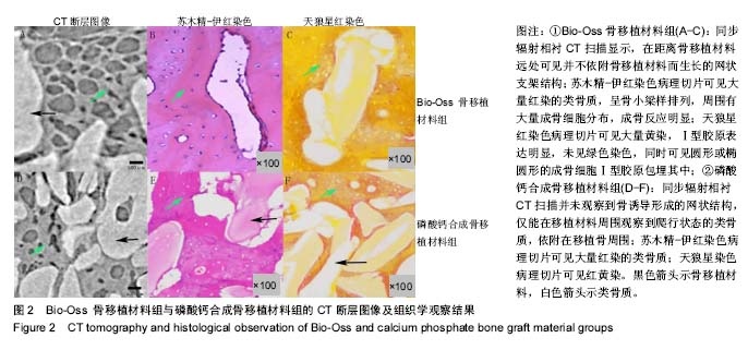
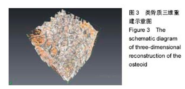
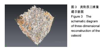
| [1]刘佳,谢志刚,鲍济波.骨移植材料有效骨再生的评价方法及应用[J].国际口腔医学杂志,2015,42(2):173-176.[2]Boskey AL.Matrix proteins and mineralization :an overview.Conn Tissue Res.1996;35(1-4):357-363.[3]Heidi A.Declercq,Ronald M.H.Verbeeck,et al.Calcification as an indicator of osteoinductive capacity of biomaterials in osteoblastic cell cultures.Biomaterials.2005;26:4964-4974.[4]Hinze MC,Wiedmann-Al-Ahmad M,Glaum R,et al.Bone engineering-vitalisation of alloplastic andallogenic bone grafts by human osteoblast-like cells.Br J Oral Maxillofac Surg.2010;48(5): 369-373.[5]Wilkins SW,Gureyev TE,Gao D,et al.Phase-contast imaging using polychromatic hard X-ray.Nature.1996;384:335-338.[6]Wu X,Liu H.Clinical implementation of X-ray phase-contrast imaging:theoretical foundationsand design considerations.Med Phys.2003;30:2169-2179.[7]简建波,杨浩,秦莉莉,等.肝细胞癌微观结构的X线相衬CT成像研究[J].中国医学影像学杂志,2016,24(16):869-872.[8]李泽键,赖仁发.骨移植材料成骨效能检测指标及方法的选择与应用[J].中国美容医学,2013,22(7):793-796.[9]李家峰,崔群,孙秀英,等.纳米羟基磷灰石/聚己内酯电纺支架修复种植体周围骨缺损[J].中国组织工程研究, 2014,18(16):2257-2562.[10]Chen RC,Rigon L,Longo R.Quantitative 3D refractive index decrement reconstruction using single-distance phase-contrast tomography data.J Phys D Appl Phys. 2011;44(49):495401-495409.[11]Iwasaki H,Fukushima K,Kawamoto A,et al. Synchrotronradiation coronarymicroangiography formorphometric andphysiological evaluation of myocardial neovascularization inducedby endothelial progenitor cell transplantation.Arterioscler Thromb Vasc Biol.2007;27:1326-1333.[12]Takeda T,Momese A,Wu J,et al.Vessel imaging byinterfemmetric phase-contrast X-ray technique.Circulation.2002;105:1708-1712.[13]Zehbe R,Haibel A,Riesemeier H,et al.Synchrotron micro-computed tomography as amethodology for biological tissue characterization:from tissuemorphology to individual cells.Royal Society Interface.2010;7:49-59.[14]姜庆军,肖湘生,刘士远,等.相干X 线相衬成像的初步研究: 离体大白鼠肝脏、肺脏成像实验[J].中国医学影像技术, 2003,19(9):1116-1117.[15]刘骏桢,林江,严福华.同步辐射X 线相衬技术在肝血管成像中的研究进展[J].中国医学影像学杂志,2011, 19(12):935-937.[16]孙莲莲,王志兴.同步辐射显微断层成像监测口腔骨植入材料置入体内后的生物相容性及定量分析:随机对照动物实验方案[J].中国组织工程研究,2017,21(6):952-956.[17]王甜,陈芬,林宏辉.一种安全有效的动物骨骼标本制作方法[J].实验科学与技术,2015,13(5):260-263.[18]宋彬妤.对制作高质量病理标本的方法研究[J].当代医药论丛, 2015, 13(23):12-13.[19]曹莉,王礼鑫,曹颖,等.盐酸化伊红溶液在病理小标本包埋操作中的应用[J].临床与实验病理学杂志,2016,32(7):829-830.[20]丛长虹,陈宁,田慧,等.同步辐射衍射增强CT应用于根管充填质量评价的实验研究[J].口腔医学研究,2017,33(3):324-327.[21]Longo R.Current studies and future perspectives of synchrotron radiation imaging trials in human patients.Nucl Instr Meth Phys Res.2016;809:13-22.[22]彭志刚,张璐,张旭.裸鼠胃癌模型的相位衬度微血管成像[J].北京生物医学工程,2017,36(2):123-127.[23]秦莉莉,简建波,赵新颜,等.利用X射线相衬CT技术观察胆管结扎诱导肝纤维化微脉管的变化[J].中国医学影像学杂志, 2017,25(3):165-168,173.[24]Zolotarev K,Kulipanov G,Levichev E,et al.Synchrotron Radiation Applications in the Siberian Synchrotron and Terahertz Radiation Center.Phys Proc.2016;84:11.[25]Chen-Chen X,Yadav AK,Kai Z,et al.Synchrotron radiation (SR) diffraction enhanced imaging (DEI) of chronic glomerulonephritis(CGN) mode.J Xray Sci Technol.2016;24(1):145-159. [26]王明,李辉,WANGMing,等.同步辐射X射线的医学应用[J].中国医学物理学杂志,2016,33(12):1253-1256.[27]夏琛琛,ARUNKumarYadav,张凯,等.同步辐射衍射增强应用于慢性肾小球肾炎模型的研究[J].同济大学学报(医学版), 2015,36(6):24-30.[28]沈富强,监悟帅,孙郁,等.乳腺癌相衬成像与病理对照[J].生物医学工程研究,2015,34(4):227-229.[29]刘成磊,郗艳,左后东,等.同步辐射显微CT的人关节软骨三维成像研究[J].CT理论与应用研究,2015,24(6):793-799.[30]陈华斌,胡建中,周京泳,等.同步辐射X线相衬成像技术在兔髌骨-髌腱连接点纤维软骨细胞的三维可视化应用研究[J].核技术, 2015, 38(11):5-12.[31]吴天定,吕红斌,曹勇,等.正常及挫伤脊髓血管的三维直视化:同步辐射相衬成像[J].中国组织工程研究, 2015,19(46):7478-7483. [32]王飞翔,邓彪,王玉丹,等.基于毛细管分光的同步辐射X射线立体成像[J].光学学报,2016,36(8):334-340.[33]姚圣坤.基于同步辐射X射线的多尺度生物成像研究[D].山东大学, 2016.[34]Garg AD,Yadav S,Kumar M,et al.Studies of longitudinal profile of electron bunches and impedance measurements at Indus-2 synchrotron radiation source.Nucl Instr Meth Phys Res. 2016; 814:66-72.[35]Abukawa T,Yamamoto S,Yukawa R,et al.Time-resolved soft X-ray core-level photoemission spectroscopy at 880 ° C using the pulsed laser and synchrotron radiation and the pulse heating current.Surf Sci.2017;656:43-47.[36]Li WT,Wang WT,Liu JS,et al.Developments in laser wakefield accelerators:From single-stage to two-stage.Chin Phys B. 2015; 24(1):76-84.[37]Hafz NAM,Li S,et al.Generation of high-quality electron beams by ionization injection in a single acceleration stage.High[38]Nakajima K,Kim HT,Jeong TM,et al.Scaling and design of high-energy laser plasma electron acceleration.High Power Laser Sci Eng.2015;3(1):81-91.[39]Kazuhisa N.Conceptual designs of a laser plasma accelerator-based EUV-FEL and an all-optical Gamma-beam source.High Power Laser Sci Eng.2014;2(4):31-38.[40]Bolton PR.The integrated laser-driven ion accelerator system and the laser-driven ion beam radiotherapy challenge.Nucl Instr Meth Phys Res.2016;809:149-155. |