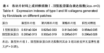
| [1]Franz MG.The biology of hernias and the abdominal wall. Hernia.2006;10(6):462-471.[2]Read RC.Recent advances in the repair of groin herniation. Curr Probl Surg.2003;40(1):13-79.[3]Deeken CR,Faucher KM,Matthews BD.A review of the composition, characteristics,and effectiveness of barrier mesh prostheses utilized for laparoscopic ventral hernia repair.Surg Endosc. 2012;26(2):566-575.[4]Bostanci O,Idiz UO,Yazar M,et al.A Rare Complication of Composite Dual Mesh: Migration and Enterocutaneous Fistula Formation.Case Rep Surg.2015;2015:293659. [5]Klinge U,Klosterhalfen B.Modified classification of surgical meshes for hernia repair based on the analyses of 1,000 explanted meshes.Hernia.2012;16(3):251-258.[6]Kassem MI,El-Haddad HM.Polypropylene-based composite mesh versus standard polypropylene mesh in the reconstruction of complicated large abdominal wall hernias: a prospective randomized study. Hernia.2016;20(5):691-700.[7]Esfahani H,Jose R,Ramakrishna S.Electrospun Ceramic Nanofiber Mats Today: Synthesis, Properties, and Applications.Materials(Basel).2017;10(11).pii: E1238.doi: 10.3390/ma10111238.[8]USP XXII 24版. Biological Tests/ Biological Reactivity Tests, In-vivo.[9]Hamer-Hodges DW,Scott NB.Surgeon's workshop. Replacement of an abdominal wall defect using expanded PTFE sheet(Gore-tex).J R Coll Surg Edinb.1985;30(1):65-67.[10]Cobb WS,Burns JM,Kercher KW,et al.Normal intraabdominal pressure in healthy adults.J Surg Res.2005;129(2):231-235.[11]Klinge U,Klosterhalfen B,Conze J,et al.Modified mesh for hernia repair that is adapted to the physiology of the abdominal wall.Eur J Surg.1998;164(12):951-960.[12]Bachman S, Ramshaw B.Prosthetic material in ventral hernia repair: how do I choose?Surg Clin North Am. 2008;88(1): 101-112.[13]Sahin-Toth G,Halasz T,Viczian C,et al.Late complication after mesh repair of incisional hernias: pseudocyst formation.Magy Seb.2007;60(6):293-296.[14]Jaiswal D,James R,Shelke NB,et al.Gelatin Nanofiber Matrices Derived from Schiff Base Derivative for Tissue Engineering Applications.J Biomed Nanotechnol. 2015;11(11): 2067-2080.[15]Tian A,Chen Y,Liao J,et al.Cytotoxicity of a new type silicone rubber for maxillofacial prosthesis: an in vitro evaluation. Sheng Wu Yi Xue Gong Cheng Xue Za Zhi. 2014;31(5): 1046-1049,1056.[16]Farah S,Anderson DG,Langer R.Physical and mechanical properties of PLA, and their functions in widespread applications-A comprehensive review.Adv Drug Deliv Rev. 2016;107:367-392.[17]Ramot Y,Haim-Zada M,Domb AJ,et al.Biocompatibility and safety of PLA and its copolymers.Adv Drug Deliv Rev.2016; 107:153-162.[18]Inkinen S,Hakkarainen M,Albertsson AC,et al.From lactic acid to poly(lactic acid) (PLA): characterization and analysis of PLA and its precursors.Biomacromolecules. 2011;12(3): 523-532.[19]刘晓军,汪海滨.聚乳酸和聚乙醇酸及其共聚物细胞毒性的评价[J].中国临床康复,2005,9(46):46-47.[20]Hoveizi E,Nabiuni M,Parivar K,et al.Functionalisation and surface modification of electrospun polylactic acid scaffold for tissue engineering.Cell Biol Int.2014;38(1):41-49.[21]Bashur CA,Shaffer RD,Dahlgren LA,et al.Effect of fiber diameter and alignment of electrospun polyurethane meshes on mesenchymal progenitor cells.Tissue Eng Part A. 2009; 15(9):2435-2445.[22]Taghiabadi E,Nasri S,Shafieyan S,et al.Fabrication and characterization of spongy denuded amniotic membrane based scaffold for tissue engineering.Cell J. 2015;16(4): 476-487.[23]Bacakova M,Musilkova J,Riedel T,et al.The potential applications of fibrin-coated electrospun polylactide nanofibers in skin tissue engineering.Int J Nanomedicine. 2016;11:771-789. [24]徐少骏.成纤维细胞接触性和密度抑制特性对异常瘢痕形成的意义[J].杭州医学高等专科学校学报, 2004,25(6):269-271.[25]Aland S,Hatzikirou H,Lowengrub J,et al.A Mechanistic Collective Cell Model for Epithelial Colony Growth and Contact Inhibition.Biophys J.2015;109(7):1347-1357.[26]Thomson RC,Yaszemski MJ,Powers JM,et al.Fabrication of biodegradable polymer scaffolds to engineer trabecular bone.J Biomater Sci Polym Ed.1995;7(1):23-38.[27]Klinge U,Dietz U,Fet N,et al.Characterisation of the cellular infiltrate in the foreign body granuloma of textile meshes with its impact on collagen deposition.Hernia.2014;18(4):571-578. |
