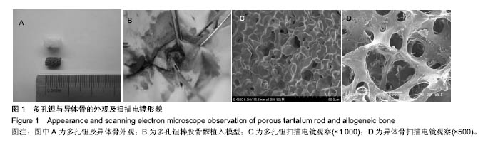
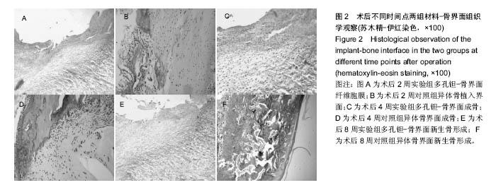
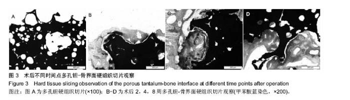
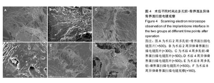
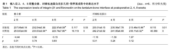
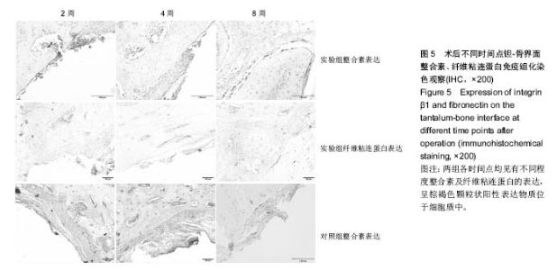
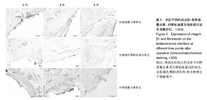
| [1]Kim DG, Huja SS, Tee BC, et al. Bone ingrowth and initial stability of titanium and porous tantalum dental implants: a pilot canine study. Implant Dent. 2013; 22(4):399-405.[2]Fernandez-Fairen M, Querales V, Jakowlew A, et al. Tantalum is a good bone graft substitute in tibial tubercle advancement. Clin Orthop Relat Res.2010; 468(5): 1284-1295.[3]李琪佳,王茜,甘洪全,等. 多孔钽材料细胞毒性、生物相容性及体内成骨性研究[J].中华骨科杂志,2014,34(9):954-961.[4]Keselowsky BG, Collard DM, García AJ. Surface chemistry modulates fibronectin conformation and directs integrin binding and specificity to control cell adhesion. J Biomed Mater Res A. 2003;66(2):247-259.[5]祁洁,张姝江,刘宗智.整合素α5β1在成骨细胞与生物衍生材料黏附过程中的表达[J]. 中国组织工程研究与临床康复, 2011, 15(47):8762-8764. [6]Rapuano BE,MacDonald DE.Surface oxide net charge of a titanium alloy:modulation of fibronectin-activated attachment and spreading of osteogenic cells.Colloids Surf B Biointerfaces. 2011;82(1):95-103.[7]甘洪全,李琪佳,王茜,等国产多孔钽材料兔髌腱内植入形态学特点及生物相容性评价[J].中国修复重建外科杂志, 2014,28(4): 452-456.[8]Anselme K. Biomaterials and interface with bone. Osteoporos Int. 2011;22(6):2037-2042.[9]Pegueroles M, Aguirre A, Engel E, et al. Effect of blasting treatment and Fn coating on MG63 adhesion and differentiation on titanium: a gene expression study using real-time RT-PCR. J Mater Sci Mater Med.2011; 22(3):617-627.[10]Stephansson SN, Byers BA, Garcia AJ. Enhanced expression of the osteoblastic phenotype on substrates that modulate fibronectin conformation and integrin receptor binding. Biomaterials. 2002;23(12):2527-2534.[11]Hosgor F, Yilmaz N, Senyurt O, et al. Effect of osteoblast cell culture on the bone implant contact . Acta Odontol Scand. 2013;71(3-4) :626-631.[12]耿丽鑫,甘洪全,王茜,等.国产多孔钽对成骨细胞生物相容性及其相关成骨基因表达的影响[J].第三军医大学学报,2014,36(11): 1163-1167.[13]Amanatullah DF, Farac R, McDonald TJ, et al.Subtrochanteric Fracture following Removal of a Porous Tantalum Implant. Case Rep Orthop. 2013; 2013:946745. .[14]Cunha A,Renz RP,Blando E, et al. Osseointegration of atmospheric plasma-sprayed titanium implants: Influence of the native oxide layer. J Biomed Mater Res A. 2014;102(1): 30-36.[15]Mrosek EH, Schagemann JC, Chung HW, et al. Porous tantalum and poly-epsilon -caprolactone biocomposites for osteochondral defect repair: preliminary studies in rabbits. Orthop Res.2010; 28(2): 141-148.[16]张辉,李亮,王茜等.骨形成蛋白-7对多孔钽-软骨细胞复合物分泌功能以及COL-II、AGG和Sox9基因表达的影响[J].北京大学学报(医学版),2015,47(1):216-222.[17]王茜,张辉,耿丽鑫,等. MG63细胞与国产多孔钽材料共培养后成骨相关因子的表达研究[J].中国修复重建外科杂志, 2014, 28(11):1422-1427.[18]Thull R. Physicochemical principles of tissue material interactions. Biomol Eng. 200;19(2-6):43-50.[19]周群,谭兆军,方铁钧,等. C/C-SiC复合材料-骨结合界面的组织学研究[J]. 口腔医学, 2012,32(2):84-87.[20]王兴,焦艳军,孙晓军,等. 整合素α5β1和纤维粘连蛋白在Ⅱ型糖尿病大鼠种植体周围骨组织的表达及意义[J]. 临床口腔医学杂志, 2011,27(9):530-532.[21]Keselowsky BG, Collard DM, García AJ. Surface chemistry modulates fibronectin conformation and directs integrin binding and specificity to control cell adhesion. J Biomed Mater Res A. 2003;66(2):247-259.[22]Knight MM, Toyoda T, Lee DA, et al. Mechanical compression and hydrostatic pressure induce reversible changes in actin cytoskeletal organisation in chondrocytes in agarose. J Biomech. 2006;39(8):1547-1551.[23]Vestermark MT. Strontium in the bone-implant interface. Dan Med Bull.2011;58(5):B4286. |