Chinese Journal of Tissue Engineering Research ›› 2013, Vol. 17 ›› Issue (24): 4553-4560.doi: 10.3969/j.issn.2095-4344.2013.24.026
Research and application progress of visual fixation component separation
Wei Wei1, Shi Geng-hu2, Li Yu-tang2, Zhang Bing2, Gao Chuang3
- 1 College of Science, Huazhong Agricultural University, Wuhan 430070, Hubei Province, China
2 School of Psychology, Central China Normal University, Wuhan 430070, Hubei Province, China
3 Key Laboratory of Adolescent Cyberpsychology and Behavior, Minstry of Education (Central China Normal University), Wuhan 430070, Hubei Province, China
-
Received:2013-03-30Revised:2013-04-06Online:2013-06-11Published:2013-06-11 -
Contact:Gao Chuang, M.D., Associate professor, Key Laboratory of Adolescent Cyberpsychology and Behavior, Minstry of Education (Central China Normal University), Wuhan 430070, Hubei Province, China gaochuang@phy.ccnu.edu.cn -
About author:Wei Wei☆, M.D., Associate professor, College of Science, Huazhong Agricultural University, Wuhan 430070, Hubei Province, China Shi Geng-hu, Studying for master’s degree, School of Psychology, Central China Normal University, Wuhan 430070, Hubei Province, China 365711146@qq.com -
Supported by:the National Key Technology Research & Development Program of China, No. 2011BAK08B03, 2012*
CLC Number:
Cite this article
Wei Wei, Shi Geng-hu, Li Yu-tang, Zhang Bing, Gao Chuang. Research and application progress of visual fixation component separation[J]. Chinese Journal of Tissue Engineering Research, 2013, 17(24): 4553-4560.
share this article

2.1 固像实验(Stabilized Retinal Image) 固视期间的眼动,是一种非常有趣的现象。一方面,为了获得物体细节信息,眼睛需要维持足够长时间处于静止状态,另一方面,微小眼动却使眼睛不能保持绝对静止,导致视网膜像由于微小移动而使细节模糊。这一表面似乎矛盾的现象所引发的问题是:固视期间的眼动,其发生的根本原因是什么?它的基本作用是什么?为此1953年Riggs等[5]设计了“固像实验”,以探求在完全没有眼动情况下的视知觉状态,从而间接推断固视眼动的起因和作用。其基本思路是:通过实验手段,将物体视象固定在视网膜特定位置,从而形成固定视象[6]。但固视期间的眼动无法用意志消除,因此,让眼睛保持完全静止是不现实的。如果使被注视的物体跟随眼睛同步运动,并保持相对静止状态,那么,物体在视网膜上投射的像的位置将保持不变,即视网膜固像。其基本做法是:在被试者所佩戴的隐形眼镜上安置一微小反光镜。刺激物投射到小镜子上,再反射到一个屏幕上,见图1。"
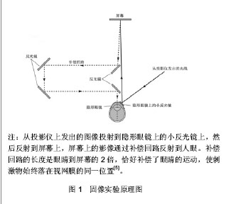

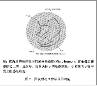
屏幕上的影像沿着光学补偿回路射入人的眼睛。该光学补偿回路的长度是眼睛到屏幕距离的2倍。当被试的眼睛运动的时候,小反光镜随着运动,反光镜投射到屏幕上的影像也随着小反光镜一起运动,影像的运动恰好补偿了眼睛的运动。这样,刺激物便始终作用在视网膜同一位置上了。当给被试者呈现一条黑线刺激的时候,结果发现,起初可以清楚地被知觉到的黑线,经过几秒钟后,却不可见了,而且,继续呈现黑线也不会再次被知觉到。 固像实验表明,长期进化的视觉系统,具有倾向于忽略静止物体,而觉察周围环境改变的能力。因为相比运动的物体,静止物体对人们产生的威胁信息要少得多。这可能是视觉系统进化出来的一个设计精巧的属性,这种属性被归结为生理的视网膜适应。而一旦视像被固定在视网膜上,视网膜适应的直接灾难性后果是知觉图像的消退[7]。因此,固视期间的微小眼动也被认为是维持视知觉的重要条件[8-9]。 2.2 固视中眼动成分 这些微小的眼动在进行一些行为任务时可以忽略不计,如在阅读或研究图像处理时眼球运动的控制[10]。但是当眼睛盯在固定物体上时,它们对于维持视知觉有重要作用[9]。由上所述,固视也往往被表述为:试图将眼睛保持在静止物体上,同时眼睛会做微小幅度运动,即固视眼动[11-13]。 到1950年代,随着眼动记录技术的成熟和精度提高,科学界进一步分离确立了眼动中的成分。它们分别是:微颤(micro tremor)、漂移(drift)和微跳视(micro saccade) [14-17]。而且发现,这些成分不但存在于人类的固视现象中,动物的固视中,同样也存在相同成分,如灵长类动物、猫、兔子、海龟、蜥蜴、猫头鹰等[18]。眼动固视成分的作用及分离方法问题开始引起关注,迄今为止,依然存在争议[1]。微颤(micro tremor),也被称为震颤,是一种非周期性,高频振荡成分(30-100 Hz)[19]。由于其振幅比较微小(只有一个锥体细胞那么大,所以很难被精确地记录,因此,微颤是最小的眼动成分[1]。因此,从微颤的特性——低振幅和高频率——出发,微颤往往被认为是眼动系统噪声[1],是双眼无关的,即单眼成分。这可能是:在产生立体视觉时,给视觉系统匹配视网膜上对应点的能力增加了一个生理极限。因为,双眼视觉的“对应点”代表的是对应的平均位置,而不是双眼锥体细胞的一对一的对应[20]。漂移与微颤同时发生,并与微颤成分叠加在一起。它是一种低速眼动(最大速度低于30角分/s)现象,发生在两次微跳视之间[21]。漂移期间,视网膜视像移动的范围可以跨越十几个感光细胞。 最初,有观点认为:可能是眼动神经系统的非稳定性,导致了眼睛的随机运动。但是,随后研究发现,在没有微跳视或者微跳视补偿作用很弱时,漂移对维持准确的固视起到了补偿作用[22]。漂移既有双眼的[23-24],也有单眼的[25-26]。与微颤一样,漂移也被认为是:脑干运动神经元神经冲动诱发系列的自由发放并产生的噪声[1]。但是,如果漂移和微颤确实是共轭的[27],也就是说它们是双眼成分,那么它们至少有部分的中枢神经系统的根源。临床观察到的脑干损伤患者的微颤减少或完全没有的事实支持了这个观点[28]。微跳视是快速、小幅度的眼动,平均发生频率是每秒1到2次[29-30]。它的幅度可以跨越几十到甚至几百个感光器[29],但很少大于30角分,持续时间大约25 ms左右[31-32]。但是微跳视的定义不只建立在幅度的基础上,跳视(saccade)的幅度可以跟微跳视(micro saccade)一样小[33-35],并且它们的幅度与峰速度都遵循主序关系[36]。所以微跳视指的是固视期间发生的非随意的跳视,也叫固视跳视[37]。 2.3 固视中眼动功能 围绕3个成分,长期以来,其基本问题是:这些成分对视知觉的贡献是什么?具体讲,可以归结为2个问题:①固视成分是否可防止知觉消退?②微小跳视是否可对视觉系统进行纠错? 防止知觉消退观点认为,微跳视和漂移对于获得最佳视知觉都是必需的[38]。由于中央凹附近的感受野太小,在没有微跳视的情况下,微颤和漂移足以防止视觉消退[39-40]。如果排除掉微颤和漂移,在固视时微跳视也足够维持视知觉。由于外围的感觉野太大,可能只有微跳视足够大,足够快(相比微颤和漂移),可以防止视觉消退[41-42],尤其是低对比度的刺激[43]。Ditchburn[44]则证实,在低对比度时,微跳视对于知觉色调的差异是必需的。Westheimer表示微跳视可以提高知觉立体的敏感度[45]。而Carpenter则认为,3种固视成分中,只有微跳视对维持视知觉的贡献最重要,因为漂移的速度太低,微颤的幅度和频率则使其有害而无益[46]。对视觉系统纠错的观点认为,由于漂移存在,会造成视轴偏离,导致注视目标物偏离中央视野,因此,微跳视的基本作用是纠正漂移造成的视轴偏离,使眼睛重返固视的目标物。 且微跳视在对抗视疲劳维持视知觉方面起重要作用[47]。这是因为在固像实验中,只有微跳视可以使人恢复视知觉[48],而微颤和漂移在防止视知觉消退方面的效果很小[1] 。 但是微跳视对视觉系统的纠错准确性仍是有限的,而且非纠错的微跳视也会发生。最近的研究显示,短时程的微跳视是对抗视网膜适应,而长时程微跳视则是纠错。与上述两种观点相反,Steinman则认为在微跳视在维持视知觉中没有任何用途。他有来自两个方面的有力证据证明自己的观点。第一,受过训练的被试可以抑制自己的眼睛不发生微跳视数秒,而不会引想知觉消退。第二,微跳视在高精度的观察任务(如射击和穿针引线)中可以被随意抑制[49]。如果纠正错误是微跳视的基本功能的话,由于漂移和微颤引起的累积偏差,在这些任务中会观察到双眼差异的增加。然而当微跳视被抑制时,漂移和微颤却也能维持固视点和双眼协调[50-51]。把这些发现放在一起,Kowler和Steinman断定“微跳视没有任何用途”[41],见图2。"
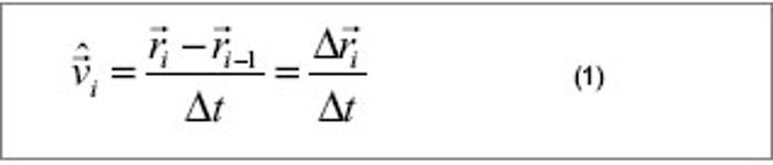
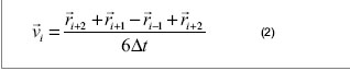
2.4 微跳视成分分离 为了分离出固视的3种成分,一些研究者试图利用信号处理的方法,从眼动记录数据中,分离这些这些成分。相比漂移和微颤,微跳视的眼动轨迹是线性的,它的峰值速度也要大得多[52]。所以微跳视可以通过对速度阈限的估计进行检测。为此,Engbert等[53]提出了微跳视的监测算法。其基本过程包含两个过程:①求出固视中所有采样点的速度。②根据微跳视成分的特性,应用多元统计方法将其从固视中分离出来。在屏幕参考系中,用一个基于时间的点序列{rn}表示固视的所有采样点,每一点的位置矢量表示为 。已知所有相邻点的时间间隔相等,根据则两点之间速度估计值为:"

可根据眼动仪的采样率而定, 近似表示为第i点的速度,因而可用序列 表示所有采样点的瞬时速度。利用这种近似的方法求得速度会存在一定的误差,另外由于眨眼、仪器等的影响,会产生大量噪声,所以,有必要通过5点相邻平均法对采集到的数据进行平滑处理"
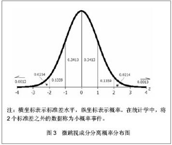
所得基于时间的速度序列即为所有采样点的速度。微跳视(micosaccade)是双眼成分,而微颤(tremor)和漂移(shift)可理解为神经系统的噪声[54]。一次微跳视的峰速度要高于微颤和漂移的峰速度[55]。另外,在采集的固视成分中,大部分的实验数据属于微颤和漂移,微跳视则是小概率事件。根据实验误差理论,噪声信号满足平均值为零的正态分布。由此,可以通过类似推断统计的方法,定义标准差水平,来区分这两类事件(噪声事件和微跳视事件),见图3。"
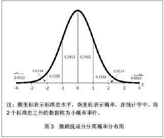
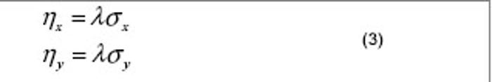
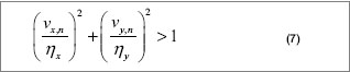
速度是个矢量,可以分解为x方向和y方向2个分量。因此,定义噪声信号(微颤、漂移)和微跳视事件的分界阈值为:"
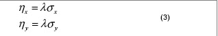

其中 , 分别表示x方向和y方向的阈值。从统计学上讲,尽管阈值的大小可能不同,却采用了同样的标准差水平,因此,也可以统一写成:"


是一个维度无关参数。而且,从这个定义式中可以看出,被试者差异和试次之间的差异都包含在其中。 在固视眼动数据中,速度分布往往是偏态的,因此,需用中位数代替平均值求标准差:"
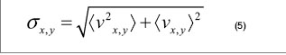
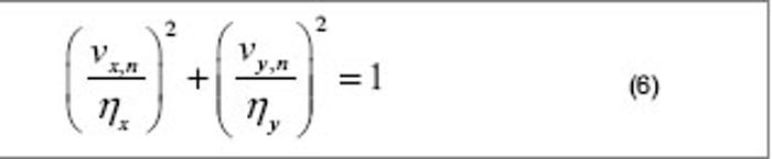
这里 表示中位数。 显然,速度的2个分量是独立的,相关系数r=0。根据多元统计理论,二维变量所确定的实验数据分布在一个椭圆区域(马氏区域),且椭球的长轴和短轴分别平行于x轴和y轴。以速度阈值确定的马氏区域可以表示为:"
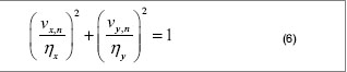

在这个区域以内的事件是噪声事件,之外的则为小概率事件,即微跳视事件。因此,可得微跳视的算法:"
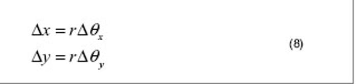
将速度序列 代入上式,分离出所有大于1的数据,其对应的点即为所要分离的微跳视成分。 微跳视分离的实验数据,一般是源于固视的数据,而对于固视而言,眼动的范围是非常微小的,因此,把屏幕坐标系的数据转化为弧度坐标时,可以采用近似计算,则眼动的弧距可以表示为:"
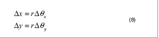

也就是说,在微小跳视情况下,弧度坐标和屏幕坐标之间满足线性关系。"

| 1 Carpenter RHS. Movements of the eyes. Pion Limited, 1988. http://www.tandfonline.com/doi/abs/10.1080/09500348914551271?journalCode=tmop202 Raghunandan A, Frasier J, Poonja S, et al. Psychophysical measurements of referenced and unreferenced motion processing using high-resolution retinal imaging. J Vis. 2008;8(14): 1-11.http://www.ncbi.nlm.nih.gov/pubmed/?term=Psychophysical+measurements+of+referenced+and+unreferenced+motion+processing+using+high-resolution+retinal+imaging.3 Hasegawa J, Yagi T. Emulation of retinal cell responses during fixational eye movements. IEICE Electronics Express. 2010; 7: 184-189. http://a.int-yt.com/detail?paper_id=1dc765f1-357b-b4e6-990a-1192e5b46eb84 Helmholtz H. Helmholtz's Treatise on Physiological Optics Gryphon Editions edn. 1985. http://www.abebooks.com/book-search/title/helmholtz's-treatise-physiological-optics/page-1/5 Riggs LA, Ratliff F, Cornsweet JC, et al. The disappearance of steadily fixated visual test objects. J Opt Soc Am. 1953;43(6):495-501http://www.ncbi.nlm.nih.gov/pubmed/?term=The+disappearance+of+steadily+fixated+visual+test+objects.6 Arathorn DW, Yang Q, Vogel CR, et al. Retinally stabilized cone-targeted stimulus delivery. Opt Express. 2007;15(21):13731-13744.http://www.ncbi.nlm.nih.gov/pubmed/?term=Retinally+stabilized+cone-targeted+stimulus+delivery.7 Martinez-Conde S, Macknik SL. Fixational eye movements across vertebrates: Comparative dynamics, physiology, and perception. J Vis. 2008;8(14):1-16.http://www.ncbi.nlm.nih.gov/pubmed/?term=Fixational+eye+movements+across+vertebrates%3A+Comparative+dynamics%2C+physiology%2C+and+perception.8 Murakami I. Fixational eye movements and motion perception. Prog Brain Res. 2006;154:193-209.http://www.ncbi.nlm.nih.gov/pubmed/170107119 Macedo AF, Crossland MD, Rubin GS. The effect of retinal image slip on peripheral visual acuity. J Vis. 2008;8(14):1-11.http://www.ncbi.nlm.nih.gov/pubmed/?term=Macedo+AF%2C+Crossland+MD%2C+Rubin+GS.+The+effect+of+retinal+image+slip+on+peripheral+visual+acuity.10 Rayner K. Eye movements in reading and information processing: 20 years of research. Psychol Bull. 1998;124(3):372-422.http://www.ncbi.nlm.nih.gov/pubmed/?term=Eye+movements+in+reading+and+information+processing%3A+20+years+of+research.+Psychol+Bull.11 Hermens F, Zanker JM, Walker R. Microsaccades and preparatory set: a comparison between delayed and immediate, exogenous and endogenous pro- and anti-saccades. Exp Brain Res. 2010;201(3):489-498. http://www.ncbi.nlm.nih.gov/pubmed/?term=Microsaccades+and+preparatory+set%3A+a+comparison+between+delayed+and+immediate%2C+exogenous+and+endogenous+pro-+and+anti-saccades.12 Otero-Millan J, Serra A, Leigh RJ, et al. Distinctive Features of Saccadic Intrusions and Microsaccades in Progressive Supranuclear Palsy. J Neurosci. 2011;31(12):4379-4387.http://www.ncbi.nlm.nih.gov/pubmed/?term=Distinctive+Features+of+Saccadic+Intrusions+and+Microsaccades+in+Progressive+Supranuclear+Palsy.13 McCamy M. The efficacy and contribution of microsaccades during visual fixation. Arizona State University, 2011.http://www.ncbi.nlm.nih.gov/pubmed/?term=McCamy+M.+The+efficacy+and+contribution+of+microsaccades+during+visual+fixation.14 Inagaki K, Hirata Y, Usui S. A modeling study on the signal transformation for the microsaccade generation. BMC Neuroscience. 2010;11:115. http://www.biomedcentral.com/1471-2202/11/S1/P11515 Murakami I. Eye movements during fixation as velocity noise in minimum motion detection. Jap Psychol Res. 2010; 52:54-66. http://onlinelibrary.wiley.com/doi/10.1111/j.1468-5884.2010.00424.x/abstract16 Kuang X, Poletti M, Victor JD, et al. Temporal encoding of spatial information during active visual fixation. Curr Biol. 2012;22(6):510-514.http://www.ncbi.nlm.nih.gov/pubmed/2234275117 Komogortsev OV, Gobert DV, Jayarathna S, et al. Standardization of automated analyses of oculomotor fixation and saccadic behaviors. IEEE Trans Biomed Eng. 2010. [Epub ahead of print]http://www.ncbi.nlm.nih.gov/pubmed/?term=Standardization+of+automated+analyses+of+oculomotor+fixation+and+saccadic+behaviors.18 Brien DC, Corneil BD, Fecteau JH, et al. The behavioral and neurophysiological modulation of microsaccades in monkeys. J Eye Mov Res. 2009; 3: 1-12. http://bmi.ssc.uwo.ca/CorneilLab.org/Corneil_Lab/Publications_files/Brien_et_al_2009.pdf19 Laurutis V, Zemblys R, Buivis L. Fixational Eye Movements: Influence on the Accuracy in the Target Pointing Tasks. Electronics and Electrical Engineering. Kaunas: Technologija, 2009:93. http://bimc.su.lt/en/pub/34-s1/47-fixational-eye-movements-influence-on-the-accuracy-in-the-target-pointing-tasks20 Martinez-Conde S. The Role of Eye Movements During Visual Fixation. Barrow Quarterly. 2005; 21: 44-48.http://smc.neuralcorrelate.com/files/publications/martinez-conde_bniq05.pdf 21 Martinez-Conde S. Fixational eye movements in normal and pathological vision. Prog Brain Res. 2006;154:151-176.http://www.ncbi.nlm.nih.gov/pubmed/1701070922 Steinman RM, Cunitz RJ, Timberlake GT, et al. Voluntary control of microsaccades during maintained monocular fixation. Science. 1967;155(3769):1577-1579.http://www.ncbi.nlm.nih.gov/pubmed/?term=Voluntary+control+of+microsaccades+during+maintained+monocular+fixation. 23 Spauschus A, Marsden J, Halliday DM, et al. The origin of ocular microtremor in man. Exp Brain Res. 1999;126(4):556-562.http://www.ncbi.nlm.nih.gov/pubmed/?term=The+origin+of+ocular+microtremor+in+man.24 Li J, Zhang X. In Imaging Systems and Techniques (IST). IEEE Int Conf. 2012: 28-33.http://ieeexplore.ieee.org/xpl/mostRecentIssue.jsp?reload=true&punumber=595540225 Krauskopf J, Cornsweet TN, Riggs LA. Analysis of eye movements during monocular and binocular fixation. J Optical Soc Am. 1960. http://www.opticsinfobase.org/josa/abstract.cfm?uri=josa-50-6-57226 Zhang X, Li J. in Proc. 4th International Conference on Bioinformatics and Biomedical Technology. 133-140.27 Moshel S, Liang J, Caspi A, et al. Phase‐Synchronization Decay of Fixational Eye Movements. Ann N Y Acad Sci. 2005;1039:484-488.http://www.ncbi.nlm.nih.gov/pubmed/?term=Phase%E2%80%90Synchronization+Decay+of+Fixational+Eye+Movements.28 Shakhnovich AR, Thomas JG. Micro-tremor of the eyes of comatose patients. Electroencephalogr Clin Neurophysiol. 1977;42(1):117-119.http://www.ncbi.nlm.nih.gov/pubmed/?term=Micro-tremor+of+the+eyes+of+comatose+patients.29 Valsecchi M, Betta E, Turatto M. Visual oddballs induce prolonged microsaccadic inhibition. Exp Brain Res. 2007;177(2):196-208.http://www.ncbi.nlm.nih.gov/pubmed/?term=Visual+oddballs+induce+prolonged+microsaccadic+inhibition.30 Pastukhov A, Braun J. Rare but precious: Microsaccades are highly informative about attentional allocation. Vision Res. 2010;50(12):1173-1184.http://www.ncbi.nlm.nih.gov/pubmed/?term=Rare+but+precious%3A+Microsaccades+are+highly+informative+about+attentional+allocation.31 Horowitz TS, Fine EM, Fencsik DE, et al. Fixational eye movements are not an index of covert attention. Psychol Sci. 2007;18(4):356-363.http://www.ncbi.nlm.nih.gov/pubmed/1747026232 Laubrock J, Kliegl R, Rolfs M, et al. When do microsaccades follow spatial attention? Atten Percept Psychophys. 2010;72(3):683-694. http://www.ncbi.nlm.nih.gov/pubmed/?term=When+do+microsaccades+follow+spatial+attention%3F33 Otero-Millan J, Troncoso XG, Macknik SL, et al. Saccades and microsaccades during visual fixation, exploration, and search: Foundations for a common saccadic generator. J Vis. 2008;8(14): 1-18.http://www.ncbi.nlm.nih.gov/pubmed/?term=Saccades+and+microsaccades+during+visual+fixation%2C+exploration%2C+and+search%3A+Foundations+for+a+common+saccadic+generator.34 Mergenthaler K, Engbert R. Microsaccades are different from saccades in scene perception. Exp Brain Res. 2010;203(4):753-757.http://www.ncbi.nlm.nih.gov/pubmed/?term=Microsaccades+are+different+from+saccades+in+scene+perception. 35 Hafed ZM, Krauzlis RJ. Similarity of superior colliculus involvement in microsaccade and saccade generation. J Neurophysiol. 2012;107(7):1904-1916.http://www.ncbi.nlm.nih.gov/pubmed/?term=Similarity+of+superior+colliculus+involvement+in+microsaccade+and+saccade+generation.36 Inagaki K, Hirata Y, Usui S. A model-based theory on the signal transformation for microsaccade generation. Neural Netw. 2011;24(9):990-997.http://www.ncbi.nlm.nih.gov/pubmed/?term=A+model-based+theory+on+the+signal+transformation+for+microsaccade+generation.37 Horwitz GD, Albright TD. Short-latency fixational saccades induced by luminance increments. J Neurophysiol. 2003;90(2):1333-1339.http://www.ncbi.nlm.nih.gov/pubmed/1290451238 Collewijn H, Kowler E. The significance of microsaccades for vision and oculomotor control. J Vis. 2008;8(14):1-21.http://www.ncbi.nlm.nih.gov/pubmed/?term=The+significance+of+microsaccades+for+vision+and+oculomotor+control.39 Guy W. The temporal and spatial limits of compensation for fixational eye movements. Vision Res. 2006;46(18):2848-2858.http://www.ncbi.nlm.nih.gov/pubmed/?term=The+temporal+and+spatial+limits+of+compensation+for+fixational+eye+movements.40 Ikuya M. In Progress in brain research. Elsevier. 2006;15: 193-209. http://www.sciencedirect.com/science/article/pii/S007961230654010541 Martinez-Conde S, Macknik SL, Troncoso XG, et al. Microsaccades Counteract Visual Fading during Fixation. Neuron. 2006;49(2):297-305.http://www.ncbi.nlm.nih.gov/pubmed/1642370242 Mergenthaler K, Engbert R. Modeling the control of fixational eye movements with neurophysiological delays. Phys Rev Lett. 2007;98(13):138104.http://www.ncbi.nlm.nih.gov/pubmed/?term=Modeling+the+control+of+fixational+eye+movements+with+neurophysiological+delays. 43 Martinez-Conde S, Macknik SL, Hubel DH. Microsaccadic eye movements and firing of single cells in the striate cortex of macaque monkeys. Nat Neurosci. 2000;3(3):251-258.http://www.ncbi.nlm.nih.gov/pubmed/1070025744 Ditchburn R. The function of small saccades. Vision Res. 1980;20(3):271-272.http://www.ncbi.nlm.nih.gov/pubmed/?term=Ditchburn+R.+The+function+of+small+saccades.45 Ralf E. In Progress in brain research. Elsevier. 2006;154: 177-192. http://www.sciencedirect.com/science/article/pii/S007961230654009946 Bruno MA, Vanhaudenhuyse A, Schnakers C, et al. Visual fixation in the vegetative state: an observational case series PET study. BMC Neurol. 2010;10:35. doi: 10.1186/1471-2377-10-35.http://www.ncbi.nlm.nih.gov/pubmed/?term=Visual+fixation+in+the+vegetative+state%3A+an+observational+case+series+PET+study.47 Ditchburn R, Fender D, Mayne S. Vision with controlled movements of the retinal image. J Physiol. 1959;145(1):98-107.http://www.ncbi.nlm.nih.gov/pubmed/1362142448 Bonneh YS, Donner TH, Sagi D, et al. Motion-induced blindness and microsaccades: Cause and effect. J Vis. 2010;10(14):22.http://www.ncbi.nlm.nih.gov/pubmed/?term=Motion-induced+blindness+and+microsaccades%3A+Cause+and+effect.49 Engbert R, Kliegl R. Microsaccades keep the eyes' balance during fixation. Psychol Sci. 2004;15(6):431-436.http://www.ncbi.nlm.nih.gov/pubmed/?term=Engbert+R%2C+Kliegl+R.+Microsaccades+keep+the+eyes'+balance+during+fixation.50 Engbert R, Mergenthaler K. Microsaccades are triggered by low retinal image slip. Proc Natl Acad Sci U S A. 2006;103(18):7192-7197.http://www.ncbi.nlm.nih.gov/pubmed/?term=ngbert+R%2C+Mergenthaler+K.+Microsaccades+are+triggered+by+low+retinal+image+slip.51 Martinez-Conde S, Macknik SL, Hubel DH. The role of fixational eye movements in visual perception. Nat Rev Neurosci. 2004;5(3):229-240.http://www.ncbi.nlm.nih.gov/pubmed/1497652252 Blignaut P, Beelders T. In Proceedings of the Symposium on Eye Tracking Research and Applications. 289-292 (ACM). http://dl.acm.org/citation.cfm?id=2168556&picked=prox53 Engbert R, Kliegl R. Binocular coordination in microsaccades. The mind’s eyes: Cognitive and applied aspects of eye movements, 2003: 103-117.54 Paladini C, Holschneider M, Mergenthaler K, et al. Microsaccade characterization using the continuous wavelet transform and principal component analysis. J Eye Movement Res. 2010; 3(5):1-14.http://www.researchgate.net/publication/233932469_Microsaccade_characterization_using_the_continuous_wavelet_transform_and_principal_component_analysis55 Engbert R, Mergenthaler K, Sinn P, et al. An integrated model of fixational eye movements and microsaccades. Proc Natl Acad Sci U S A. 2011;108(39):E765-770. http://www.ncbi.nlm.nih.gov/pubmed/?term=An+integrated+model+of+fixational+eye+movements+and+microsaccades.56 Macknik SL, Livingstone MS. Neuronal correlates of visibility and invisibility in the primate visual system. Nat Neurosci. 1998;1(2):144-149.http://www.ncbi.nlm.nih.gov/pubmed/?term=Neuronal+correlates+of+visibility+and+invisibility+in+the+primate+visual+system.57 Hass CA, Horwitz GD. Effects of microsaccades on contrast detection and V1 responses in macaques. J Vis. 2011;11(3):1-17. http://www.ncbi.nlm.nih.gov/pubmed/?term=Effects+of+microsaccades+on+contrast+detection+and+V1+responses+in+macaques.58 Hafed ZM, Krauzlis RJ. Microsaccadic suppression of visual bursts in the primate superior colliculus. J Neurosci. 2010;30(28):9542-9547. http://www.ncbi.nlm.nih.gov/pubmed/?term=Microsaccadic+suppression+of+visual+bursts+in+the+primate+superior+colliculus.59 Martinez-Conde S, Macknik SL, Troncoso XG, et al. Microsaccades: a neurophysiological analysis. Trends Neurosci. 2009;32(9):463-475.http://www.ncbi.nlm.nih.gov/pubmed/1971618660 Greschner M, Bongard M, Rujan P, et al. Retinal ganglion cell synchronization by fixational eye movements improves feature estimation. Nat Neurosci. 2002;5(4):341-347.http://www.ncbi.nlm.nih.gov/pubmed/?term=Retinal+ganglion+cell+synchronization+by+fixational+eye+movements+improves+feature+estimation.61 Martinez-Conde S, Macknik SL, Hubel DH. The function of bursts of spikes during visual fixation in the awake primate lateral geniculate nucleus and primary visual cortex. Proc Natl Acad Sci U S A. 2002;99(21):13920-13925.http://www.ncbi.nlm.nih.gov/pubmed/?term=The+function+of+bursts+of+spikes+during+visual+fixation+in+the+awake+primate+lateral+geniculate+nucleus+and+primary+visual+cortex.62 Reppas JB, Usrey WM, Reid RC. Saccadic eye movements modulate visual responses in the lateral geniculate nucleus. Neuron. 2002; 35: 961-974.http://www.ncbi.nlm.nih.gov/pubmed/12372289 63 Tse PU, Baumgartner FJ, Greenlee MW. Event-related functional MRI of cortical activity evoked by microsaccades, small visually-guided saccades, and eyeblinks in human visual cortex. Neuroimage. 2010;49(1):805-816.http://www.ncbi.nlm.nih.gov/pubmed/?term=Event-related+functional+MRI+of+cortical+activity+evoked+by+microsaccades%2C+small+visually-guided+saccades%2C+and+eyeblinks+in+human+visual+cortex 64 Melloni L, Schwiedrzik CM, Rodriguez E, et al. (Micro)Saccades, corollary activity and cortical oscillations. Trends Cogn Sci. 2009;13(6):239-245.http://www.ncbi.nlm.nih.gov/pubmed/?term=(Micro)Saccades%2C+corollary+activity+and+cortical+oscillations. |
| [1] | Wu Guang-wen, Ye Jin-xia, Zheng Chun-song, Chen Wen-lie, Liu Xian-xiang, Ye Hong-zhi . Effect of Tougu Xiaotong capsule on articular cartilage changes in rat models of osteoarthritis [J]. Chinese Journal of Tissue Engineering Research, 2014, 18(49): 7924-7929. |
| [2] | Lu Shou-ming, Lu Shou-liang, Sun Tian-wei, Zhang Hang, Wang Qi-ming. Estrogen effects on serum interleukin-8 and interleukin-10 expression in ovariectomized rats with osteoporosis [J]. Chinese Journal of Tissue Engineering Research, 2013, 17(24): 4394-4400. |
| [3] | Adila•Azhati, Zhao Long, Wang Qian, Ma Yi-tong. Cardiac function and electrophysiological characteristics in myocardial infarction rats after tissue engineered cardiac patch transplantation [J]. Chinese Journal of Tissue Engineering Research, 2013, 17(24): 4401-4408. |
| [4] | Shi Yu-lu, Li Xiao-yuan, Cao Mei-na, Yu Shu-yuan, Wang Ping. Simultaneous isolation of myocardial cells and cardiac fibroblasts from neonatal rats [J]. Chinese Journal of Tissue Engineering Research, 2013, 17(24): 4414-4420. |
| [5] | Wang Qi-feng, Wang Zhi-gang, Huang Jing. Left ventricular papillary muscle ablation in canines by ultrasound ablation catheter [J]. Chinese Journal of Tissue Engineering Research, 2013, 17(24): 4409-4413. |
| [6] | Chen Li-ying, Fan Kun-wu, Wang Jin-shui, Yu Xu-ming, Xu Cheng. Gene expression of the skin and sweat glands in different development stages of embryo [J]. Chinese Journal of Tissue Engineering Research, 2013, 17(24): 4421-4428. |
| [7] | Li Wei-hua, Gao Yu-wei, Li De-shui, Xing Yu-xi. Botulinum toxin type A induces apoptosis of human hypertrophic scar fibroblasts [J]. Chinese Journal of Tissue Engineering Research, 2013, 17(24): 4429-4435. |
| [8] | Zhao Gang, Wei Jia-jia, Zhang Xiao-ping, Yuan Xu, Shen Jian-xuan, Zhang Wei. Two kinds of intermaxillary traction influence the mandibular micro-implant and its surrounding: Three-dimensional finite element analysis [J]. Chinese Journal of Tissue Engineering Research, 2013, 17(24): 4444-4450. |
| [9] | Liu Gang, Ma Xin-long, Deng Shu-cai, Chen Si. Expression and significance of stromal cell derived factor-1 in the intervertebral disk after lumbar disc degeneration [J]. Chinese Journal of Tissue Engineering Research, 2013, 17(24): 4488-4494. |
| [10] | Zhu Jian-hua, Tang Chun-ling, Zhang Yan-qiu, Liu Ji-guang. Number of mast cells and expression of transforming growth factor beta 1 in the submandibular gland of diabetes mellitus rats [J]. Chinese Journal of Tissue Engineering Research, 2013, 17(24): 4457-4464. |
| [11] | Zhen Lei, Wang Xiao, Miu Huang-tai, Qiao Shi-bin, Wu Xing-xin, Qiao Yan, Liu Bai-qiu, Liu Xin-min, Que Bin, Nie Shao-ping. In vivo concentration gradient of basic fibroblast growth factor after coronary venous retrograde perfusion [J]. Chinese Journal of Tissue Engineering Research, 2013, 17(24): 4473-4480. |
| [12] | Wang Jing, Dong Fang-fang, Li Xiao-feng, Xu Jin-hai, Shu Bing, Shi Qi, Wang Yong-jun, Zhou Chong-jian. 低氧诱导因子1α基因敲除小鼠椎间盘退变与益气化瘀方的干预 [J]. Chinese Journal of Tissue Engineering Research, 2013, 17(24): 4481-4487. |
| [13] | Ning Chang, Yu Chang-lin, Hu Kai-xun. Establishing a mouse model of radiation-induced thymus injury [J]. Chinese Journal of Tissue Engineering Research, 2013, 17(24): 4465-4472. |
| [14] | Chen Dan-dan, Zhou Da-xin, Guan Li-hua, Chen Fa-dong, Dong Li-li, Qian Ju-ying, Ge Jun-bo. Application of continuous thermodilution method in beagle models with pulmonary arterial hypertension [J]. Chinese Journal of Tissue Engineering Research, 2013, 17(24): 4509-4514. |
| [15] | Dong Gang, Zheng Jian-jin, Li Tao, Xu Xin, Lu Shu-lai. A Wistar rat model of radiation-induced masseter injury [J]. Chinese Journal of Tissue Engineering Research, 2013, 17(24): 4515-4520. |
| Viewed | ||||||
|
Full text |
|
|||||
|
Abstract |
|
|||||