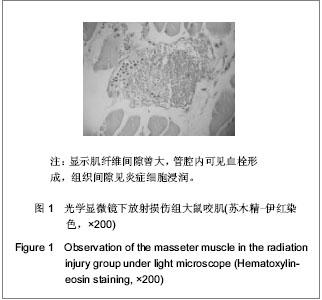
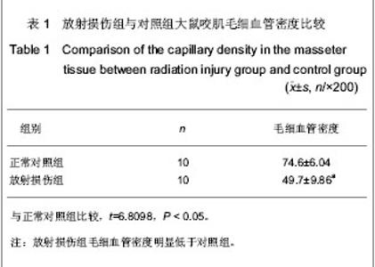
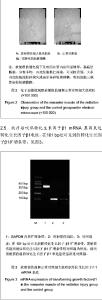
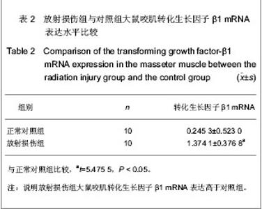
| [1] Czarnota GJ, Karshafianc R, Burnsc PN,et al.Tumor radiation response enhancement by acoustical stimulation of the vasculature. Proceedings of the National Academy of Sciences. 2012;109(30):2033-2041.http://www.pnas.org/content/early/2012/06/29/1200053109 [2] Williams JP, Brown SL, Georges GE, et al. Animal models for medical countermeasures to radiation exposure.Radiat Res. 2010;173(4):557-78.http://www.ncbi.nlm.nih.gov/pubmed/20334528 [3] Zhang LZ, Cao YD, Tang TY, et al. Zhonghua Fangshe Yixue yu Fanghu Zazhi.2006;26(2):148-150.张龙珍,曹远东,唐天友,等.全脑常规照射开放血脑屏障动物模型的建立[J].中华放射医学与防护杂志,2006,26(2):148-150.http://www.cqvip.com/QK/95691X/200602/21671656.html [4] Pohlers D, Brenmoehl J, Loffler I, et al. TGF-β and fibrosis in different organs- molecular pathway imprints. Biochim Biophys Acta. 2009;1792(8):746-756.http://www.sciencedirect.com/science/article/pii/S0925443909001355 [5] Liu K,Kudo K,Abe Y,et al.Inhibition of Transforming Growth Factor-β, Hypoxia-inducibleFactor-1α and Vascular Endothelial Growth Factor Reduced Late Rectal Injury Induced by Irradiation. J Radiat Res. 2009; 50(3):233-239.http://www.ncbi.nlm.nih.gov/pubmed/19346676 [6] Liao CT, Chang JT, Wang HM, et al. Does adjuvant radiation therapy improve outcomes in Pt1 - 3N0 oral cavity cancer with tumor–free margins and perineural invasion? . Int J Radiat Oncol Biol Phys.2008;71(2):371-376.http://www.ncbi.nlm.nih.gov/pubmed/18474310 [7] Nichol AM, Smith SL, D’yachkova Y, et al. Quantification of masticatory muscle atrophy after high-dose radiotherapy. Int J Radiat Onco Biol Phys.2003; 56(4):1170-1179. http://www.ncbi.nlm.nih.gov/pubmed/12829156 [8] Tolentino Ede S, Centurion BS, Ferreira LH, et al. Oral adverse effects of head and neck radiotherapy: literature review and suggestion of a clinical oral care guideline for irradiated patients. J Appl Oral Sci. 2011; 19(5):448-454.http://www.ncbi.nlm.nih.gov/pubmed/21986648 [9] Sciubba JJ, Goldenberg D, et al. Oral complications of radiotherapy. Lancet Oncol. 2006;7:175-183.http://www.ncbi.nlm.nih.gov/pubmed/16455482 [10] Dewhirst MW. Relationships between cycling hypoxia, HIF-1, angiogenesis and oxidative stress. Radiat Res. 2009;172(6): 653–665.http://www.ncbi.nlm.nih.gov/pubmed/?term=Relationships+between+cycling+hypoxia%2C+HIF-1%2C+angiogenesis+and+oxidative+stress [11] Liu J,Gao QG.Linchuang Yiyao Shijian Zazhi.2006;15(1):3-5.刘军,高庆国.放射性骨骼肌损伤[J].临床医药实践杂志, 2006, 15(1):3-5.http://www.cnki.com.cn/Article/CJFDTotal-SXLC200601001.htm [12] Zhang S,Lei CX,Chu ZY,et al.Zhongguo Fushe Yixue.2009; 18(1) :87-88.张术,雷呈祥,储智勇,等. 辐射剂量的快速生物学估算[J]. 中国辐射卫生, 2009, 18(1):87-88.http://www.cnki.com.cn/Article/CJFDTotal-REDI200901054.htm [13] He K, Feng YH, Luo HM, et al. Shiyan Dongwuxue. Zazhi 2008;25(1):12-14.何康,冯有辉,罗红梅,等.不同厂家的戊巴比妥钠对大鼠和家兔麻醉效果的比较[J]. 实验动物学,2008,25(1):12-14.http://www.cnki.com.cn/Article/CJFDTotal-SYDG200801005.htm [14] Li ZM,Qu MX,Xu SP,et al.Xiandai Kouqiang Yixue Zazhi.2009; 23(4):532-535.李志民,曲民兴,徐寿平,等.电力辐射对大鼠咬肌细胞超微结构及糖原含量的影响[J]. 现代口腔医学杂志.2009, 23(5):532-535.http://www.cnki.com.cn/Article/CJFDTotal-XDKY200905032.htm [15] Boncompagni S, Amelio L, Fulle S, et al. Progressive disorganization of the excitation-contraction coupling apparatus in aging human skeletal muscle as revealed by electron microscopy: a possible role in the decline of muscle performance. J Gerontol A Biol Sci Med Sci. 2006;61(10): 995-1008.http://www.ncbi.nlm.nih.gov/pubmed/17077192 [16] Zhao L, Ji W, Zhang L, et al.Changes of circulating transforming growth factor-beta1 level during radiation therapy are correlated with the prognosis of locally advanced non-small cell lung cancer. J Thorac Oncol. 2010;5(4): 521-525.http://www.ncbi.nlm.nih.gov/pubmed/20130485 [17] Liu H,Xiong M,Rong TH,et al.Aizheng.2008;27(1):18-24. 刘慧,熊迈,戎铁华,等.大鼠心脏组织TGF-beta1表达水平与放射性损伤关系的实验研究[J].癌症,2008;27(1):18-24.http://www.cnki.com.cn/Article/CJFDTOTAL-AIZH200801008.htm [18] Gervaz P, Morel P, Vozenin-Broton MC. Molecular aspects of intestinal radiation-induced fibrosis. Curr Mol Med.2009;9(3): 273-280.http://www.ncbi.nlm.nih.gov/pubmed/19355909 |