Chinese Journal of Tissue Engineering Research ›› 2017, Vol. 21 ›› Issue (2): 260-267.doi: 10.3969/j.issn.2095-4344.2017.02.018
Previous Articles Next Articles
Preparation of span-poly(ethylene glycol) ultrasound contrast agent microbubbles combined with folate-carbon nano tube-paclitaxel
- 1Pharmaceutical Research Institute of Heilongjiang Province, Jiamusi University, Jiamusi 154007, Heilongjiang Province, China; 2Department of Neurology, First Affiliated Hospital of Harbin Medical University, Harbin 150081, Heilongjiang Province, China; 3Department of Pharmacy, Heilongjiang General Hospital of Arm Police Force, Harbin 150070, Heilongjiang Province, China
-
Received:2016-12-09Online:2017-01-18Published:2017-02-27 -
Contact:Zhang Jie, M.D., Lecturer, Pharmaceutical Research Institute of Heilongjiang Province, Jiamusi University, Jiamusi 154007, Heilongjiang Province, China; Department of Neurology, First Affiliated Hospital of Harbin Medical University, Harbin 150081, Heilongjiang Province, China Zhang Yu, Master, Professor, Pharmaceutical Research Institute of Heilongjiang Province, Jiamusi University, Jiamusi 154007, Heilongjiang Province, China -
About author:Liu Jun-xi, Studying for master’s degree, Pharmaceutical Research Institute of Heilongjiang Province, Jiamusi University, Jiamusi 154007, Heilongjiang Province, China -
Supported by:the National Natural Science Foundation of China, No. 81601616; the Post Doctoral Foundation of Heilongjiang Province, No. LBH-Z15164; the Natural Science Foundation of Heilongjiang Province, No. H2016086; the Scientific and Technologic Innovation Program for the Graduates of Jiamusi University, No. LM2015-091; the Scientific Innovation Group of Jiamusi University, No. CXTD-2013-05
CLC Number:
Cite this article
Liu Jun-xi, Zhang Jie, Zhang Yu, Zhao Yue, Wan Guo-jing, Li Guo-zhong.
share this article

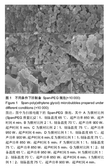
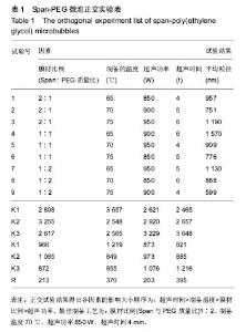
2.1 优化Span-PEG微泡的制备工艺 采用声振空化法制备Span-PEG微泡,不同膜材比例、制备温度、超声功率、超声时间条件下所制备 Span-PEG微泡形态见图1。各组微泡的粒径见表1。各因素的影响大小顺序为:超声时间>制备温度>膜材比例>超声功率。最佳制备工艺为:膜材比例(Span与PEG质量比)1∶2、制备温度70 ℃、超声功率850 W、超声时间4 min。"
"
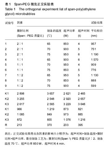
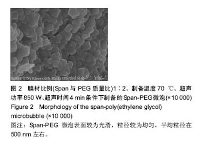
膜材比例(Span : PEG质量比)1∶2、制备温度70 ℃、超声功率850 W、超声时间4 min条件下所制备的Span-PEG微泡见图2。这种Span-PEG微泡表面较为光滑,粒径较为均匀,平均粒径在500 nm左右。"


2.2 复合物中间体 2.2.1 FA-CS偶合物 壳聚糖的红外光谱中在3 345 cm-1附近存在较宽的一段吸收峰,这是由于壳聚糖中O-H和N-H的伸缩振动叠加造成的,在2 920 cm-1 和2 864 cm-1 处出现的是C-H伸缩振动吸收峰,在1 655 cm-1和1 597 cm-1吸收峰分别是C-N伸缩振动和氨基(-NH2)的特征峰(图3a)。叶酸的红外光谱中在3 547 cm-1处出现叶酸中羟基(O-H)的吸收峰,在3 419 cm-1和3 321 cm-1处吸收峰是由叶酸中氨基、-NH的伸缩振动引起的(图3c)。FA-CS偶合物红外光谱中在3 436 cm-1附近只有一个吸收峰,为仲酰胺的特征峰,这是由于壳聚糖上的氨基基团与叶酸上的羧基基团(-COOH)发生静电自组装所致;在2 923 cm-1和2 868 cm-1 出现了壳聚糖中C-H伸缩振动吸收峰;在1 655 cm-1和 1 597 cm-1处的吸收峰明显减弱,在1 597 cm-1 处的N-H变形振动峰移动到了高频的1 628 cm-1[35],这是由于FA-CS偶合物中壳聚糖氨基被叶酸部分取代(图3b)。"
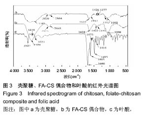

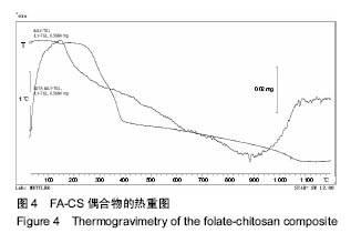
FA-CS偶合物热分析如图4。由图4可见,偶合物在240-400 ℃温度范围存在失重[35-36],这是由于FA-CS不稳定,偶合物中壳聚糖在240 ℃左右开始出现分解,至 400 ℃左右分解完全,FA-CS中壳聚糖的质量分数为35%。 图3,4说明采用静电自组装成功合成了FA-CS偶合物。"

2.2.2 羧基化碳纳米管 由图5可见,羧基化前后的碳纳米管均在波数1 550 cm-1处存在碳碳双键(C=C)的伸缩振动,这是碳纳米管自身结构的振动吸收所致。羧基化前3 625 cm-1处吸收峰为-OH吸收峰(图5a),是由于碳纳米管表面所吸附少量水分子所造成。羧基化后3 303 cm-1附近的强宽峰谱带为羧基中的-OH的伸缩振动产生的,在1 710 cm-1处出现了羧基中的C=O特征吸收峰,1 207 cm-1处存在(C-O)伸缩振动吸收峰[35](图5b),这是由于在混酸等强氧化剂作用下,碳纳米管表面带上了一定数量的羟基所引起,由此可知,经过混酸处理后的碳纳米管上连接上了羧基基团。"


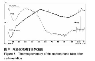
羧基化碳纳米管热分析见图6。由图6可知,在100 ℃左右发生失重,这是碳纳米管表面的水挥发所致。在400-600 ℃范围发生的重量损失,失重率为26.6%左右,并且在500-600 ℃范围存在放热峰,这是由于碳纳米管上存在羧基引起。"
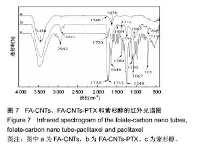
2.2.3 静电自组装产物(FA-CNTs)负载紫杉醇 由图3b,7a可知,静电自组装产物FA-CNTs在3 438 cm-1处出现了FA-CS中仲酰胺的特征峰;在1 740 cm-1处出现了一个小峰,此为酯键上的C=O伸缩振动峰;在1 630 cm-1处为羧基的C=O伸缩振动,这是由于FA-CS在1 628 cm-1处的N-H变形振动峰所致。这说明已成功将FA-CS静电自组装至碳纳米管上。 紫杉醇的红外光谱中,在3 500 cm-1附近的O-H伸缩振动吸收峰与N-H伸缩振动峰重叠;在2 942 cm-1处为苯环上的C-H伸缩振动峰,在1 734 cm-1和1 713 cm-1处出现特征吸收峰为酮羰基C=O的2个裂分峰,在1 645 cm-1处为酰胺基的羰基峰,在1 500 cm-1附近为苯环的骨架振动所产生的吸收峰;在1 380 cm-1处为甲基产生的C-H面内弯曲振动, 1 248 cm-1处为C-N键伸缩振动,在1 067 cm-1、 1 110 cm-1处为仲醇和叔醇中的C-O伸缩振动产生的吸收峰;在900-690 cm-1处为苯环C-H的面外弯曲振动所产生的吸收峰,其中715 cm-1为紫杉醇单取代苯的特征峰(图7c)。 复合物FA-CNTs-PTX在3 438 cm-1处的一个单峰,是图7(a)FA-CNTs中仲酰胺的特征峰,在2 921 cm-1苯环上的C-H伸缩振动峰;在1 372 cm-1处为紫杉醇甲基产生的C-H面内弯曲振动吸收峰,在1 241 cm-1处为紫杉醇的C-N键伸缩振动吸收峰,在1 067 cm-1处为紫杉醇的叔醇中的C-O伸缩振动吸收峰;在713 cm-1为紫杉醇单取代苯的特征峰产生的吸收峰(图7b)。这些说明已成功将紫杉醇负载到FA-CNTs上。 综上可知,成功得到复合物FA-CNTs-PTX。 2.3 复合FA-CNTs-PTX的微泡的制备结果"


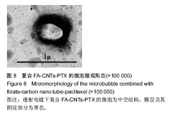
2.3.1 复合微泡形貌 图8为透射电镜下检测到的复合FA-CNTs-PTX的微泡形貌,内部为中空结构,膜层及其附近部分为黑色阴影区。"
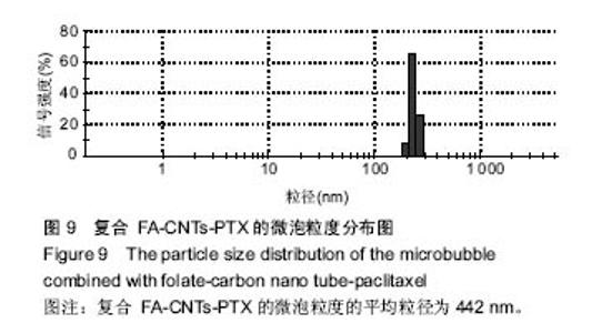
2.3.2 复合微泡的粒径分布 图9为复合FA-CNTs-PTX的微泡粒度分布图,复合微泡平均粒径为442 nm,粒径分布较为集中,肿瘤微环境中丰富的小血管壁通常其结构是不完整的,其血管壁间隙通常在700 nm附近,可见复合 FA-CNTs-PTX的微泡尺寸具有靶向肿瘤的条件,为后期抗肿瘤实验提供了理论依据。 "

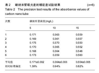
2.3.3 复合微泡中碳纳米管负载率 测得3组不同浓度的碳纳米管吸光度值见表2,通过计算得碳纳米管低中高浓度的相对标准偏差分别为1.39%,0.94%和0.82%,说明紫外分光光度法用于碳纳米管含量测定的精密度良好。"
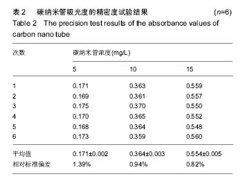
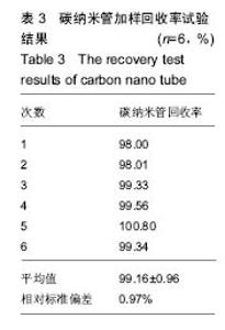
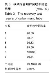
表3为碳纳米管加样回收率试验结果。根据加标样前后测得的复合微泡中碳纳米管总质量差与加入碳纳米管标样质量的比值得到碳纳米管加样回收率,即相对标准偏差为0.97%,说明用紫外分光光度法测得的碳纳米管含量准确度高、方法可行,符合测试的相关要求。"
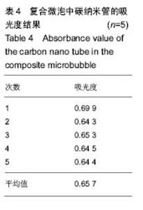
根据表4复合微泡中碳纳米管吸光度结果,得出复合微泡中碳纳米管浓度为0.0169 g/L,复合微泡干粉中碳纳米管的负载率为1.69%。"


2.3.4 复合微泡中紫杉醇负载率 测得3组不同质量浓度的紫杉醇的吸光度值见表5,通过计算得低中高浓度的相对标准偏差分别为2.17%,2.04%和1.84%,说明紫外分光光度法用于紫杉醇含量的测定精密度良好。"
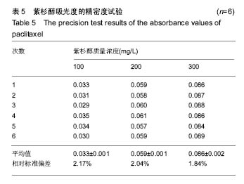

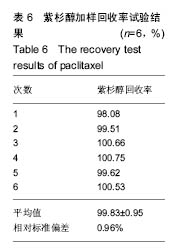
表6为紫杉醇的加样回收率试验结果,根据测得的加标样前后复合微泡中紫杉醇总质量差与加入紫杉醇标样质量的比值得紫杉醇加样回收率,即相对标准偏差为0.96%,说明用紫外分光光度法测得的紫杉醇含量准确度高、方法可行,符合测试的相关要求。"
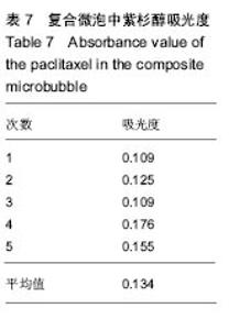
根据表7复合微泡中紫杉醇吸光度结果,得出复合微泡中紫杉醇质量浓度0.479 g/L,复合微泡冻干粉中紫杉醇负载率为47.9%。 综上可知,复合微泡中紫杉醇的负载率(47.9%)大于碳纳米管负载率(1.69%),即紫杉醇在复合微泡中的载药量远大于碳纳米管在复合微泡中的负载量,这是因为紫杉醇不仅可以通过π-π吸附负载于碳纳米管表面,而且还可以通过碳纳米管独特的中空管状结构进入碳纳米管内部。"

| [1]Gramiak R, Shah PM. Echocardiography of the aortic root. Invest Radiol. 1968;3(5):356-366.[2]Sheeran PS, Luois S, Dayton PA, et al. Formulation and acoustic studies of a new phase-shift agent for diagnostic and therapeutic ultrasound. Langmuir. 2011;27(17):10412-10420.[3]Eisenbrey JR, Burstein OM, Kambhampati R, et al. Development and optimization of a doxorubicin loaded poly(lactic acid) contrast agent for ultrasound directed drug delivery. J Control Release. 2010;143(1):38-44.[4]Yan F, Li X, Jin Q, et al. Therapeutic ultrasonic microbubbles carrying paclitaxel and LyP-1 peptide: preparation, characterization and application to ultrasound-assisted chemotherapy in breast cancer cells. Ultrasound Med Biol. 2011;37(5):768-779.[5]Unger EC, Hersh E, Vannan M, et al. Local drug and gene delivery through microbubbles. Prog Cardiovasc Dis. 2001; 44(1):45-54.[6]Eisenbrey JR, Burstein OM, Kambhampati R, et al. Development and optimization of a doxorubicin loaded poly(lactic acid) contrast agent for ultrasound directed drug delivery. J Control Release. 2010;143(1):38-44.[7]Wicki A, Rochlitz C, Orleth A, et al. Targeting tumor-associated endothelial cells: anti-VEGFR2 immunoliposomes mediate tumor vessel disruption and inhibit tumor growth. Clin Cancer Res. 2012;18(2):454-464.[8]Gao ZG, Fain HD, Rapoport N. Controlled and targeted tumor chemotherapy by micellar-encapsulated drug and ultrasound. J Control Release. 2005;102(1):203-222.[9]Wang L, Li L, Guo Y, et al. Construction and in vitro/in vivo targeting of PSMA-targeted nanoscale microbubbles in prostate cancer. Prostate. 2013;73(11):1147-1158.[10]Yang K, Hu L, Ma X, et al. Multimodal imaging guided photothermal therapy using functionalized graphene nanosheets anchored with magnetic nanoparticles. Adv Mater. 2012;24(14):1868-1872.[11]Janib SM, Moses AS, MacKay JA. Imaging and drug delivery using theranostic nanoparticles. Adv Drug Deliv Rev. 2010; 62(11):1052-1063.[12]Barua A, Yellapa A, Bahr JM, et al. Interleukin 16- (IL-16-) Targeted Ultrasound Imaging Agent Improves Detection of Ovarian Tumors in Laying Hens, a Preclinical Model of Spontaneous Ovarian Cancer. Biomed Res Int. 2015;2015: 567459.[13]Mancini M, Greco A, Salvatore G, et al. Imaging of thyroid tumor angiogenesis with microbubbles targeted to vascular endothelial growth factor receptor type 2 in mice. BMC Med Imaging. 2013;13:31.[14]Mannaris C, Averkiou MA. Investigation of microbubble response to long pulses used in ultrasound-enhanced drug delivery. Ultrasound Med Biol. 2012;38(4):681-691.[15]Rapoport N, Nam KH, Gupta R, et al. Ultrasound-mediated tumor imaging and nanotherapy using drug loaded, block copolymer stabilized perfluorocarbon nanoemulsions. J Control Release. 2011;153(1):4-15.[16]Tong R, Cheng J. Paclitaxel-initiated, controlled polymerization of lactide for the formulation of polymeric nanoparticulate delivery vehicles. Angew Chem Int Ed Engl. 2008;47(26):4830-4834.[17]Singh RK, Kim TH, Patel KD, et al. Biocompatible magnetite nanoparticles with varying silica-coating layer for use in biomedicine: physicochemical and magnetic properties, and cellular compatibility. J Biomed Mater Res A. 2012;100(7): 1734-1742.[18]Wu H, Liu G, Zhuang Y, et al. The behavior after intravenous injection in mice of multiwalled carbon nanotube / Fe3O4 hybrid MRI contrast agents. Biomaterials. 2011;32(21): 4867-4876.[19]Lacerda L, Russier J, Pastorin G, et al. Translocation mechanisms of chemically functionalised carbon nanotubes across plasma membranes. Biomaterials. 2012;33(11): 3334-3343.[20]Jiang H. Chemical preparation of graphene-based nanomaterials and their applications in chemical and biological sensors. Small. 2011;7(17):2413-2427.[21]Singh RK, El-Fiqi AM, Patel KD, et al. A novel preparation of magnetic hydroxyapatite nanotubes. Mater Lett. 2012;75(1): 130-133.[22]Prakash S, Malhotra M, Shao W, et al. Polymeric nanohybrids and functionalized carbon nanotubes as drug delivery carriers for cancer therapy. Adv Drug Deliv Rev. 2011;63(14-15): 1340-1351.[23]Kesharwani P, Tekade RK, Gajbhiye V, et al. Cancer targeting potential of some ligand-anchored poly(propylene imine) dendrimers: a comparison. Nanomedicine. 2011;7(3):295-304.[24]Pruthi J, Mehra NK, Jain NK. Macrophages targeting of amphotericin B through mannosylated multiwalled carbon nanotubes. J Drug Target. 2012;20(7):593-604.[25]Biju V. Chemical modifications and bioconjugate reactions of nanomaterials for sensing, imaging, drug delivery and therapy. Chem Soc Rev. 2014;43(3):744-764.[26]Madani SY, Naderi N, Dissanayake O, et al. A new era of cancer treatment: carbon nanotubes as drug delivery tools. Int J Nanomedicine. 2011;6:2963-2979.[27]Rapoport N, Gao Z, Kennedy A. Multifunctional nanoparticles for combining ultrasonic tumor imaging and targeted chemotherapy. J Natl Cancer Inst. 2007;99(14):1095-1106.[28]Liu Z, Yan K g, Lee ST. Single-walled carbon nanotubes in biomedical imaging. J Mater Chem. 2010;21(3):586-598.[29]Wang X, Wang C, Cheng L, et al. Noble metal coated single-walled carbon nanotubes for applications in surface enhanced Raman scattering imaging and photothermal therapy. J Am Chem Soc. 2012;134(17):7414-7422.[30]Tucker-Schwartz JM, Hong T, Colvin DC, et al. Dual-modality photothermal optical coherence tomography and magnetic-resonance imaging of carbon nanotubes. Opt Lett. 2012;37(5):872-874.[31]de la Zerda A, Liu Z, Bodapati S, et al. Ultrahigh sensitivity carbon nanotube agents for photoacoustic molecular imaging in living mice. Nano Lett. 2010;10(6):2168-2172.[32]El-Sayed R, Eita M, Barrefelt A, et al. Thermostable luciferase from Luciola cruciate for imaging of carbon nanotubes and carbon nanotubes carrying doxorubicin using in vivo imaging system. Nano Lett. 2013;13(4):1393-1398.[33]Lee N, Cho HR, Oh MH, et al. Multifunctional Fe3O4/TaO(x) core/shell nanoparticles for simultaneous magnetic resonance imaging and X-ray computed tomography. J Am Chem Soc. 2012;134(25):10309-10312.[34]Delogu LG, Vidili G, Venturelli E, et al. Functionalized multiwalled carbon nanotubes as ultrasound contrast agents. Proc Natl Acad Sci U S A. 2012;109(41):16612-16617.[35]Zhou M, Li PW, Wang G, et al. Preparation of nanoparticles by crosslinking folate conjugated chitosan with vanillin and its characterization. Adv Mater Res. 2012;466-467: 454-457.[36]Kuang SP, Wang ZZ, Liu J, et al. Preparation of triethylene-tetramine grafted magnetic chitosan for adsorption of Pb(II) ion from aqueous solutions. J Hazard Mater. 2013; 260:210-219.[37]Leamon CP, Reddy JA. Folate-targeted chemotherapy. Adv Drug Deliv Rev. 2004;56(8):1127-1141. |
| [1] | Yao Xiaoling, Peng Jiancheng, Xu Yuerong, Yang Zhidong, Zhang Shuncong. Variable-angle zero-notch anterior interbody fusion system in the treatment of cervical spondylotic myelopathy: 30-month follow-up [J]. Chinese Journal of Tissue Engineering Research, 2022, 26(9): 1377-1382. |
| [2] | Zhang Jinglin, Leng Min, Zhu Boheng, Wang Hong. Mechanism and application of stem cell-derived exosomes in promoting diabetic wound healing [J]. Chinese Journal of Tissue Engineering Research, 2022, 26(7): 1113-1118. |
| [3] | An Weizheng, He Xiao, Ren Shuai, Liu Jianyu. Potential of muscle-derived stem cells in peripheral nerve regeneration [J]. Chinese Journal of Tissue Engineering Research, 2022, 26(7): 1130-1136. |
| [4] | He Yunying, Li Lingjie, Zhang Shuqi, Li Yuzhou, Yang Sheng, Ji Ping. Method of constructing cell spheroids based on agarose and polyacrylic molds [J]. Chinese Journal of Tissue Engineering Research, 2022, 26(4): 553-559. |
| [5] | He Guanyu, Xu Baoshan, Du Lilong, Zhang Tongxing, Huo Zhenxin, Shen Li. Biomimetic orientated microchannel annulus fibrosus scaffold constructed by silk fibroin [J]. Chinese Journal of Tissue Engineering Research, 2022, 26(4): 560-566. |
| [6] | Chen Xiaoxu, Luo Yaxin, Bi Haoran, Yang Kun. Preparation and application of acellular scaffold in tissue engineering and regenerative medicine [J]. Chinese Journal of Tissue Engineering Research, 2022, 26(4): 591-596. |
| [7] | Kang Kunlong, Wang Xintao. Research hotspot of biological scaffold materials promoting osteogenic differentiation of bone marrow mesenchymal stem cells [J]. Chinese Journal of Tissue Engineering Research, 2022, 26(4): 597-603. |
| [8] | Shen Jiahua, Fu Yong. Application of graphene-based nanomaterials in stem cells [J]. Chinese Journal of Tissue Engineering Research, 2022, 26(4): 604-609. |
| [9] | Zhang Tong, Cai Jinchi, Yuan Zhifa, Zhao Haiyan, Han Xingwen, Wang Wenji. Hyaluronic acid-based composite hydrogel in cartilage injury caused by osteoarthritis: application and mechanism [J]. Chinese Journal of Tissue Engineering Research, 2022, 26(4): 617-625. |
| [10] | Li Hui, Chen Lianglong. Application and characteristics of bone graft materials in the treatment of spinal tuberculosis [J]. Chinese Journal of Tissue Engineering Research, 2022, 26(4): 626-630. |
| [11] | Gao Cangjian, Yang Zhen, Liu Shuyun, Li Hao, Fu Liwei, Zhao Tianyuan, Chen Wei, Liao Zhiyao, Li Pinxue, Sui Xiang, Guo Quanyi. Electrospinning for rotator cuff repair [J]. Chinese Journal of Tissue Engineering Research, 2022, 26(4): 637-642. |
| [12] | Guan Jian, Jia Yanfei, Zhang Baoxin , Zhao Guozhong. Application of 4D bioprinting in tissue engineering [J]. Chinese Journal of Tissue Engineering Research, 2022, 26(3): 446-455. |
| [13] | Liu Jiali, Suo Hairui, Yang Han, Wang Ling, Xu Mingen. Influence of lay-down angles on mechanical properties of three-dimensional printed polycaprolactone scaffolds [J]. Chinese Journal of Tissue Engineering Research, 2022, 10(16): 2612-2617. |
| [14] | Huang Bo, Chen Mingxue, Peng Liqing, Luo Xujiang, Li Huo, Wang Hao, Tian Qinyu, Lu Xiaobo, Liu Shuyun, Guo Quanyi . Fabrication and biocompatibility of injectable gelatin-methacryloyl/cartilage-derived matrix particles composite hydrogel scaffold [J]. Chinese Journal of Tissue Engineering Research, 2022, 10(16): 2600-2606. |
| [15] | Li Xuan, Sun Yimin, Li Longbiao, Wang Zhenming, Yang Jing, Wang Chenglin, Ye Ling. Manufacturing of nano-modified polycaprolactone microspheres and its biological effects in dental pulp cells [J]. Chinese Journal of Tissue Engineering Research, 2022, 26(10): 1530-1536. |
| Viewed | ||||||
|
Full text |
|
|||||
|
Abstract |
|
|||||