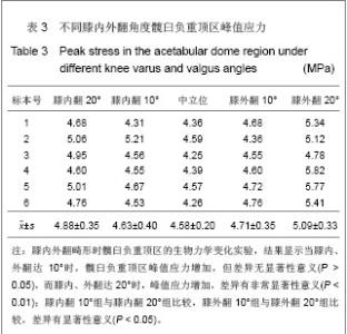
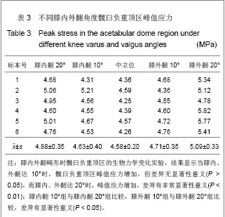
| [1] Bay BK, Hamel AJ, Olson SA,et al.Statically equivalent load and support conditions produce different hip joint contact pressures and periacetabular strains.J Biomech. 1997;30(2): 193-196.[2] Sparks DR, Beason DP, Etheridge BS, et al.Contact pressures in the flexed hip joint during lateral trochanteric loading.J Orthop Res. 2005;23(2):359-366.[3] Tannast M, Siebenrock KA.Operative treatment of T-type fractures of the acetabulum via surgical hip dislocation or Stoppa approach.Oper Orthop Traumatol. 2009;21(3): 251-269.[4] Wang YC.Beijing:Renmin Weisheng Chubanshe. 1999:334.王亦璁. 膝关节外科基础和临床[M].北京:人民卫生出版社. 1999:334.[5] Lv HS,Sun TZ,Liu ZH.Zhongguo Guzhi Shusong Zazhi.2004; 10(1):7-22.吕厚山, 孙铁铮,刘忠厚. 骨关节炎的诊治与研究进展[J].中国骨质疏松杂志,2004,10(1):7-22.[6] Wang F,Zhang Q.Hebei Yiyao.2006; 28(2):83-85.王飞,张奇.胫骨高位截骨对膝关节接触应力影响的生物力学研究[J].河北医药,2006,28(2):83-85. [7] Wang T,Wang KZ,Wang L,et al.Guangdong Yixue.2010; 31(8): 1038-1040.王涛,王坤正,王磊,等.膝内翻外翻与膝骨性关节炎发生和发展的关系[J].广东医学,2010,31(8):1038-1040.[8] Matta JM, Ferguson TA.Total hip replacement after acetabular fracture.Orthopedics. 2005;28(9):959-960.[9] Gu DY,Dai KR,Wang Y,et al.Shengwu Yixue Gongchengxue Zazhi.2003; 20(4):618-621.顾冬云,戴尅戎,王友,等.髋臼骨关节面形态特征的研究[J].生物医学工程学杂志,2003,20(4):618-621.[10] Dong JD,Wang Y,Zhu ZA,et al.Zhonghua Waike Zahzi.2005; 55(24):1583-1586.董建东,王友,朱振安,等.成人髋臼骨计算机三维重建及形态学测量[J].中华外科杂志,2005,55(24):1583-1586.[11] Menschik F.The hip joint as a conchoid shape.J Biomech. 1997;30(9):971-973.[12] Jiang Y, Jose G. Finite element modeling of in vivo knee joint contact under axial loading. http://www.me.rochester.edu/jiyao/preors2003.pdf [pubmed][13] Olson SA, Bay BK, Pollak AN,et al.The effect of variable size posterior wall acetabular fractures on contact characteristics of the hip joint.J Orthop Trauma. 1996;10(6):395-402.[14] Olson SA, Kadrmas MW, Hernandez JD,et al.Augmentation of posterior wall acetabular fracture fixation using calcium-phosphate cement: a biomechanical analysis.J Orthop Trauma. 2007;21(9):608-616.[15] Olson SA, Bay BK, Chapman MW,et al.Biomechanical consequences of fracture and repair of the posterior wall of the acetabulum.J Bone Joint Surg Am. 1995;77(8):1184-1192.[16] Zhang MC,Liao JM,Li M,et al.Diyi Junyi Daxue Xuebao.2004; 21(7):756-757.张美超,廖进民,李敏,等.激光三维扫描系统重建下颌骨[J].第一军医大学学报,2004,21(7):756-757.[17] Fan JH,Li JY,Liu C,et al.Jiupouxue Zazhi.2006; 29(4): 452-452,483.樊继宏,李鉴轶,刘畅, 等.用激光扫描三维重建骨的图像[J].解剖学杂志,2006,29(4):452-452,483.[18] von Eisenhart-Rothe R, Witte H, Steinlechner M,et al.Quantitative determination of pressure distribution in the hip joint during the gait cycle.Unfallchirurg. 1999;102(8): 625-631.[19] Raimondi MT, Santambrogio C, Pietrabissa R, et al.Improved mathematical model of the wear of the cup articular surface in hip joint prostheses and comparison with retrieved components.Proc Inst Mech Eng H. 2001;215(4):377-391.[20] Wang XS,Duan YX,Ma J,et al.Qingdao Li Gong Daxue Xuebao.2006; 27(6):8-12.王西十,段以详,马健, 等.一个评估髋关节生物力学属性的三维静力模型[J].青岛理工大学学报,2006,27(6):8-12.[21] A L,ji SJ,Fan CF.Zhonghua Xiaoeer Waike Zazhi.2000; 20(6): 327-330.阿良,吉士俊,范慈方.有限元分析先天性髋关节半脱位及髋臼发育不良的应力变化[J].中华小儿外科杂志,2000,20(6):327-330.[22] Shan SG,Wang LP,Zhang JB,et al.Zhongguo Linchuang Jiepouxue Zazhi.2007; 25(6):650-655.单石官,王连鹏,张金波,等.股胫关节面的形态与扣锁机制的解剖学基础[J].中国临床解剖学杂志,2007,25(6):650-655.[23] Domazet N, Starovi? D, Nedeljkovi? R.Biomechanical significance of the acetabular roof and its reaction to mechanical injury.Srp Arh Celok Lek. 1999;127(11-12): 359-364.[24] Matta JM, Anderson LM, Epstein HC,et al.Fractures of the acetabulum. A retrospective analysis.Clin Orthop Relat Res. 1986;(205):230-240.[25] Kummer B.The clinical relevance of biomechanical analysis of the hip area.Z Orthop Ihre Grenzgeb. 1991;129(4):285-294.[26] Ma CX,Liu GL,Fang LG,et al.Zhonghua Xiaoer Waike Zazhi. 1986; 7(1):31-33.马承宣,刘贵林,房论光,等.儿童髋关节表面应力分布状态及与髋臼指数的关系[J].中华小儿外科杂志,1986,7(1):31-33.[27] Soames RW. Biomechanics of the Locomotor Apparatus. Contributions on the Functional Anatomy of the Locomotor Apparatus.J Anat. 1982; 135(Pt 2): 442-443. |