Chinese Journal of Tissue Engineering Research ›› 2013, Vol. 17 ›› Issue (16): 2994-3001.doi: 10.3969/j.issn.2095-4344.2013.16.021
Previous Articles Next Articles
Preparation and characterization of hepatocyte-specific macroporous microcarrier
Guo Wei, Gong Du-hui, Hu Zhi-wei, Cheng Yuan, Gao Yi, Pan Ming-xin
- Second Department of Hepatobiliary Surgery, Zhujiang Hospital, Southern Medical University, Guangzhou 510282, Guangdong Province, China
-
Received:2013-03-15Revised:2013-03-19Online:2013-04-16Published:2013-05-24 -
Contact:Pan Ming-xin, Doctor, Chief physician, Professor, Second Department of Hepatobiliary Surgery, Zhujiang Hospital, Southern Medical University, Guangzhou 510282, Guangdong Province, China pmxwxy@sohu.com -
About author:Guo Wei★, Studying for master’s degree, Physician, Second Department of Hepatobiliary Surgery, Zhujiang Hospital, Southern Medical University, Guangzhou 510282, Guangdong Province, China 921329794@qq.com -
Supported by:he National High Technology Research and Development Program of China (863 Program), No. 2006AA02A141*; the Science and Technology Plan of Guangdong Province, No. 2007B010600009*

CLC Number:
Cite this article
Guo Wei, Gong Du-hui, Hu Zhi-wei, Cheng Yuan, Gao Yi, Pan Ming-xin. Preparation and characterization of hepatocyte-specific macroporous microcarrier[J]. Chinese Journal of Tissue Engineering Research, 2013, 17(16): 2994-3001.
share this article
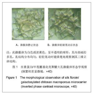
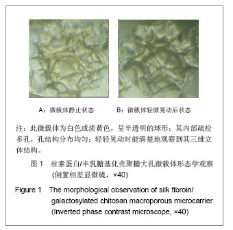
2.1 丝素蛋白/半乳糖基化壳聚糖大孔微载体的吸水率和密度 经冷冻干燥处理得到的丝素蛋白/半乳糖基化壳聚糖大孔微载体,完全保持了其三维网络结构, 这种结构使其容易溶胀,吸水率可达5.4 mL/g以上,随着半乳糖基化壳聚糖所占比例的增加而略有增加。通过此方法制备出的微载体结构疏松,湿态微载体密度较小,经反复多次测量,其浓度为1.02-1.05 g/mL,适合用于旋转微重力培养及搅拌培养。 2.2 丝素蛋白/半乳糖基化壳聚糖大孔微载体的形态 见图1。 图1为倒置显微镜下观察丝素蛋白/半乳糖基化壳聚糖大孔微载体:该大孔微载体为白色或淡黄色,呈半透明的球形;其内部疏松多孔,孔结构分布均匀;轻轻晃动时能清楚地观察到其三维立体结构。该大孔微载体内部疏松多孔的结构扩大了比表面积,并提供了较大的空体积,能将细胞固定在孔内生长,且便于观察,适合用于肝细胞高密度培养。"
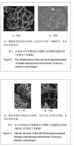
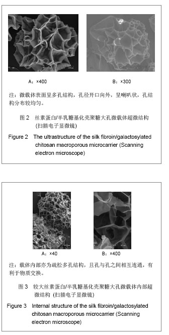
2.3 丝素蛋白/半乳糖基化壳聚糖大孔微载体的超微结构 扫描电镜结果如图2,3所示,微载体表面呈多孔结构,孔径为40-80 µm,开口向外,呈喇叭状,孔结构分布较均匀。相比于市售的大孔微载体,该微载体孔径更大,且开口向外;利于细胞黏附生长,并有足够大的孔隙使细胞能够长入微载体内部。"
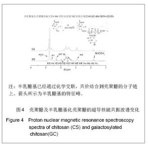
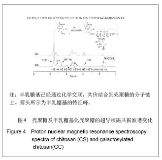
2.4 半乳糖基化壳聚糖超导核磁共振波谱分析结果 图4为半乳糖基化壳聚糖的1HNMR谱分析,其1HNMR归属如下: 1HNMR(300 MHz,D2O/H3CCOOH)δ: 1. 952 (-COCH3中的H),4.755 (H1),4. 131,4. 415 (H1′,Hc),3. 3-4. 0 (H3,H4, H5, H6, H2′, H3′, H4′, H5′, H6′, Ha, Hb, Hd, He), 3. 066 (H2)。 结果证实,半乳糖基已经通过化学交联,共价结合到壳聚糖的分子链上。"
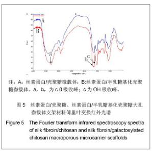
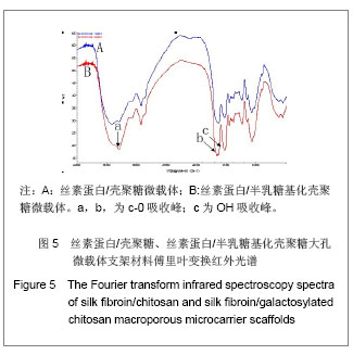
2.5 丝素蛋白/半乳糖基化壳聚糖大孔微载体材料的傅里叶变换红外光谱图像 见图5。 图5为丝素蛋白/壳聚糖丝素蛋白/半乳糖基化壳聚糖大孔微载体材料的傅里叶变换红外光谱图像。由于乳糖酸的羧酸基团和壳聚糖的氨基反应形成酰胺键,从而酰胺键的吸收峰Ⅰ和Ⅱ发生了化学位移,且峰值增宽,1 070.7 cm-1为半乳糖糖基的C-O伸展骨架振动的吸收峰,见图5b,c。另外,由于壳聚糖接枝半乳糖基后,使OH的数目大大增加,所以OH峰变宽变强,见图5a。"
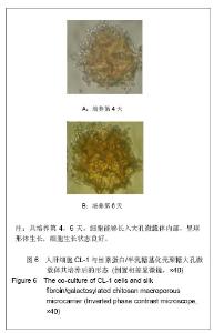
2.6 人肝细胞CL-1与丝素蛋白/半乳糖基化壳聚糖大孔微载体共培养后的形态 共培养第4,6天;于倒置显微镜下观察,细胞能够长入大孔微载体内部,呈多细胞聚集的团块,细胞呈球形体生长。肝细胞能克服重力的作用,而与材料特异性黏附、聚集生长,细胞生长状态良好。见图6。"
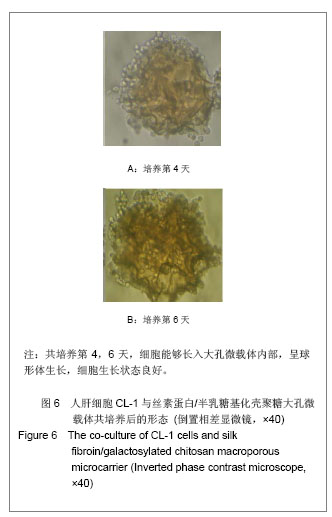
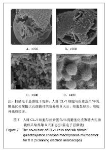
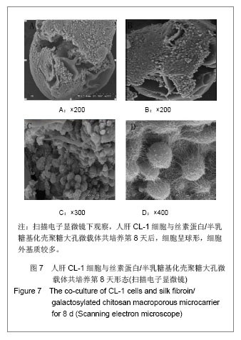
图7为利用丝素蛋白/半乳糖基化壳聚糖大孔微载体在RCCS生物反应器中高密度培养CL-1细胞,细胞能够黏附在大孔微载体上生长,呈多细胞聚集的团块,由于肝细胞表面ASGPR和支架上半乳糖基之间的分子识别功能,细胞附着牢固。且随着培养时间的延长微载体之间开始发生团聚,细胞浓度接近1010 L-1。通过扫描电镜观察,细胞可进入微载体内部,细胞呈球形,聚集生长,分泌较多的细胞外基质[17] 。"
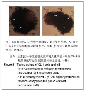
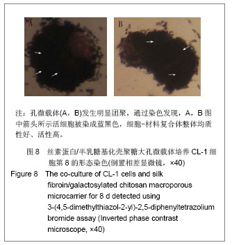
MTT染色法检测肝细胞活力结果显示,在倒置显微镜下观察,随着培养时间的延长,培养后期大孔微载体发生明显团聚,通过染色发现,图8中箭头所示活细胞被染成蓝黑色,细胞-材料复合体整体均质性好、活性高。"
| [1]Hoekstra R, Nibourg GA, van der Hoeven TV, et al. The HepaRG cell line is suitable for bioartificial liver application. Int J Biochem Cell Biol. 2011;43(10):1483-1489.[2]Pan XP, Li LJ. Advances in cell sources of hepatocytes for bioartificial liver. Hepatobiliary Pancreat Dis Int. 2012;11(6): 594-605.[3]Barakat O, Abbasi S, Rodriguez G, et al. Use of Decellularized Porcine Liver for Engineering Humanized Liver Organ. J Surg Res. 2012;173(1):e11-25. [4]Wu C, Pan J, Bao Z, et al. Fabrication and characterization of chitosan microcarrier for hepatocyte culture. J Mater Sci Mater Med. 2007;18(11):2211-2214.[5]Park Y, Subramanian K, Verfaillie CM, et al. Expansion and hepatic differentiation of rat multipotent adult progenitor cells in microcarrier suspension culture. J Biotechnol. 2010;150(1): 131-139. [6]Eibes G, dos Santos F, Andrade PZ, et al. Maximizing the ex vivo expansion of human mesenchymal stem cells using a microcarrier-based stirred culture system. J Biotechnol. 2010; 146(4):194-197.[7]Rustad KC, Wong VW, Sorkin M, et al. Enhancement of mesenchymal stem cell angiogenic capacity and stemness by a biomimetic hydrogel scaffold. Biomaterials. 2012;33(1): 80-90.[8]Bhardwaj N, Nguyen QT, Chen AC, et al. Potential of 3-D tissue constructs engineered from bovine chondrocytes/silk fibroin-chitosan for in vitro cartilage tissue engineering. Biomaterials. 2011;32(25):5773-5781. [9]Rigopoulou EI, Roggenbuck D, Smyk DS, et al. Asialoglycoprotein receptor (ASGPR) as target autoantigen in liver autoimmunity: lost and found. Autoimmun Rev. 2012; 12(2): 260-269.[10]Selman L, Hansen S. Structure and function of collectin liver 1 (CL-L1) and collectin 11 (CL-11, CL-K1). Immunobiology. 2012;217(9):851-863.[11]Wen X, Peng X, Fu H, et al. Preparation and in vitro evaluation of silk fibroin microspheres produced by a novel ultra-fine particle processing system. Int J Pharm. 2011; 416(1):195-201. [12]Zheng D, Duan C, Zhang D, et al. Galactosylated chitosan nanoparticles for hepatocyte-targeted delivery of Oridonin. Int J Pharm. 2012;436(1-2):379-386.[13]Li L, Qian Y, Jiang C, et al. The use of hyaluronan to regulate protein adsorption and cell infiltration in nanofibrous scaffolds. Biomaterials. 2012;33(12):3428-3445. [14]Park IK, Yang J, Jeong HJ, et al. galactosylated chitosan as a synthetic extracellular matrix for hepatocytes attachment. Biomaterials. 2003;24(13):2331-2337.[15]Park TG. Perfusion culture of hepatocytes within galactose derivatized biodegradable poly (lactide-co-glycolide) scaffolds prepared by gas foaming of effervescent salts. J Biomed Mater Res. 2002;59(1):127-135.[16]Jin H J, Chen JS, Karageorgiou V, et al. Human bone marrow stromal cell responses on electrospun silk fibroin mats. Biomaterials. 2004;25(6):1039-1047.[17]Wu XB, Peng CH, Huang F, et al. Preparation and characterization of chitosan porous microcarriers for hepatocyte culture. Hepatobiliary Pancreat Dis Int. 2011; 10(5):509-515.[18]Duan B, Wang M, Zhou WY, et al. Three-dimensional nanocomposite scaffolds fabricated via selective laser sintering for bone tissue engineering. Acta Biomater. 2010; 6(12):4495-505. [19]Barakat O, Abbasi S, Rodriguez G, et al. Use of decellularized porcine liver for engineering humanized liver organ. J Surg Res. 2012;173(1):e11-25. [20]Wei X, Zhang Z, Qian Z, et al. Pharmacokinetics and in vivo fate of drug loaded chitosan nanoparticles. Curr Drug Metab. 2012;13(4):364-371.[21]Bhardwaj N, Kundu SC. Chondrogenic differentiation of rat MSCs on porous scaffolds of silk fibroin/chitosan blends. Biomaterials. 2012;33(10):2848-2857.[22]Wang Y, Bella E, Lee CS, et al. The synergistic effects of 3-D porous silk fibroin matrix scaffold properties and hydrodynamic environment in cartilage tissue regeneration. Biomaterials. 2010;31(17):4672-4681.[23]Wen X, Peng X, Fu H, et al. Preparation and in vitro evaluation of silk fibroin microspheres produced by a novel ultra-fine particle processing system. Int J Pharm. 2011; 16(1):195-201. [24]Lu Q, Huang Y, Li M, et al. Silk fibroin electrogelation mechanisms. Acta Biomater. 2011;7(6):2394-400. [25]Waldert M, Klatte T, Haitel A, et al. Hybrid renal cell carcinomas containing histopathologic features of chromophobe renal cell carcinomas and oncocytomas have excellent oncologic outcomes. Eur Urol. 2010;57(4):661-665. [26]Silva GA, Coutinho OP, Ducheyne P, et al. The efect of starch and starch-bioactive glass composite microparticles on the adhesion and expression of the osteoblastic phenotype of a bone cell line. Biomaterials. 2007;28(2):326-334. [27]Harada K, Mitaka T, Miyamoto S, et al. Rapid formation of hepatic organoid in collagen sponge by rat small hepatocytes and hepatic nonparenchymal cells. J Hepatol. 2003;39(5): 716-723.[28]Chung HJ, Kim IK, Kim TG, et al.Highly open porous biodegradable microcarriers: in vitro cultivation of chondrocytes for injectable delivery. Tissue Eng Part A. 2008;14(5):607-615.[29]Ohashi K, Yokoyama T, Yamato M, et al. Engineering functional two- and three-dimensional liver systems in vivo using hepatic tissue sheets. Nat Med. 2007;13(7):880-885. |
| [1] | Liu Hongjie, Mu Qiuju, Shen Yuxue, Liang Fei, Zhu Lili. Metal organic framework/carboxymethyl chitosan-oxidized sodium alginate/platelet-rich plasma hydrogel promotes healing of diabetic infected wounds [J]. Chinese Journal of Tissue Engineering Research, 2026, 30(8): 1929-1939. |
| [2] | Guo Yuchao, Ni Qianwei, Yin Chen, Jigeer·Saiyilihan, Gao Zhan . Quaternized chitosan hemostatic materials: synthesis, mechanism, and application [J]. Chinese Journal of Tissue Engineering Research, 2026, 30(8): 2091-2100. |
| [3] | Yu Shiyu, Yu Sutong, Xu Yang, Zhen Xiangyan, Han Fengxuan. Advances in research and application of tissue engineering therapeutic strategies in oral submucous fibrosis [J]. Chinese Journal of Tissue Engineering Research, 2026, 30(4): 936-948. |
| [4] | Wang Quan, Tang Zhenglong. Visualization analysis of literature on medication-related osteonecrosis of the jaw [J]. Chinese Journal of Tissue Engineering Research, 2026, 30(35): 9327-9335. |
| [5] | Jin Yuan, Zhou Jiabing. Applications and advances of hydrogels in bone tissue engineering repair related to sports injuries [J]. Chinese Journal of Tissue Engineering Research, 2026, 30(32): 8512-8520. |
| [6] | Sun Jingshan, Deng Jingqian, Qi Liya, Zhao Xiaohuan, Hou Dandan. Material selection and manufacture method of non-porous adherent cell microcarriers [J]. Chinese Journal of Tissue Engineering Research, 2026, 30(32): 8536-8543. |
| [7] | Xia Jinyan, Su Meifang, Yang Wenyu, Hu Qilan, Gong Li. Application of novel materials in tissue repair of diabetic foot wounds [J]. Chinese Journal of Tissue Engineering Research, 2026, 30(32): 8563-8568. |
| [8] | Li Xuanze, Fang Hanhong, Xu Zhe. Preparation and cytocompatibility of sodium alginate-hydroxyapatite-graphene oxide hydrogels [J]. Chinese Journal of Tissue Engineering Research, 2026, 30(32): 8420-8426. |
| [9] | Yang Hu, Zheng Yu, Jia Chengming, Wang Tong, Zhang Guangfei, Ji Yaoyao. Immune microenvironment regulates bone regeneration [J]. Chinese Journal of Tissue Engineering Research, 2026, 30(3): 701-710. |
| [10] | Yang Guang, Yin Zhitao, Xu Yan. Three-dimensional printed isoniazid liposome photothermal composite scaffolds and their performance evaluation [J]. Chinese Journal of Tissue Engineering Research, 2026, 30(26): 6701-6709. |
| [11] | Zhao Qinglan, Zhou Xinting, Wang Huajun, Zhong Jiaxuan, Zheng Liheng, Tan Wencheng, Yang Xinchun, Huang Shusen, Wu Tingting, Zheng Xiaofei, Hong Jinsong. Ginsenoside Rg3-loaded liposome hydrogel promotes chondrogenic differentiation of stem cells [J]. Chinese Journal of Tissue Engineering Research, 2026, 30(26): 6760-6767. |
| [12] | Zhang Yibo, Li Jian, Wang Peng, Jiang Qing. Curcumin-loaded chitosan/beta-glycerophosphate sodium thermoresponsive hydrogel promotes tendon healing in rats [J]. Chinese Journal of Tissue Engineering Research, 2026, 30(26): 6779-6798. |
| [13] | Chen Gang, Ge Caijun, Chen Jianpeng, Wang Yuanbin, Wang Qianliang. Mechanism of ferrostatin-1 hydrogel in treatment of lumbar disc herniation [J]. Chinese Journal of Tissue Engineering Research, 2026, 30(26): 6807-6813. |
| [14] | Yu Jinye, Jiang Nan, Zhao Yixun, Huang Mengjing, Yang Jie, Sun Rui, Feng Suolan, Jiang Hui, Yang Jun. Ready-to-use sodium alginate@paper material for three-dimensional cell culture [J]. Chinese Journal of Tissue Engineering Research, 2026, 30(26): 6873-6879. |
| [15] | Wang Jieyan, Yao Jiayi, Xin Yingtong, Zhang Xinwen, Li Riwang, Liu Dahai. Chitosan hydrogel drug delivery system is a safer and more effective solution for treating oral ulcers [J]. Chinese Journal of Tissue Engineering Research, 2026, 30(20): 5331-5340. |
| Viewed | ||||||
|
Full text |
|
|||||
|
Abstract |
|
|||||