Chinese Journal of Tissue Engineering Research ›› 2026, Vol. 30 ›› Issue (21): 5376-5385.doi: 10.12307/2026.778
Previous Articles Next Articles
Finite element analysis of the effect of morphological differences in endplate defects on biomechanics of lumbar intervertebral discs
Yang Yiting, Li Zheng, Yang Yong, Fan Chunsun, Lu Yonggang#br#
- Affiliated Qidong Hospital of Nantong University, Qidong People's Hospital (Qidong Liver Cancer Institute), Nantong 226200, Jiangsu Province, China
-
Accepted:2025-09-02Online:2026-07-28Published:2026-03-03 -
Contact:Li Zheng, Attending physician, Affiliated Qidong Hospital of Nantong University, Qidong People's Hospital (Qidong Liver Cancer Institute), Nantong 226200, Jiangsu Province, China Lu Yonggang, Chief physician, Affiliated Qidong Hospital of Nantong University, Qidong People's Hospital (Qidong Liver Cancer Institute), Nantong 226200, Jiangsu Province, China -
About author:Yang Yiting, MS, Affiliated Qidong Hospital of Nantong University, Qidong People's Hospital (Qidong Liver Cancer Institute), Nantong 226200, Jiangsu Province, China -
Supported by:Nantong Municipal Science and Technology Program Guidance Project, No. MSZ2024151 (to YYT); Nantong Municipal Health Commission Youth Directive Research Project, No. QN2024073 (to YYT); Youth Project of Clinical Medicine Special Research Fund of Nantong University, No. 2024JQ044 (to YYT); Key Project of Clinical Medicine Special Research Fund of Nantong University, No. 2023LZ003 (to LYG)
CLC Number:
Cite this article
Yang Yiting, Li Zheng, Yang Yong, Fan Chunsun, Lu Yonggang. Finite element analysis of the effect of morphological differences in endplate defects on biomechanics of lumbar intervertebral discs [J]. Chinese Journal of Tissue Engineering Research, 2026, 30(21): 5376-5385.
share this article
Add to citation manager EndNote|Reference Manager|ProCite|BibTeX|RefWorks

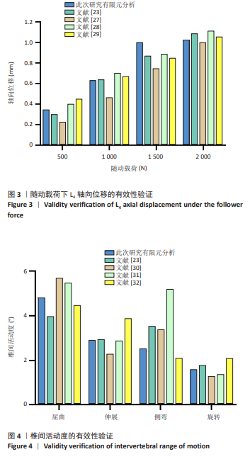
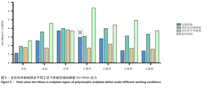
2.1 腰椎有限元模型有效性验证 在500,1 000,1 500,2 000 N随动载荷下,L4轴向位移为0.35,0.63,1.00和1.02 mm,与既往体外实验和有限元研究的结果相近(图3)[23,27-29]。在500 N随动载荷下,L4上方参考点施加10 N·m力矩,模拟屈伸、侧弯与旋转工况,获取腰椎间活动度分别为4.82°,2.919°,2.528°和1.582°,结果提示其与既往尸体标本实验和有限元研究的数据接近,处于合理范围内(图4)[23,30-32]。"

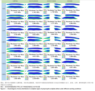
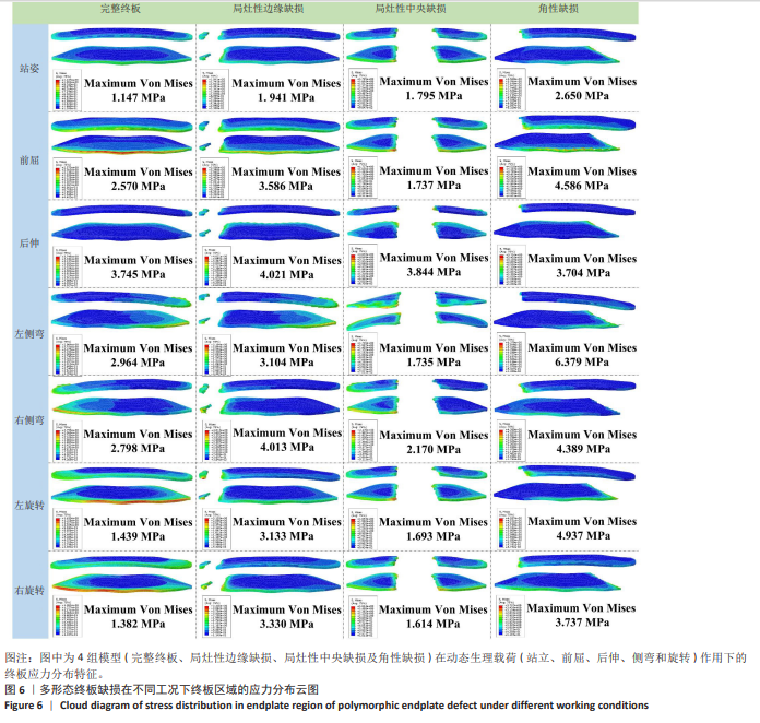
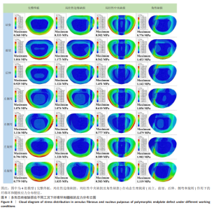
2.2 终板区域Von Mises应力分布 3种不同形态的终板缺损在不同工况下(站姿、前屈、伸展、侧弯和旋转)终板区域均表现出显著的Von Mises应力峰值和应力分布差异(图5,6)。直立站姿状态下健康完整终板呈现均匀应力分布,峰值应力较低(1.147 MPa),而缺损模型均出现不同程度的应力集中现象,其中角形缺损峰值应力最高(2.650 MPa)。局灶性边缘缺损在前屈时产生明显的应力集中(峰值3.586 MPa),较完整模型前屈工况增加28.3%。局灶性中央缺损在各工况下的应力增幅较完整健康终板较小。角性缺损在左侧弯运动时表现出最显著的应力集中(6.379 MPa),为完整终板的5.6倍,该缺损形态在所有动态载荷下均呈现广泛的高应力区分布,其中旋转运动时应力峰值达4.937 MPa,且应力集中区域与缺损的不规则边缘高度吻合(图4)。值得注意的是,缺损边缘处的应力梯度变化显著大于完整终板区域,提示这些部位可能存在更高的微损伤风险。"
"

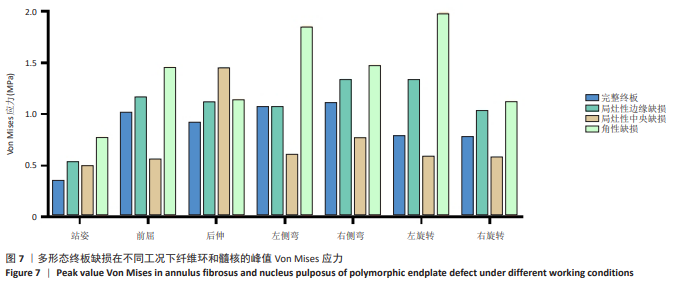
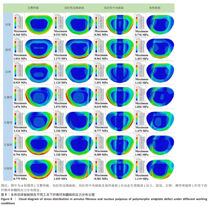
2.3 纤维环髓核Von Mises应力分布 3种不同形态终板缺损在不同工况下(站姿、前屈、伸展、侧弯和旋转)椎间盘组织(纤维环和髓核区域)的Von Mises应力峰值和应力分布云图可见图7,8,终板缺损形态显著影响纤维环和髓核的应力分布模式。完整终板模型在各类载荷下椎间盘应力分布均匀,站姿状态下的峰值应力为0.36 MPa;相比之下,缺损模型均表现出不同程度的应力异常,增幅为40%-111%。局灶性边缘缺损在前屈运动时导致纤维环外侧应力显著升高(1.173 MPa)。局灶性中央缺损在后伸动作中引起纤维环两侧后方应力集中,应力高达1.451 MPa。角性缺损在左旋转运动时产生最显著的应力异常,纤维环峰值应力达1.982 MPa,为完整模型的2.5倍。旋转运动时,所有缺损模型均呈现不对称的应力分布,其中角性缺损模型的应力梯度变化最为剧烈。特别值得注意的是,缺损模型在动态载荷下的应力峰值均出现在与缺损位置相对应的椎间盘区域,这种空间对应关系提示终板缺损可能通过改变局部力学环境直接影响椎间盘的载荷传递路径。此外,角性缺损模型在所有工况下均表现出最高的应力集中系数,进一步证实此类缺损形态对椎间盘力学环境的破坏最为严重。"
"
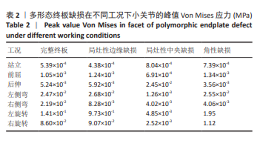
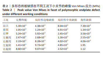
2.4 小关节Von Mises峰值应力 腰椎L4-5小关节的应力数据结果列于表2,终板缺损形态和运动方向共同影响小关节峰值应力。 健康完整终板和局灶性中央缺损在各工况下小关节受力均较低,角性缺损模型在旋转运动时表现出较高的应力集中现象。局灶性边缘缺损和角性缺损的终板边缘形态造成了左右旋转运动呈现明显不对称性,局灶性边缘缺损左右旋转时小关节应力峰值分别为9.73×10⁻¹ MPa和9.07×10⁻² MPa,角性缺损左右旋转时小关节应力峰值分别为1.95 MPa和 1.12 MPa。 "
| [1] AKTER F, KOTTER M. Pathobiology of Degenerative Cervical Myelopathy. Neurosurg Clin North Am. 2018;29(1):13-19. [2] BAUCHER G, TASKOVIC J, TROUDE L, et al. Risk factors for the development of degenerative cervical myelopathy: a review of the literature. Neurosurg Rev. 2022;45(2):1675-1689. [3] MUFTULER LT, JARMAN JP, YU HJ, et al. Association between intervertebral disc degeneration and endplate perfusion studied by DCE-MRI. Eur Spine J. 2015;24(4):679-685. [4] DIN RU, CHENG X, YANG H. Diagnostic Role of Magnetic Resonance Imaging in Low Back Pain Caused by Vertebral Endplate Degeneration. J Magn Reson Imaging. 2022;55(3):755-771. [5] LAWAN A, CRITES VIDEMAN J, BATTIÉ MC. The association between vertebral endplate structural defects and back pain: A systematic review and meta-analysis. Eur Spine J. 2021;30(9):2531-2548. [6] KUHN A, HUBER-LANG M, WECKBACH S, et al. Analysis of Intervertebral Disc Degeneration Induced by Endplate Drilling or Needle Puncture in Complement C6-Sufficient and C6-Deficient Rabbits. Biomedicines. 2024;12(8):1692. [7] WANG Y, BATTIÉ MC, BOYD SK, et al. The osseous endplates in lumbar vertebrae: Thickness, bone mineral density and their associations with age and disk degeneration. Bone. 2011;48(4):804-809. [8] MA Y, YU X, LI W, et al. Animal Models of Internal Endplate Injury-Induced Intervertebral Disc Degeneration: A Systematic Review. J Invest Surg. 2024;37(1):2400478. [9] RODRIGUEZ AG, SLICHTER CK, ACOSTA FL, et al. Human disc nucleus properties and vertebral endplate permeability. Spine. 2011;36(7): 512-520. [10] ZEHRA U, CHEUNG JPY, BOW C, et al. Multidimensional vertebral endplate defects are associated with disc degeneration, modic changes, facet joint abnormalities, and pain. J Orthop Res. 2019;37(5): 1080-1089. [11] ZEHRA U, BOW C, LOTZ JC, et al. Structural vertebral endplate nomenclature and etiology: A study by the ISSLS spinal phenotype focus group. Eur Spine J. 2018;27(1): 2-12. [12] ZHANG C, LIU S. The advancement of MRI in differentiating Modic type I degenerative changes from early spinal infections. Br J Radiol. 2023; 96(1152):20230551. [13] ZEHRA U, BOW C, LOTZ JC, et al. Structural vertebral endplate nomenclature and etiology: A study by the ISSLS spinal phenotype focus group. Eur Spine J. 2018;27(1):2-12. [14] ADAMS MA, FREEMAN BJ, MORRISON HP, et al. Mechanical initiation of intervertebral disc degeneration. Spine. 2000;25(13):1625-1636. [15] HOLM S, HOLM AK, EKSTRÖM L, et al. Experimental disc degeneration due to endplate injury. J Spinal Disord Tech. 2004;17(1):64-71. [16] KERTTULA LI, SERLO WS, TERVONEN OA, et al. Post-traumatic findings of the spine after earlier vertebral fracture in young patients: Clinical and MRI study. Spine. 2000;25(9):1104-1108. [17] FENG Z, LIU Y, YANG G, et al. Lumbar Vertebral Endplate Defects on Magnetic Resonance Images: Classification, Distribution Patterns, and Associations with Modic Changes and Disc Degeneration. Spine. 2018; 43(13):919-927. [18] RAJASEKARAN S, VENKATADASS K, NARESH BABU J, et al. Pharmacological enhancement of disc diffusion and differentiation of healthy, ageing and degenerated discs: Results from in-vivo serial post-contrast MRI studies in 365 human lumbar discs. Eur Spine J. 2008; 17(5):626-643. [19] APPLEBAUM A, NESSIM A, CHO W. Modic Change: An Emerging Complication in the Aging Population. Clin Spine Surg. 2022;35(1): 12-17. [20] MOSTOFI K, MOGHADDAM BG, PEYRAVI M. Late appearance of low back pain relating to Modic change after lumbar discectomy. J Craniovertebr Junction Spine. 2018;9(2):93-95. [21] ZHANG J, CHEN W, WENG R, et al. Biomechanical effect of endplate defects on the intermediate vertebral bone in consecutive two-level anterior cervical discectomy and fusion: a finite element analysis. BMC Musculoskelet Disord. 2023;24(1):407. [22] 葛志林, 何嘉辉, 程招军, 等. 内镜下不同入路腰椎融合术对腰椎生物力学效能影响的有限元分析[J]. 中国脊柱脊髓杂志,2024, 34(6):620-628. [23] 王宏卫, 刘新宇, 万熠. 人体腰椎L4-L5段有限元模型建立及力学有效性验证[J]. 医学与哲学,2017,38(5):50-53. [24] SINGH A, LU Y, CHEN C, et al. Mechanical properties of spinal nerve roots subjected to tension at different strain rates. J Biomech. 2006; 39(9):1669-1676. [25] POLAK K, CZYŻ M, ŚCIGAŁA K, et al. Biomechanical characteristics of the porcine denticulate ligament in different vertebral levels of the cervical spine-preliminary results of an experimental study. J Mech Behav Biomed Mater. 2014;34:165-170. [26] FENG Z, LIU Y, YANG G, et al. Lumbar Vertebral Endplate Defects on Magnetic Resonance Images: Classification, Distribution Patterns, and Associations with Modic Changes and Disc Degeneration. Spine. 2018; 43(13):919-927. [27] 颜文涛,赵改平,方新果, 等.人体腰椎L4-5节段有限元建模及分析[J]. 生物医学工程学杂志,2014,31(3):612-618. [28] MARKOLF KL, MORRIS JM. The structural components of the intervertebral disc. A study of their contributions to the ability of the disc to withstand compressive forces. J Bone Joint Surg Am. 1974; 56(4):675-687. [29] MOMENI SHAHRAKI N, FATEMI A, GOEL VK, et al. On the use of biaxial properties in modeling annulus as a holzapfel-gasser-ogden material. Front Bioeng Biotechnol. 2015;3:69. [30] 方新果, 赵改平, 王晨曦, 等. 基于ct图像腰椎L4L5节段有限元模型建立与分析[J]. 中国生物医学工程学报,2014,33(4):487-492. [31] NACHEMSON AL, SCHULTZ AB, BERKSON MH. Mechanical properties of human lumbar spine motion segments. Influence of age, sex, disc level, and degeneration. Spine. 1979;4(1):1-8. [32] CHEN CS, CHENG CK, LIU CL, et al. Stress analysis of the disc adjacent to interbody fusion in lumbar spine. Med Eng Phys. 2001;23(7): 483-491. [33] WANG Y, VIDEMAN T, BATTIÉ MC. Morphometrics and lesions of vertebral end plates are associated with lumbar disc degeneration: Evidence from cadaveric spines. J Bone Joint Surg Am. 2013;95(5):e26. [34] WANG Y, VIDEMAN T, BATTIÉ MC. ISSLS prize winner: Lumbar vertebral endplate lesions: associations with disc degeneration and back pain history. Spine. 2012;37(17):1490-1496. [35] FAUL J, UMOH J, HOLDSWORTH DW, et al. Thoracolumbar Vertebral Endplate Defect Morphology: A Descriptive Study of Human Cadaveric Spines Using Micro-Computed Tomography. Spine. 2023;48(19): 1397-1404. [36] LI R, WANG Z, MA L, et al. Lumbar vertebral endplate defects on magnetic resonance imaging in degenerative spondylolisthesis: Novel classification, characteristics, and correlative factor analysis. World Neurosurg. 2020;141:e423-e430. [37] ZEHRA U, FLOWER L, ROBSON-BROWN K, et al. Defects of the vertebral end plate: Implications for disc degeneration depend on size. Spine J. 2017;17(5):727-737. [38] WANG Y, VIDEMAN T, BATTIÉ MC. Lumbar vertebral endplate lesions: Prevalence, classification, and association with age. Spine. 2012; 37(17):1432-1439. [39] 杨泽. 退行性腰椎滑脱患者腰椎终板缺损的MRI特征及相关因素分析[D]. 湛江:广东医科大学,2023. [40] MÄÄTTÄ JH, RADE M, FREIDIN MB, et al. Strong association between vertebral endplate defect and modic change in the general population. Sci Rep. 2018;8:16630. [41] RAJASEKARAN S, RAMACHANDRAN K, KS SVA, et al. From modic to disc endplate bone marrow complex - the natural course and clinical implication of vertebral endplate changes. Global Spine J. 2025;15(1): 196-209. [42] MOK FPS, SAMARTZIS D, KARPPINEN J, et al. ISSLS prize winner: Prevalence, determinants, and association of schmorl nodes of the lumbar spine with disc degeneration: a population-based study of 2449 individuals. Spine. 2010;35(21):1944-1952. [43] COLOMBINI A, GALBUSERA F, CORTESE MC, et al. Classification of endplate lesions in the lumbar spine and association with risk factors, biochemistry, and genetics. Eur Spine J. 2021;30(8):2231-2237. [44] IMRAN S, LATIF R, AHMAD I, et al. Vertebral Endplate Defects Induced Mechanical Alterations and Disc Calcification. Global Spine J. 2025; 15(3):1564-1571. [45] LI R, WANG LF, WANG F, et al. The relationship between endplate defect scores and lumbar sagittal translation stability in lumbar spondylolisthesis patients. World Neurosurg. 2024;181:e938-e946. [46] SAMARTZIS D, CHEUNG JPY, RAJASEKARAN S, et al. Critical values of facet joint angulation and tropism in the development of lumbar degenerative spondylolisthesis: An international, large-scale multicenter study by the AOSpine asia pacific research collaboration consortium. Global Spine J. 2016;6(5):414-421. [47] WILLIAMS R, CHEUNG JPY, GOSS B, et al. An international multicenter study assessing the role of ethnicity on variation of lumbar facet joint orientation and the occurrence of degenerative spondylolisthesis in asia pacific: A study from the AOSpine asia pacific research collaboration consortium. Global Spine J. 2016;6(1):35-45. |
| [1] | Chen Huiting, Zeng Weiquan, Zhou Jianhong, Wang Jie, Zhuang Congying, Chen Peiyou, Liang Zeqian, Deng Weiming. Tail anchoring technique of vertebroplasty in treatment of osteoporotic vertebral compression fractures with intravertebral cleft: a finite element analysis [J]. Chinese Journal of Tissue Engineering Research, 2026, 30(9): 2145-2152. |
| [2] | Cheng Qisheng, Julaiti·Maitirouzi, Xiao Yang, Zhang Chenwei, Paerhati·Rexiti. Finite element analysis of novel variable-diameter screws in modified cortical bone trajectory of lumbar vertebrae [J]. Chinese Journal of Tissue Engineering Research, 2026, 30(9): 2162-2171. |
| [3] | Liu Jiafu, Ren Ruxia, Liao Zhouwei, Zhou Xiali, Wu Yihong, Zhang Shaoqun. Three-dimensional finite element analysis of cervical spine biomechanical characteristics in a rat model of cervical vertigo [J]. Chinese Journal of Tissue Engineering Research, 2026, 30(9): 2182-2190. |
| [4] | Zhang Zizheng, Luo Wang, Liu Changlu. Application value of finite element analysis on unicompartmental knee arthroplasty for medial knee compartmental osteoarthritis [J]. Chinese Journal of Tissue Engineering Research, 2026, 30(9): 2313-2322. |
| [5] | Liu Wenlong, Dong Lei, Xiao Zhengzheng, Nie Yu. Finite element analysis of tibial prosthesis loosening after fixed-bearing unicompartmental knee arthroplasty for osteoporosis [J]. Chinese Journal of Tissue Engineering Research, 2026, 30(9): 2191-2198. |
| [6] | Zheng Wangyang, Fei Ji, Yang Di, Zhao Lang, Wang Lingli, Liu Peng, Li Haiyang. Finite element analysis of the force changes of the supraspinatus tendon and glenohumeral joint during the abduction and flexion of the humerus [J]. Chinese Journal of Tissue Engineering Research, 2026, 30(9): 2199-2207. |
| [7] | Cai Qirui, Dai Xiaowei, Zheng Xiaobin, Jian Sili, Lu Shaoping, Liu Texi, Liu Guoke, Lin Yuanfang. Mechanical effects of Long’s traction orthopedic method on cervical functional units: quantitative analysis of biomechanical model of head and neck [J]. Chinese Journal of Tissue Engineering Research, 2026, 30(9): 2208-2216. |
| [8] | Rao Jingcheng, Li Yuwan, Zheng Hongbing, Xu Zhi, Zhu Aixiang, Shi Ce, Wang Bing, Yang Chun, Kong Xiangru, Zhu Dawei. Biomechanical differences between the new proximal femoral stable intramedullary nail and traditional intramedullary nail#br# [J]. Chinese Journal of Tissue Engineering Research, 2026, 30(9): 2217-2225. |
| [9] | Chen Long, Wang Xiaozhen, Xi Jintao, Lu Qilin. Biomechanical performance of short-segment screw fixation combined with expandable polyetheretherketone vertebral body replacement in osteoporotic vertebrae [J]. Chinese Journal of Tissue Engineering Research, 2026, 30(9): 2226-2235. |
| [10] | Zheng Xuying, Hu Hongcheng, Xu Libing, Han Jianmin, Di Ping. Stress magnitude and distribution in two-piece cement-retained zirconia implants under different loading conditions and with varying internal connection shapes [J]. Chinese Journal of Tissue Engineering Research, 2026, 30(8): 1979-1987. |
| [11] | Zhong Caihong, Xiao Xiaoge, Li Ming, Lin Jianhong, Hong Jing. Biomechanical mechanism of sports-related patellar tendinitis [J]. Chinese Journal of Tissue Engineering Research, 2026, 30(6): 1417-1423. |
| [12] | Xu Hao, Ding Lu, Li Xiao. Mechanical effect of mechanical wear of abutment screws on the Morse taper connection implant system: a three-dimensional finite element analysis [J]. Chinese Journal of Tissue Engineering Research, 2026, 30(6): 1375-1383. |
| [13] | Shang Depeng, Wei Haiyu, Yang Fan. Finite element analysis for three different types of internal screw fixation in treatment of severe lumbar 1 vertebral body fractures [J]. Chinese Journal of Tissue Engineering Research, 2026, 30(3): 537-545. |
| [14] | Yu Xinlin, Chen Huiyu, Wang Yingying, Guo Weizhong, Feng Bin Lin Chengshou, Lin Wang. Finite element analysis of internal fixation with new retrograde intramedullary nail on lateral femur condyle for distal type A2 femur fractures [J]. Chinese Journal of Tissue Engineering Research, 2026, 30(3): 546-552. |
| [15] | Zhao Jingang, Liu Liping, Chen Jianwei, . Finite element analysis comparing lumbar fusion and artificial intervertebral disc replacement [J]. Chinese Journal of Tissue Engineering Research, 2026, 30(3): 553-560. |
| Viewed | ||||||
|
Full text |
|
|||||
|
Abstract |
|
|||||