Chinese Journal of Tissue Engineering Research ›› 2026, Vol. 30 ›› Issue (3): 537-545.doi: 10.12307/2026.579
Finite element analysis for three different types of internal screw fixation in treatment of severe lumbar 1 vertebral body fractures
Shang Depeng, Wei Haiyu, Yang Fan
- Department of Orthopedics, Zhongshan Hospital Affiliated to Dalian University, Dalian 116001, Liaoning Province, China
-
Received:2024-12-11Accepted:2025-03-14Online:2026-01-28Published:2025-07-01 -
Contact:Yang Fan, MD, Attending physician, Department of Orthopedics, Zhongshan Hospital Affiliated to Dalian University, Dalian 116001, Liaoning Province, China -
About author:Shang Depeng, MS, Attending physician, Department of Orthopedics, Zhongshan Hospital Affiliated to Dalian University, Dalian 116001, Liaoning Province, China -
Supported by:Liaoning Provincial Science and Technology Joint Program, No. 2024011521-JH3/4700 (to YF); Clinical Application Project of Dalian Municipal Health Commission, No. 2111038 (to YF); Dalian Science and Technology Bureau Life and Health Field Guidance Program, No. DKSN(2023)243 (to YF)
CLC Number:
Cite this article
Shang Depeng, Wei Haiyu, Yang Fan. Finite element analysis for three different types of internal screw fixation in treatment of severe lumbar 1 vertebral body fractures[J]. Chinese Journal of Tissue Engineering Research, 2026, 30(3): 537-545.
share this article
Add to citation manager EndNote|Reference Manager|ProCite|BibTeX|RefWorks

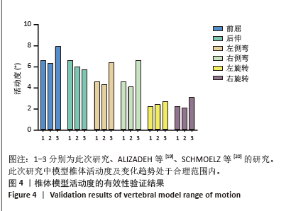
2.1 模型有效性的验证 为了验证椎体模型的合理性,模型T11-L3在施加7.5 Nm扭矩时,测量其在前屈、后伸、左侧弯、右侧弯和左右旋转等6种运动状态下的运动范围,并与以往方法的实验数据进行比较[19-20]。结果表明,虽然模型之间存在差异,但差异很小,模型的活动度值与其他学者的研究结果吻合较好(图4),证明了该实验模型的有效性。"
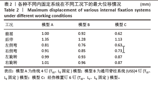
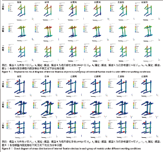
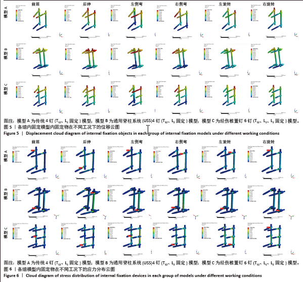
2.2 内固定位移情况 3种螺钉内固定方式中内固定均发生不同程度的位移,其中后伸状态下内固定物位移最明显,通过观察位移云图(图5),前屈、后伸、左侧弯、右侧弯、左旋转、右旋转6种加载模式下,传统4钉固定(模型A)的最大位移均大于其他两组,而经伤椎6钉内固定(模型C)组最大位移最小,具体内固定物最大位移情况见表2。"
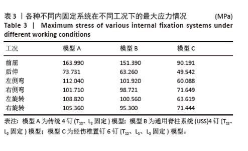
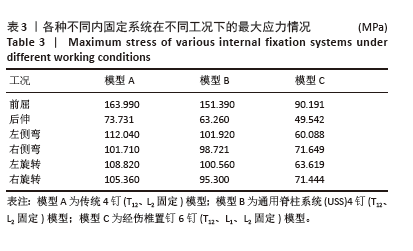
2.3 内固定最大应力 通过观察应力云图(图6),总结3种模型的最大应力集中于钉棒连接处和螺钉根部以及螺钉前端处,螺钉应力的最大值出现在前屈,最小值出现在后伸时,与螺钉位移云图相对应,在不同加载条件下,经伤椎内固定组(模型C)最大应力最小,而传统4钉组(模型A)在不同加载条件下最大应力均大于其他两组,分析原因是固定在伤椎的2枚螺钉将集中于下位螺钉根部和连接棒处的应力部分向上转移,分散了整体内固定系统的应力,见表3。"
"
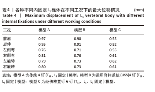
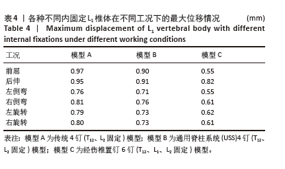
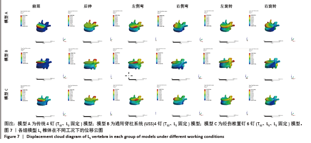
2.4 L1椎体最大位移情况 通过观察L1椎体最大位移云图(图7),前屈、后伸、左侧弯、右侧弯、左旋转、右旋转6种加载模式下,模型C < 模型B < 模型A。在前屈状态下,3组的L1椎体最大位移差异最大;后伸状态下3组L1椎体最大位移差异最小;通过观察位移云图,前屈、后伸、左侧弯、右侧弯、左旋转、右旋转6种加载模式下,传统4钉固定(模型A)的L1椎体最大位移均大于其他两组,而经伤椎6钉内固定(模型C)组L1椎体最大位移最小,见表4。"
"
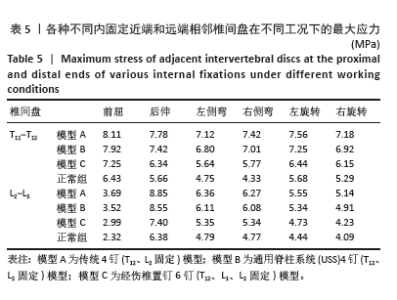
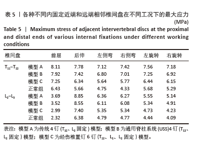
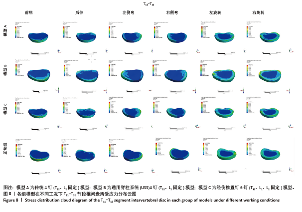
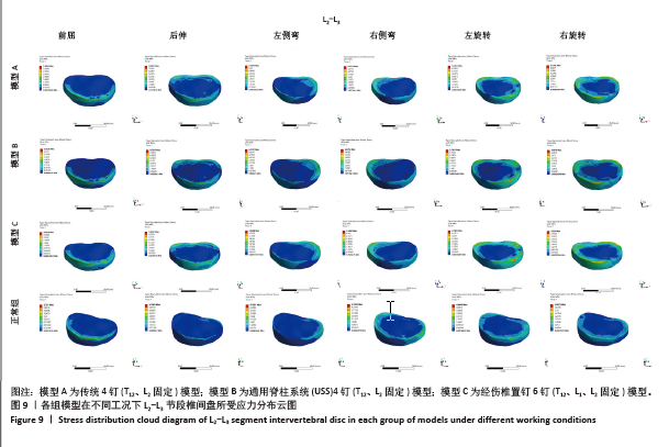
2.5 内固定近端和远端相邻椎间盘的应力情况 在前屈、后伸、左右侧弯、左右旋转6个方向上,近端(T11-T12节段)及远端(L2-L3节段)相邻椎间盘应力云图分别见图8,9,显示椎间盘应力主要集中于外周的纤维环,中间的髓核应力则最小,近端和远端相邻椎间盘所受的最大应力经伤椎6钉组(模型C)最小,且3组最大应力均大于无骨折无内固定的正常椎体模型组,各工况最大应力数值见表5。"

"

"
| [1] 苏林涛,余秋宇,马俊,等. 胸腰椎骨折椎体水肿对经皮椎弓钉固定疗效的影响[J]. 中国矫形外科杂志,2024,32(20):1852-1857. [2] HUANG L, XIONG C, GUO Z, et al. Comparison of monoplanar and polyaxial screw fixation systems in percutaneous intermediate fixation for thoracolumbar fractures. BMC Musculoskelet Disord. 2022;23(1): 172. [3] 程明煌,胡侦明,江维,等. 胸腰椎爆裂骨折治疗的研究进展[J]. 临床骨科杂志,2023,26(4):598-602. [4] LI WJ, GUO LX. Influence of different postures under vertical impact load on thoracolumbar burst fracture. Med Biol Eng Comput. 2020; 58(11):2725-2736. [5] SHEN J, YANG Z, FU M, et al. The influence of topical use of tranexamic acid in reducing blood loss on early operation for thoracolumbar burst fracture: A randomized double-blinded controlled study. Eur Spine J. 2021;30(10):3074-3080. [6] YU D, DA F, JUN C, et al. Anonymous, Anterior, posterior and anterior-posterior approaches for the treatment of thoracolumbar burst fractures: a network meta-analysis of randomized controlled trials. J Invest Surg. 2024;37(1):2301794. [7] XU L, LIN X, WU C, et al. Is unilateral pedicle screw fixation as effective as bilateral pedicle screw fixation in transforaminal lumbar interbody fusion: A meta-analysis of randomized controlled trials. Eur Spine J. 2023;32(2):700-711. [8] ALIMOHAMMADI E, BAGHERI SR, JOSEPH B, et al. Analysis of factors associated with the failure of treatment in thoracolumbar burst fractures treated with short-segment posterior spinal fixation. J Orthop Surg. 2023;18(1):690. [9] YANG S, SHANG DP, LU JM, et al. Modified Posterior Short-Segment Pedicle Screw Instrumentation for Lumbar Burst Fractures with Incomplete Neurological Deficit. World Neurosurg. 2018;119:977-985. [10] ALANAY A, ACAROGLU E, YAZICI M, et al. Short-segment pedicle instrumentation of thoracolumbar burst fractures: does transpedicular intracorporeal grafting prevent early failure? Spine. 2001;26(2): 213-217. [11] CHOU KN, WANG PW, CHUNG MH, et al. Hybrid kyphoplasty with short-versus intermediate- and long-segment pedicle screw fixations for the management of thoracolumbar burst fractures. BMC Musculoskelet Disord. 2024;25(1):203. [12] LI C, ZHOU Y, WANG H, et al. Treatment of unstable thoracolumbar fractures through short segment pedicle screw fixation techniques using pedicle fixation at the level of the fracture: a finite element analysis. PloS One. 2014;9(6):99156. [13] 赵豪,高山,陈文恒,等. 经伤椎与跨伤椎固定胸腰椎爆裂骨折的比较[J]. 中国矫形外科杂志,2022,30(22):2039-2044. [14] 王越,李良生,陈建泉,等.后路短节段经皮椎弓根螺钉内固定术治疗胸腰椎骨折术中应用横连接的必要性[J]. 脊柱外科杂志, 2024,22(3):170-175. [15] WU W, HAN Z, HU B, et al. A graphical guide for constructing a finite element model of the cervical spine with digital orthopedic software. Ann Transl Med. 2021;9(2):169. [16] LIU J, YANG S, ZHOU F, et al. The feasibility of short-segment Schanz screw implanted in an oblique downward direction for the treatment of lumbar 1 burst fracture: a finite element analysis. J Orthop Surg. 2020;15(1):537. [17] MCCORMACK T, KARAIKOVIC E, GAINES RW. The load sharing classification of spine fractures. Spine. 1994;19(15):1741-1744. [18] XU G, FU X, DU C, et al. Biomechanical comparison of mono-segment transpedicular fixation with short-segment fixation for treatment of thoracolumbar fractures: a finite element analysis. Proc Inst Mech Eng. 2014;228(10):1005-1013. [19] ALIZADEH M, KADIR MRA, FADHLI MM, et al. The use of X-shaped cross-link in posterior spinal constructs improves stability in thoracolumbar burst fracture: a finite element analysis. J Orthop Res Off Publ Orthop Res Soc. 2013;31(9):1447-1454. [20] SCHMOELZ W, SCHASER KD, KNOP C, et al. Extent of corpectomy determines primary stability following isolated anterior reconstruction in a thoracolumbar fracture model. Clin. Biomech. Bristol Avon. 2010;25(1):16-20. [21] CHOU TY, TSUANG FY, HSU YL, et al. Surgical versus non-surgical treatment for thoracolumbar burst fractures without neurological deficit: A systematic review and meta-analysis. Glob Spine J. 2024; 14(2):740-749. [22] DVORAK MF, ÖNER CF, SCHNAKE K, et al. From Radiographic Evaluation to Treatment Decisions in Neurologically Intact Patients With Thoraco-lumbar Burst Fractures. Glob Spine J. 2024;14:4-7. [23] GREINER PERTH AK, WILKE HJ, LIEBSCH C. Which spinal fixation technique achieves which degree of stability after thoracolumbar trauma? A systematic quantitative review. Spine J Off J North Am Spine Soc. 2024;S1529-9430(24)1095-7. [24] HASHIMURA T, ONISHI E, OTA S, et al. Correction loss following short-segment posterior fixation for traumatic thoracolumbar burst fractures related to endplate and intervertebral disc destruction. BMC Musculoskelet Disord. 2023;24(1):174. [25] YADAV SK, RAJNISH RK, KANTIWAL P, et al. Short segment posterior fixation of unstable thoracolumbar vertebral fractures with fractured vertebra augmentation with intermediate pedicle screw - a clinicoradiological analysis. Am J Neurodegener Dis. 2024;13(1):1-6. [26] RISPOLI R, ABOUSAYED M, HAMED AA, et al. Long versus short segment with intermediate screw fixation for burst fractures of thoracolumbar junction: Radiological and clinical results. J Neurosurg Sci. 2024;68(5): 567-573. [27] ZHANG G, LI J, ZHANG L, et al. Biomechanical effect of different posterior fixation techniques on stability and adjacent segment degeneration in treating thoracolumbar burst fracture with osteoporosis: A finite element analysis. Spine. 2024;49(15):229-238. [28] ZHANG G, DU Y, JIANG G, et al. Biomechanical evaluation of different posterior fixation techniques for treating thoracolumbar burst fractures of osteoporosis old patients: A finite element analysis. Front Bioeng Biotechnol. 2023;11:1268557. [29] LIAO JC, CHEN WP, WANG H. Treatment of thoracolumbar burst fractures by short-segment pedicle screw fixation using a combination of two additional pedicle screws and vertebroplasty at the level of the fracture: a finite element analysis. BMC Musculoskelet Disord. 2017; 18(1):262. [30] AONO H, TOBIMATSU H, ARIGA K, et al. Surgical outcomes of temporary short-segment instrumentation without augmentation for thoracolumbar burst fractures. Injury. 2016;47(6):1337-1344. [31] CAI P, XI Z, DENG C, et al. Fixation-induced surgical segment’s high stiffness and the damage of posterior structures together trigger a higher risk of adjacent segment disease in patients with lumbar interbody fusion operations. J Orthop Surg. 2023;18(1):371. [32] CUI X, ZHU J, YANG W, et al. Finite element study of sagittal fracture location on thoracolumbar fracture treatment. Front Bioeng Biotechnol. 2023;11:1229218. [33] XILONG C, JUNHUN Z, YULIANG S, et al. Biomechanical comparison of different treatment strategies for thoracolumbar burst fracture: A finite element study. World Neurosurg. 2023;180:429-439. [34] LI Q, LI X, LIU Y, et al. Treatment of thoracolumbar fracture with pedicle screws at injury level: a biomechanical study based on three-dimensional finite element analysis. Eur J Orthop Surg Traumatol Orthop Traumatol. 2013;23(7):775-780. [35] MAHAR A, KIM C, WEDEMEYER M, et al. Short-segment fixation of lumbar burst fractures using pedicle fixation at the level of the fracture. Spine. 2007;32(14):1503-1507. [36] DICK JC, JONES MP, ZDEBLICK TA, et al. A biomechanical comparison evaluating the use of intermediate screws and cross-linkage in lumbar pedicle fixation. J Spinal Disord. 1994;7(5):402-407. [37] MULLER U, BERLEMANN U, SLEDGE J, et al. Treatment of thoracolumbar burst fractures without neurologic deficit by indirect reduction and posterior instrumentation: Bisegmental stabilization with monosegmental fusion. Eur Spine J. 1999;8(4):284-289. [38] SCHOMIG F, PALMOWSKI Y, NIKIFOROV I, et al. Burst fractures lead to a fracture-associated intervertebral vacuum phenomenon: a case series of 305 traumatic fractures of the thoracolumbar spine. Eur Spine J. 2021;30(10):3068-3073. |
| [1] | Zhang Di, Zhao Jun, Ma Guangyue, Sun Hui, Jiang Rong. Mechanism of depression-like behavior in chronic social defeat stress mice based on high-throughput sequencing [J]. Chinese Journal of Tissue Engineering Research, 2026, 30(5): 1139-1146. |
| [2] | Wen Fan, Xiang Yang, Zhu Huan, Tuo Yanfang, Li Feng. Exercise improves microvascular function in patients with type 2 diabetes [J]. Chinese Journal of Tissue Engineering Research, 2026, 30(5): 1225-1235. |
| [3] | Guo Jiachen, Gao Jun, Dai Wenhao, Liao Huayuan, Jiang You, Zhang Xi . Effect of compressive stress microenvironment on cytokines during fracture healing [J]. Chinese Journal of Tissue Engineering Research, 2026, 30(4): 908-916. |
| [4] | Yang Xiao, Bai Yuehui, Zhao Tiantian, Wang Donghao, Zhao Chen, Yuan Shuo. Cartilage degeneration in temporomandibular joint osteoarthritis: mechanisms and regenerative challenges [J]. Chinese Journal of Tissue Engineering Research, 2026, 30(4): 926-935. |
| [5] | Wang Peng, Li Zhijun, Zhang Shaojie, Wu Yimin. Intervertebral disc rehydration after posterior lumbar dynamic internal fixation [J]. Chinese Journal of Tissue Engineering Research, 2026, 30(3): 711-720. |
| [6] | Yu Xinlin, Chen Huiyu, Wang Yingying, Guo Weizhong, Feng Bin Lin Chengshou, Lin Wang. Finite element analysis of internal fixation with new retrograde intramedullary nail on lateral femur condyle for distal type A2 femur fractures [J]. Chinese Journal of Tissue Engineering Research, 2026, 30(3): 546-552. |
| [7] | Zhao Jingang, Liu Liping, Chen Jianwei, . Finite element analysis comparing lumbar fusion and artificial intervertebral disc replacement [J]. Chinese Journal of Tissue Engineering Research, 2026, 30(3): 553-560. |
| [8] | Wang Meng, Lu Tan, Li Minjie, Liu Zhicheng, Guo Xiaoyong. Finite element analysis of stress distribution of anchors at different implantation depths under different bone density conditions in rotator cuff tears [J]. Chinese Journal of Tissue Engineering Research, 2026, 30(3): 561-569. |
| [9] | Ma Jingbo, Yang Guangnan, Liu Jiang, Jiang Qiang, Zhang Hanshuo, Han Jiaheng, Ding Yu. Endoscopic lumbar canal decompression for upper lumbar spinal stenosis: a comparison of biomechanical stability of three surgical models [J]. Chinese Journal of Tissue Engineering Research, 2026, 30(3): 577-585. |
| [10] | Abudusalamu·Tuoheti, Xiao Yang, Wang Yixi, Musitapa·Mijiti, Chen Qihao, Maimaitiming·Saiyiti, Guo Hailong, Paerhati·Rexiti. Effects of three internal fixation techniques on biomechanics of adjacent segment degeneration in lumbar interbody fusion [J]. Chinese Journal of Tissue Engineering Research, 2026, 30(3): 586-595. |
| [11] | Dong Chao, Zhao Mohan, Liu Yunan, Yang Zeli, Chen Leqin, Wang Lanfang. Effects of magnetic nano-drug carriers on exercise-induced muscle injury and inflammatory response in rats [J]. Chinese Journal of Tissue Engineering Research, 2026, 30(2): 345-353. |
| [12] | Cheng Yanan, Yu Jiazhi, Liu Yinchang, Wu Jie, Yu Tong, Wang Lu, Li Xiaoguang. Three-dimensional finite element analysis of molar distalization with clear aligners with different thicknesses and edges [J]. Chinese Journal of Tissue Engineering Research, 2026, 30(2): 310-318. |
| [13] | Kang Zirui, Wu Yang, Song Hailong, Yang Qiaoyun, Zang Lixiang, Xu Dongliang. Finite element analysis of implants with different crown-to-implant ratios under different bone conditions [J]. Chinese Journal of Tissue Engineering Research, 2026, 30(2): 319-328. |
| [14] | Wu Zhijing, Li Jiali, Zhang Jiaxin, Wang Tangrong, Zheng Yuzhou, Sun Zixuan. Alpha-ketoglutarate engineered small extracellular vesicles delay skin aging [J]. Chinese Journal of Tissue Engineering Research, 2026, 30(1): 120-129. |
| [15] | Xu Hao, Ding Lu, Li Xiao. Investigating the effect of the mechanical wear on abutment screw in Morse taper connection implant implant system by using finite element analysis [J]. Chinese Journal of Tissue Engineering Research, 2025, 29(在线): 1-9. |
| Viewed | ||||||
|
Full text |
|
|||||
|
Abstract |
|
|||||