Chinese Journal of Tissue Engineering Research ›› 2026, Vol. 30 ›› Issue (9): 2208-2216.doi: 10.12307/2026.535
Previous Articles Next Articles
Mechanical effects of Long’s traction orthopedic method on cervical functional units: quantitative analysis of biomechanical model of head and neck
Cai Qirui1, Dai Xiaowei2, Zheng Xiaobin1, Jian Sili1, Lu Shaoping2, Liu Texi1, Liu Guoke1, Lin Yuanfang1, 2
- 1Shenzhen Traditional Chinese Medicine Hospital, Shenzhen 518033, Guangdong Province, China; 2The Fourth Clinical Medical College of Guangzhou University of Chinese Medicine, Shenzhen 518033, Guangdong Province, China
-
Received:2024-11-14Accepted:2025-01-20Online:2026-03-28Published:2025-09-05 -
Contact:Lin Yuanfang, MS, Chief physician, Master’s supervisor, Shenzhen Traditional Chinese Medicine Hospital, Shenzhen 518033, Guangdong Province, China; The Fourth Clinical Medical College of Guangzhou University of Chinese Medicine, Shenzhen 518033, Guangdong Province, China -
About author:Cai Qirui, Attending physician, Shenzhen Traditional Chinese Medicine Hospital, Shenzhen 518033, Guangdong Province, China -
Supported by:Guangdong Province Shenzhen Science and Technology Plan Project Basic Research Project (Natural Science Foundation), No. JCYJ20210324111212035 (to LYF); Guangdong Province Shenzhen Government Medical and Health Three Engineering Project, No. SZZYSM202311006 (to LYF)
CLC Number:
Cite this article
Cai Qirui, Dai Xiaowei, Zheng Xiaobin, Jian Sili, Lu Shaoping, Liu Texi, Liu Guoke, Lin Yuanfang. Mechanical effects of Long’s traction orthopedic method on cervical functional units: quantitative analysis of biomechanical model of head and neck[J]. Chinese Journal of Tissue Engineering Research, 2026, 30(9): 2208-2216.
share this article
Add to citation manager EndNote|Reference Manager|ProCite|BibTeX|RefWorks
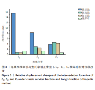
2.1 头颈-肌肉-脊柱有限元模型验证 原始模型已根据作者之前研究获得的数据进行了全方位的验证[13],具体来说,从单一组织或脊柱功能单元再到整体节段逐步获得的解剖测量值以及整个头颈模型运动[27-33],包括准静态响应和动态响应[34]。 2.2 C5、C6、C7椎间孔相对位移改变 如图3所示,经典颈椎牵引与龙氏牵引下正骨手法对于椎间孔的相对位移改变差异显著。其中,推正法作用下C5椎间孔相对位移的改变最显著,高达15.6 mm,在所有手法、所有节段中最高;推正法作用下椎间孔相对位移改变随着节段的下移改变越小,至C7位移仅为3.8 mm。扳按法和摇正法作用下椎体椎间盘相对位移改变的结果相近,不同节段之间的差异较小;且随着节段的下移,椎间盘相对位移逐渐变大。而在经典颈椎牵引中,C5椎间孔相对位移改变较明显,但仅为1.3 mm。"


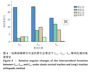
2.3 C4-5、C5-6、C6-7椎间孔相对角度变化 经典颈椎牵引与龙氏牵引下正骨法在数值和变化趋势上表现出较大差异。经典颈椎牵引下椎间孔相对角度的改变较小,且随着节段的下移,影响能力递减。而椎间孔相对角度在龙氏牵引下正骨手法实施前后的整体变化趋势与龙氏牵引下正骨手法下对相对位移的改变相似,但也有其特异性。如图4所示,推正法作用下C4-5、C5-6椎间孔相对角度的改变最大,且二者结果相近,分别为22.5°,22.9°;极差为3个手法中最小,仅为7.767°。扳按法作用下椎间孔相对角度的改变随着节段的下移逐渐增大,至C6-7最大,达到11.9°。摇正法对不同椎体椎间孔相对角度改变的数值最为集中。"
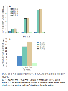
2.4 椎体侧屈的相对位移改变 2.4.1 单个椎体侧屈的相对位移改变 经典颈椎牵引给予的是向上的牵引力,使椎体产生侧屈运动的能力有限,相比之下复杂的正骨手法带来截然不同的结果,且不同手法之间的结果也差异悬殊。如图5A所示,对所有工况下单个椎体侧屈所产生的相对位移改变进行分析发现,扳按法和摇正法对椎体侧屈的相对位移改变最大,且数值和变化趋势极为相近,摇正法的整体结果均高于扳按法;随着节段的下移,侧屈的活动范围逐渐增大,椎体侧屈的相对位移同步增加,到C7分别达到峰值,分别为11.9,12.4 mm。而经典颈椎牵引和推正法对椎体侧屈产生的位移影响最大仅为1.05 mm,与摇正法的极差分别达到12.14,12.02 mm。 2.4.2 C4-7整体节段椎体侧屈的相对位移改变 选取单侧椎体从C4-7的相同节点作为参考点,测量其整体节段椎体侧屈的相对位移。扳按法的施力方式是直接使椎体产生单侧的侧屈运动,而摇正法是通过对椎体产生旋转作用而使椎体产生侧屈运动,如图5B可以看到,椎体受到旋转的作用力时产生的生物响应更大。摇正法作用下C4-7整体节段椎体侧屈相对位移的改变明显大于扳按法。经典颈椎牵引作用下整体节段侧屈的相对位移为1.9 mm,约为摇正法的八分之一。"

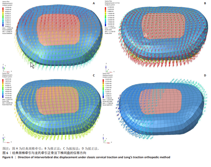
2.5 椎间盘位移方向 图6中椎间盘上不同颜色的箭头及方向代表了椎间盘位移的大小及方向。在正骨手法的椎间盘位移方向图中,箭头指向前方,表明椎间盘在外力作用下产生了向前的应变。这种前向的形变趋势可能对椎间盘的后方结构(如神经根及颈髓)产生减压作用,从而缓解其受到的压迫。推正法对椎间盘位移的改变集中且数值最大(图6B),扳按法对椎间盘的改变整体均匀(图6C),而摇正法在旋转的作用下使椎间盘发生左前右后的改变(图6D);正骨手法在 Y 轴方向的膨出位移最明显,分别为11.56,9.94,7.73 mm。而经典颈椎牵引(图6A)则使椎间盘位移向后。这4种手法通过不同的力学作用模式来影响颈椎结构,图中不同的矢量方向和强度分布为评价其生物力学效果提供了数据支持。"
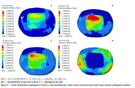
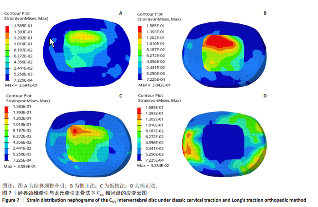
2.6 椎间盘内在应变 对C4/5椎间盘的应变云图分析如图7所示,各图的颜色表示不同的应变大小,从蓝色(最低应变)到红色(最高应变)逐渐递增,用于直观地展示不同方法对椎间盘的力学影响。在推正法中,可以观察到高应变区域明显集中于椎间盘髓核的后缘,提示此处髓核发生了显著的形变;扳按法则在髓核的右后缘出现了高应变区,这与向左下方施加的拉力导致右侧椎间孔间隙增大密切相关,椎间盘因此向左侧回缩;在摇正法中,椎间盘的应变呈现出环绕分布,这表明椎间盘在躲避外力压力的过程中发生了一定程度的回缩,尤其以手法治疗的侧面最为明显;在这3种手法中,推正法引起的椎间盘应变最为显著。而经典颈椎牵引下椎间盘产生的应变最小,分布均匀。"
| [1] YAO S, OUYANG B, LU T, et al. Treatment of cervical spondylotic radiculopathy with posterior percutaneous endoscopic cervical discectomy: Short-term outcomes of 24 cases. Medicine. 2020;99(20): e20216. [2] ZHANG Y, DONG L, ZHANG Y, et al. The effects of modified Guizhi plus Gegen decoction combined with the blade needle therapy on TCM syndromes, cervical curvature and levels of inflammatory factors in patients with cervical spondylotic radiculopathy. Am J Transl Res. 2023;15(8):5347-5355. [3] WANG C, GU Z, YU J, et al. Clinical observation of Long chiropractic treatment on patients with neurogenic cervical spondylosis: Study protocol for a randomized controlled trial .Medicine. 2022;101(9): e28861. [4] SHERROD BA, DAILEY AT, MAZUR MD. Closed cervical traction techniques: moving into the 21st century. J Neurosurg Spine. 2023; 39(5):618-627. [5] LIN T, SHANGGUAN Z, XIAO Z, et al. Whether the potential degree of cervical instability and cervical muscle degeneration in patients with cervical spondylosis radicular affect the efficacy of cervical traction. Sci Rep. 2024;14(1):20467. [6] CHENG ZJ, ZHANG SP, GU YJ, et al. Effectiveness of Tuina Therapy Combined With Yijinjing Exercise in the Treatment of Nonspecific Chronic Neck Pain: A Randomized Clinical Trial. JAMA Netw Open. 2022;5(12):e2246538. [7] KANG D, YIM J. A Study of 45 Patients with Chronic Neck Pain to Compare the Effects on Pain and Disability of 4 Weeks of Physical Therapy with the Active Release Technique and Strain-Counterstrain Manipulation with Massage Alone, Before and After Treatment. Med Sci Monit. 2023;29:e942027. [8] TU TH, WANG CY, CHEN YC, et al. Multilevel cervical disc arthroplasty: a review of optimal surgical management and future directions. J Neurosurg Spine. 2023;38(3):372-381. [9] 李德成,张彦敏,高冕,等.龙氏整脊疗法配合简易牵引治疗寰枢关节半脱位的临床疗效观察[J/OL].河北医药,1-4[2025-03-12].https://kns-cnki-net.webvpn.gzarts.edu.cn/kcms/detail/13.1090.r.20231215.1409.002.html. [10] 张其云, 赵永华, 陈亚锋. 龙氏牵引下正骨法治疗神经根型颈椎病的临床观察[J]. 河北中医,2019,41(7):1072-1075. [11] 欧志文. 龙氏牵引下正骨法治疗退行性下颈椎失稳症的临床观察[D]. 广州:广州中医药大学,2021. [12] LV G, CHEN X, WU H, et al. Finite element analysis of the use of two new types of internal fixation for acetabular fractures . J Orthop Surg Res. 2023;18(1):841. [13] LIANG Z, MO F, ZHENG Z, et al. Quantitative cervical spine injury responses in whiplash loading with a numerical method of natural neural reflex consideration. Comput Methods Programs Biomed. 2022;219:106761. [14] PANJABI MM, CHEN NC, SHIN EK, et al. The Cortical Shell Architecture of Human Cervical Vertebral Bodies. Spine. 2001;26(22):2478-2484. [15] PANZER MB, CRONIN DS. C4-C5 segment finite element model development, validation, and load-sharing investigation. J Biomech. 2009;42(4):480-490. [16] ÖSTH J, BROLIN K, SVENSSON MY, et al. A Female Ligamentous Cervical Spine Finite Element Model Validated for Physiological Loads. J Biomech Eng. 2016;138(6):061005. [17] SHEN TWD. Investigation of Whiplash Associated Disorders using Finite Element Neck Models with Active Musculature in Frontal, Rear and Lateral Impact. University of Waterloo, 2020. [18] LIU YT, DONG RC, LIU Z, et al. Finite element analysis of the cervical spine: dynamic characteristics and material property sensitivity study. Comput Methods Biomech Biomed Engin. 2024;1-15. doi: 10.1080/10255842.2024.2304285. [19] ÖSTH J, BROLIN K, CARLSSON S, et al. The occupant response to autonomous braking: a modeling approach that accounts for active musculature. Traffic Inj Prev. 2012;13(3):265-277. [20] CORRALES MA, CRONIN DS. Sex, age and stature affects neck biomechanical responses in frontal and rear impacts assessed using finite element head and neck models. Front Bioeng Biotechnol. 2021:9:681134. [21] 李婵, 李娟, 毛志涛, 等. 龙氏牵引下正骨法治疗颈源性肩周炎的临床疗效观察[J]. 广州中医药大学学报,2023,40(9):2225-2230. [22] 范德辉, 龙层花. 龙氏治脊疗法[M]. 广州: 广东科技出版社,2019. [23] SINGHAL I, HARINATHAN B, WARRAICH A, et al. Finite element modeling of the human cervical spinal cord and its applications: A systematic review . N Am Spine Soc J. 2023;15:100246. [24] HAN C, FENG M, WEN H, et al. Rotation-traction manipulation induced intradiskal pressure changes in cervical spine-an in vitro study . Front Bioeng Biotechnol. 2024;12:1322212. [25] LIN D, HE Z, WENG R, et al. Comparison of biomechanical parameters of two Chinese cervical spine rotation manipulations based on motion capture and finite element analysis. Front Bioeng Biotechnol. 2023;11: 1195583. [26] HUANG X, YE L, WU Z, et al. Biomechanical Effects of Lateral Bending Position on Performing Cervical Spinal Manipulation for Cervical Disc Herniation: A Three-Dimensional Finite Element Analysis. Evid Based Complement Alternat Med. 2018;2018:2798396. [27] FOURNELY M, PETIT Y, WAGNAC E, et al. Effect of experimental, morphological and mechanical factors on the murine spinal cord subjected to transverse contusion: A finite element study. PLoS One. 2020;15(5):e0232975. [28] FRANTSUZOV R, MONDAL S, WALSH CM, et al. A finite element model of contusion spinal cord injury in rodents. J Mech Behav Biomed Mater. 2023;142:105856. [29] SHEA M, EDWARDS WT, WHITE AA, et al. Variations of stiffness and strength along the human cervical spine. J Biomech. 1991;24(2):95-107. [30] NIGHTINGALE RW, WINKELSTEIN BA, KNAUB KE, et al. Comparative strengths and structural properties of the upper and lower cervical spine in flexion and extension. J Biomech. 2002;35(6):725-732. [31] WHEELDON JA, PINTAR FA, KNOWLES S, et al. Experimental flexion/extension data corridors for validation of finite element models of the young, normal cervical spine. J Biomech. 2006;39(2):375-380. [32] MANICKAM PS, ROY S. The biomechanical study of cervical spine: A Finite Element Analysis. Int J Artif Organs. 2022;45(1):89-95. [33] VASAVADA AN, DANARAJ J, SIEGMUND GP. Head and neck anthropometry, vertebral geometry and neck strength in height-matched men and women. J Biomech. 2008;41(1):114-121. [34] BEAUSéJOUR MH, PETIT Y, WAGNAC É, et al. Cervical spine injury response to direct rear head impact. Clin Biomech (Bristol). 2022;92: 105552. [35] WANG X, JIN Z, FENG T, et al. The immediate effect of cervical rotation-traction manipulation on cervical paravertebral soft tissue: a study using soft tissue tension cloud chart technology. BMC Musculoskelet Disord. 2024;25(1):184. [36] ZEHRA U, TRYFONIDOU M, IATRIDIS JC, et al. Mechanisms and clinical implications of intervertebral disc calcification. Nat Rev Rheumatol. 2022;18(6):352-362. [37] ROESCH ZK, TADI P. Anatomy, Head and Neck, Neck. StatPearls Publishing, 2024. [38] WANG H, LI N, HUANG H, et al. Biomechanical effect of intervertebral disc degeneration on the lower lumbar spine . Comput Methods Biomech Biomed Engin. 2023;26(14):1669-1677. [39] FAKHOURY J, DOWLING TJ. Cervical Degenerative Disc Disease. StatPearls Publishing, 2024. [40] 孙金水, 李云, 刘金鹏. 龙氏正骨牵引椅治疗颈椎病疗效的观察[J] 当代医学,2016,22(33):40-41. |
| [1] | Chen Huiting, Zeng Weiquan, Zhou Jianhong, Wang Jie, Zhuang Congying, Chen Peiyou, Liang Zeqian, Deng Weiming. Tail anchoring technique of vertebroplasty in treatment of osteoporotic vertebral compression fractures with intravertebral cleft: a finite element analysis [J]. Chinese Journal of Tissue Engineering Research, 2026, 30(9): 2145-2152. |
| [2] | Zeng Xuan, Weng Rui, Ye Shicheng, Tang Jiadong, Mo Ling, Li Wenchao. Two lumbar rotary manipulation techniques in treating lumbar disc herniation: a finite element analysis of biomechanical differences [J]. Chinese Journal of Tissue Engineering Research, 2026, 30(9): 2153-2161. |
| [3] | Cheng Qisheng, Julaiti·Maitirouzi, Xiao Yang, Zhang Chenwei, Paerhati·Rexiti. Finite element analysis of novel variable-diameter screws in modified cortical bone trajectory of lumbar vertebrae [J]. Chinese Journal of Tissue Engineering Research, 2026, 30(9): 2162-2171. |
| [4] | Wu Hongxu, Liu Xuanyu, Wang Taoyu, Wang Shiyao, Cheng Jingyi, Zhang Mingwen, Zhang Yinxia, Liu Zhihua, Wang Xiaojie. Finite element simulation of scoliosis with muscle unit introduction: verification of correction effect under bidirectional load [J]. Chinese Journal of Tissue Engineering Research, 2026, 30(9): 2172-2181. |
| [5] | Liu Jiafu, Ren Ruxia, Liao Zhouwei, Zhou Xiali, Wu Yihong, Zhang Shaoqun. Three-dimensional finite element analysis of cervical spine biomechanical characteristics in a rat model of cervical vertigo [J]. Chinese Journal of Tissue Engineering Research, 2026, 30(9): 2182-2190. |
| [6] | Zhou Daobin, Wang Kehao, Xie Yang, Ning Rende. Biomechanical characteristics of volar locking plate only versus combined dorsal mini-plate fixation of distal radius fractures with dorsal ulnar fragment [J]. Chinese Journal of Tissue Engineering Research, 2026, 30(9): 2255-2261. |
| [7] | Li Zhifei, Han Bin, Liu Qiuli, Zhang Zhanming, Wei Haokai, Zuo Kuangshi, Zhang Yisheng. Cervical motion characteristics in patients with cervical spondylotic radiculopathy based on motion capture technology [J]. Chinese Journal of Tissue Engineering Research, 2026, 30(9): 2286-2293. |
| [8] | Li Sa, Sun Ning, Sun Zhaozhong, Feng Zhimeng, Li Xuedong. Evaluation parameters and specific region of C6 nerve oppression by uncinate process degeneration [J]. Chinese Journal of Tissue Engineering Research, 2026, 30(9): 2294-2302. |
| [9] | Liu Wenlong, Dong Lei, Xiao Zhengzheng, Nie Yu. Finite element analysis of tibial prosthesis loosening after fixed-bearing unicompartmental knee arthroplasty for osteoporosis [J]. Chinese Journal of Tissue Engineering Research, 2026, 30(9): 2191-2198. |
| [10] | Zheng Wangyang, Fei Ji, Yang Di, Zhao Lang, Wang Lingli, Liu Peng, Li Haiyang. Finite element analysis of the force changes of the supraspinatus tendon and glenohumeral joint during the abduction and flexion of the humerus [J]. Chinese Journal of Tissue Engineering Research, 2026, 30(9): 2199-2207. |
| [11] | Rao Jingcheng, Li Yuwan, Zheng Hongbing, Xu Zhi, Zhu Aixiang, Shi Ce, Wang Bing, Yang Chun, Kong Xiangru, Zhu Dawei. Biomechanical differences between the new proximal femoral stable intramedullary nail and traditional intramedullary nail#br# [J]. Chinese Journal of Tissue Engineering Research, 2026, 30(9): 2217-2225. |
| [12] | Chen Long, Wang Xiaozhen, Xi Jintao, Lu Qilin. Biomechanical performance of short-segment screw fixation combined with expandable polyetheretherketone vertebral body replacement in osteoporotic vertebrae [J]. Chinese Journal of Tissue Engineering Research, 2026, 30(9): 2226-2235. |
| [13] | Yan Xiangning, Chen Lei, Chen Yonghuan, Wang Chao, Li Xiaosheng. Influence of different depths and loads on knee joint mechanics and peripheral muscle force characteristics during squatting [J]. Chinese Journal of Tissue Engineering Research, 2026, 30(9): 2236-2247. |
| [14] | Zheng Xuying, Hu Hongcheng, Xu Libing, Han Jianmin, Di Ping. Stress magnitude and distribution in two-piece cement-retained zirconia implants under different loading conditions and with varying internal connection shapes [J]. Chinese Journal of Tissue Engineering Research, 2026, 30(8): 1979-1987. |
| [15] | Zhong Caihong, Xiao Xiaoge, Li Ming, Lin Jianhong, Hong Jing. Biomechanical mechanism of sports-related patellar tendinitis [J]. Chinese Journal of Tissue Engineering Research, 2026, 30(6): 1417-1423. |
| Viewed | ||||||
|
Full text |
|
|||||
|
Abstract |
|
|||||