Chinese Journal of Tissue Engineering Research ›› 2025, Vol. 29 ›› Issue (27): 5795-5801.doi: 10.12307/2025.184
Previous Articles Next Articles
Quantitative evaluation of knee laxity after partial anterior cruciate ligament injury with a novel digital arthrometer
Zhu Zheyue, Zhang Chen, Ge Ying, Xue Han, Li Ruochen, Wu Guangwei, Ma Rui
- Joint and Foot Ankle Ward of Orthopedic Center, Second Affiliated Hospital of Xi’an Jiaotong University, Xi’an 710004, Shaanxi Province, China
-
Received:2024-03-11Accepted:2024-05-18Online:2025-09-28Published:2025-03-05 -
Contact:Ma Rui, MD, Associate researcher, Joint and Foot Ankle Ward of Orthopedic Center, Second Affiliated Hospital of Xi’an Jiaotong University, Xi’an 710004, Shaanxi Province, China -
About author:Zhu Zheyue, Master candidate, Joint and Foot Ankle Ward of Orthopedic Center, Second Affiliated Hospital of Xi’an Jiaotong University, Xi’an 710004, Shaanxi Province, China
CLC Number:
Cite this article
Zhu Zheyue, Zhang Chen, Ge Ying, Xue Han, Li Ruochen, Wu Guangwei, Ma Rui. Quantitative evaluation of knee laxity after partial anterior cruciate ligament injury with a novel digital arthrometer[J]. Chinese Journal of Tissue Engineering Research, 2025, 29(27): 5795-5801.
share this article
Add to citation manager EndNote|Reference Manager|ProCite|BibTeX|RefWorks
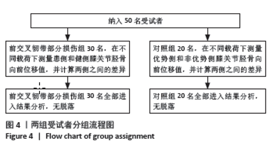
2.1 参与者数量分析 此次研究纳入30名前交叉韧带部分损伤患者及20名健康志愿者,所有受试者均参与试验流程并进入结果分析,无脱落。 2.2 试验流程图 见图4。"
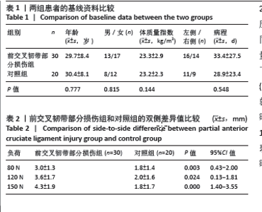
2.3 一般资料 比较基线资料发现,两组在年龄、性别、体质量指数和病程方面差异均无显著性意义(P > 0.05),见表1。 2.4 双侧差异值比较 前交叉韧带部分损伤组和对照组在不同负荷下的双侧差异值比较结果见表2。在80,120,150 N时,前交叉韧带部分损伤组的双侧差异值明显高于对照组,差异均有显著性意义(P < 0.05)。 "
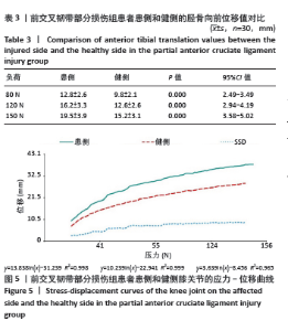
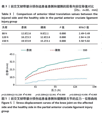
2.5 前交叉韧带部分损伤组患侧和健侧的胫骨向前位移值对比 表3显示了前交叉韧带部分损伤组患者患侧和健侧在不同负荷下的胫骨向前位移值对比结果。在80,120,150 N时,前交叉韧带部分损伤组患者患侧的胫骨向前位移值明显高于健侧,且差异均有显著性意义(P < 0.05)。图5展示了前交叉韧带部分损伤组患者患侧和健侧膝关节的应力-位移曲线。从图中可以看出,与健侧相比,患侧的应力-位移曲线更陡峭,说明患侧的松弛程度更大。另外,患侧和健侧的胫骨向前位移值随着应力的增加都有逐渐升高的趋势,双侧差异值也在随着应力的增加而缓慢升高。"
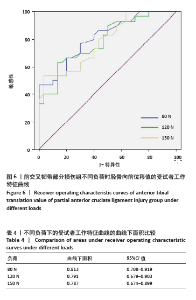
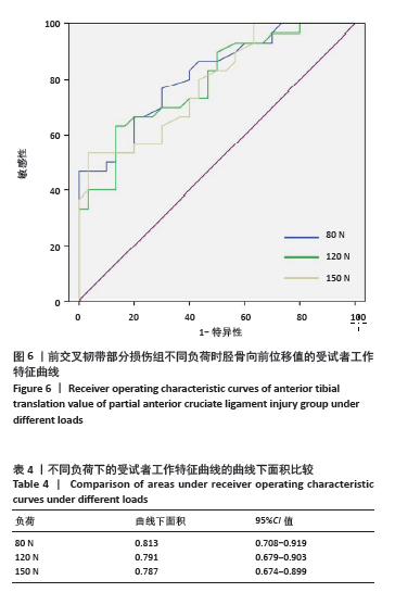
2.6 诊断准确性评价 通过软件绘制了80,120,150 N应力时的受试者工作特征曲线(图6)。统计学上可将不同曲线下面积的诊断值分为0 (曲线下面积 = 0.5)、信息量较差(0.5 <曲线下面积≤0.7)、信息量相当(0.7 <曲线下面积≤0.9)、信息量大(0.9 <曲线下面积< 1)和完美(曲线下面积 = 1),即曲线下面积越大,其代表的信息量就越大,诊断价值就越高。结果显示,此次研究中80 N时的受试者工作特征曲线的曲线下面积最大(cutoff值=10.45 mm),曲线下面积=0.813 (95%CI:0.708-0.919),见表4,敏感性为0.767,特异性为0.700。基于此得出80 N时的诊断准确性最高的结论。 2.7 不良事件 此次研究过程中所有患者及健康志愿者未出现任何不耐受及后续不良反应。"
| [1] 吴秀霖,徐俊杰,叶梓鹏,等.前交叉韧带损伤合并髌骨不稳研究进展[J].国际骨科学杂志,2024,45(1):11-14,18. [2] KHAN T, ALVAND A, PRIETO-ALHAMBRA D, et al. ACL and meniscal injuries increase the risk of primary total knee replacement for osteoarthritis: a matched case-control study using the Clinical Practice Research Datalink (CPRD). Br J Sports Med. 2019;53(15):965-968. [3] STONE AV, MARX S, CONLEY CW, et al. Management of Partial Tears of the Anterior Cruciate Ligament: A Review of the Anatomy, Diagnosis, and Treatment. J Am Acad Orthop Surg. 2021;29(2):60-70. [4] DALLO I, CHAHLA J, MITCHELL JJ, et al. Biologic approaches for the treatment of partial tears of the anterior cruciate ligament: A current concepts review. Orthop J Sports Med. 2017;5(1):2325967116681724. [5] 吕琦,马春辉,王培军,等. 3.0T MRI评估青年人群膝关节前交叉韧带(ACL)损伤的危险相关因素[J]. 复旦学报(医学版),2014, 41(5):667-672. [6] FILBAY SR, GRINDEM H. Evidence-based recommendations for the management of anterior cruciate ligament (ACL) rupture. Best Pract Res Clin Rheumatol. 2019;33(1):33-47. [7] RUNER A, SARSINA TRD, STARKE V, et al. The evaluation of Rolimeter, KLT, KiRA and KT-1000 arthrometer in healthy individuals shows acceptable intra-rater but poor inter-rater reliability in the measurement of anterior tibial knee translation. Knee Surg Sports Traumatol Arthrosc. 2021;29(8):2717-2726. [8] LI JQ, TANG JX, YAO L, et al. The validity of the Ligs digital arthrometer at different loads to evaluate complete ACL ruptures. Front Bioeng Biotechnol. 2023;11:1049100. [9] CHEN Y, CAO S, WANG C, et al. Quantitative analysis with load–displacement ratio measured via digital arthrometer in the diagnostic evaluation of chronic ankle instability: a cross-sectional study. J Orthop Surg Res. 2022;17(1):287. [10] KAEDING CC, LÉGER-ST-JEAN B, MAGNUSSEN RA. Epidemiology and diagnosis of anterior cruciate ligament injuries. Clin Sports Med. 2017;36:1-8. [11] WHITTAKER JL, WOODHOUSE LJ, NETTEL-AGUIRRE A, et al. Outcomes associated with early post-traumatic osteoarthritis and other negative health consequences 3–10 years following knee joint injury in youth sport. Osteoarthritis Cartilage. 2015;23:1122-1129. [12] JOHNSON VL, ROE JP, SALMON LJ, et al. Does age influence the risk of incident knee osteoarthritis after a traumatic anterior cruciate ligament injury? Am J Sports Med. 2016;44:2399-2405. [13] FILBAY SR, ACKERMAN IN, RUSSELL TG, et al. Return to sport matters—longer-term quality of life after ACL reconstruction in people with knee difficulties. Scand J Med Sci Sports. 2017;27:514-524. [14] SANDERS TL, MARADIT KREMERS H, BRYAN AJ, et al. Incidence of anterior cruciate ligament tears and reconstruction. Am J Sports Med. 2016;44:1502-1507. [15] ARDERN CL, EKÅS G, GRINDEM H, et al. 2018 International Olympic Committee consensus statement on prevention, diagnosis and management of paediatric anterior cruciate ligament (ACL) injuries. Knee Surg Sports Traumatol Arthrosc. 2018;26:989-1010. [16] MUSAHL V, KARLSSON J. Anterior Cruciate Ligament Tear. N Engl J Med. 2019;380:2341-2348. [17] MAGNUSSEN RA, PEDROZA AD, DONALDSON CT, et al. Time from ACL injury to reconstruction and the prevalence of additional intra-articular pathology: is patient age an important factor? Knee Surg Sports Traumatol Arthrosc. 2013;21:2029-2034. [18] SRI-RAM K, SALMON LJ, PINCZEWSKI LA, et al. The incidence of secondary pathology after anterior cruciate ligament rupture in 5086 patients requiring ligament reconstruction. Bone Joint J. 2013;95-B: 59-64. [19] MULLIGAN EP, MCGUFFIE DQ, COYNER K, et al. The reliability and diagnostic accuracy of assessing the translation endpoint during the Lachman test. Int J Sports Phys Ther. 2015;10:52-61. [20] LEBLANC MC, KOWALCZUK M, ANDRUSZKIEWICZ N, et al. Diagnostic accuracy of physical examination for anterior knee instability: a systematic review. Knee Surg Sports Traumatol Arthrosc. 2015;23(10): 2805-2813. [21] SOKAL PA, NORRIS R, MADDOX TW, et al. The diagnostic accuracy of clinical tests for anterior cruciate ligament tears are comparable but the Lachman test has been previously overestimated: a systematic review and meta-analysis. Knee Surg Sports Traumatol Arthrosc. 2022; 30(10):3287-3303. [22] LELLI A, TURI RPD, SPENCINER DB, et al. The “Lever Sign”: A new clinical test for the diagnosis of anterior cruciate ligament rupture. Knee Surg Sports Traumatol Arthrosc. 2016;24(9):2794-2797. [23] COOPERMAN JM, RIDDLE DL, ROTHSTEIN JM. Reliability and validity of judgments of the integrity of the anterior cruciate ligament of the knee using the Lachman’s test. Phys Ther. 1990;70(4):225-233. [24] GUNAYDIN B, SAHIN GG, SARI A, et al. Corrigendum to “A new method for diagnosis of anterior cruciate ligament tear: MRI with maximum flexion of knee in the prone position: A case control study” [Int. J. Surg. 68 (2019) 142-147]. Int J Surg. 2020:73:123. [25] VAN DYCK P, DE SMET E, VERYSER J, et al. Partial tear of the anterior cruciate ligament of the knee: injury patterns on MR imaging. Knee Surg Sports Traumatol Arthrosc. 2012;20(2):256-261. [26] TORZILLI PA, PANARIELLO RA, FORBES A, et al. Measurement reproducibility of two commercial knee test devices. J Orthop Res. 1991;9:730-737. [27] BACH BR, WARREN RF, FLYNN WM, et al. Arthrometric evaluation of knees that have a torn anterior cruciate ligament. J Bone Joint Surg Am. 1990;72:1299-1306. [28] BALLANTYNE BT, FRENCH AK, HEIMSOTH SL, et al. Influence of examiner experience and gender on interrater reliability of KT-1000 arthrometer measurements. Phys Ther. 1995;75:898-906. [29] ROBNETT NJ, RIDDLE DL, KUES JM. Intertester reliability of measurements obtained with the KT-1000 on patients with reconstructed anterior cruciate ligaments. J Orthop Sports Phys Ther. 1995;21(2):113-119. [30] WIERTSEMA SH, VAN HOOFF HJ, MIGCHELSEN LA, et al. Reliability of the KT1000 arthrometer and the Lachman test in patients with an ACL rupture. Knee. 2008;15(2):107-110. [31] BOYER P, DJIAN P, CHRISTEL P, et al. Reliability of the KT-1000 arthrometer (Medmetric) for measuring anterior knee laxity: comparison with Telos in 147 knees. Rev Chir Orthopédique Réparatrice Appar Mot. 2004;90(8):757-764. [32] SERNERT N, HELMERS J, KARTUS C, et al. Knee-laxity measurements examined by a left-hand- and a right-hand-dominant physiotherapist, in patients with anterior cruciate ligament injuries and healthy controls. Knee Surg Sports Traumatol Arthrosc. 2007;15(10): 1181-1186. [33] PAPANDREOU MG, ANTONOGIANNAKIS E, KARABALIS C, et al. Inter-rater reliability of Rolimeter measurements between anterior cruciate ligament injured and normal contra lateral knees. Knee Surg Sports Traumatol Arthrosc. 2005;13(7):592-597. [34] HATCHER J, HATCHER A, ARBUTHNOT J, et al. An investigation to examine the inter-tester and intra-tester reliability of the Rolimeter knee tester, and its sensitivity in identifying knee joint laxity. Randomized Controlled Trial. 2005;23(6):1399-1403. [35] ERICSSON D, ÖSTENBERG AH, ANDERSSON E, et al.Test-retest reliability of repeated knee laxity measurements in the acute phase following a knee trauma using a Rolimeter. J Exerc Rehabil. 2017;13(5):550-558. [36] ROHMAN EM, MACALENA JA. Anterior cruciate ligament assessment using arthrometry and stress imaging. Curr Rev Musculoskelet Med. 2016;9(2):130-138. [37] RYU SM, NA HD, SHON OJ, et al. Diagnostic Tools for Acute Anterior Cruciate Ligament Injury: GNRB, Lachman Test, and Telo. Knee Surg Relat Res. 2018;30(2):121-127. [38] KIM SH, PARK YB, HAM DW, et al. Stress radiography at 30° of knee flexion is a reliable evaluation tool for high-grade rotatory laxity in complete ACL-injured knees. Knee Surg Sports Traumatol Arthrosc. 2020;28(7):2233-2244. [39] RAGGI F, SARSINA TRD, SIGNORELLI C, et al. Triaxial accelerometer can quantify the Lachman test similarly to standard arthrometers. Knee Surg Sports Traumatol Arthrosc. 2019;27:2698-2703. [40] MASSEY PK, HARRIS JD, WINSTON LA, et al. Critical Analysis of the Lever Test for Diagnosis of Anterior Cruciate Ligament Insufficiency. Arthroscopy. 2017;33(8):1560-1566. [41] WU D, WANG DH, HAN YJ, et al. A novel digital arthrometer to measure anterior tibial translation. J Orthop Surg Res. 2023;18(1):101. |
| [1] | Zhang Shuang, Huang Zishuai, Wang Jian. Functional impacts of bone bruise and combined injuries in anterior cruciate ligament injuries [J]. Chinese Journal of Tissue Engineering Research, 2026, 30(10): 2491-2502. |
| [2] | Wang Juan, Wang Guanglan, Zuo Huiwu. Efficacy of exercise therapy in the treatment of anterior cruciate ligament reconstruction patients: #br# a network meta-analysis #br# [J]. Chinese Journal of Tissue Engineering Research, 2025, 29(8): 1714-1726. |
| [3] | Wang Changbing, Zhao Lilian, Fu Chuying, Li Yanjin. Modified double-bundle arthroscopic repair of the anterior cruciate ligament after Sherman type I injury [J]. Chinese Journal of Tissue Engineering Research, 2025, 29(6): 1192-1198. |
| [4] | Xiong Bohan, Wang Guoliang, Yu Yang, Xue Wenqiang, Yu Hong, Liu Jinrui, Ruan Zhaohui, Li Yajuan, Liu Haolong, Dong Kaiyan, Long Dan, Chen Zhao. Internal tension relieving technique assisted anterior cruciate ligament reconstruction to promote ligamentization of Achilles tendon grafts in small ear pigs in southern Yunnan province [J]. Chinese Journal of Tissue Engineering Research, 2025, 29(4): 713-720. |
| [5] | Wang Feng, Cao Chunfeng, He Chao, Zhang Tao, Zhou Zixian, Zhu Fengchen. All-inside versus traditional techniques of anterior cruciate ligament reconstruction: meta-analysis of therapeutic efficacy and radiological outcomes [J]. Chinese Journal of Tissue Engineering Research, 2025, 29(35): 7629-7638. |
| [6] | Liu Mengling, Li Yongjie, Liu Hongju. Effect of increased stride length on knee kinematics and dynamics of asymmetric gait after anterior cruciate ligament reconstruction [J]. Chinese Journal of Tissue Engineering Research, 2025, 29(33): 7109-7115. |
| [7] | Wei Zhiheng, Guan Tianmin, Liu Qing, Gong Jue, Xiang Xianxiang. Application of 3D printing accurate osteotomy guide combined with the revision of anterior cruciate ligament with abnormally increased posterior slope of tibial plateau [J]. Chinese Journal of Tissue Engineering Research, 2025, 29(33): 7130-7136. |
| [8] | Wang Xia, Xue Boshi, Yang Chen, Zhou Zhipeng, Zheng Liangliang. Influence of neuromuscular function on the risk of biomechanical injury in landing manoeuvres in patients undergoing anterior cruciate ligament reconstruction [J]. Chinese Journal of Tissue Engineering Research, 2025, 29(26): 5556-5562. |
| [9] | Zhang Sizhuo, Liu Xiaoqian, Wang Guanglan. Effects of visual interference on biomechanical characteristics of lower limbs in athletes after anterior cruciate ligament reconstruction [J]. Chinese Journal of Tissue Engineering Research, 2025, 29(20): 4223-4229. |
| [10] | Wei Mengli, Zhong Yaping, Yu Tingting, Tan Xilin, Cao Sijia. Correlation between muscle synergy characteristics of the affected leg and gait stability during walking in patients undergoing anterior cruciate ligament reconstruction [J]. Chinese Journal of Tissue Engineering Research, 2025, 29(14): 2899-2906. |
| [11] | Chen Wenhan, Men Jie, Yang Wei, Zhao Xiaoyu. Effect of vibration therapy combined with suspension training on movement and knee joint function after anterior cruciate ligament reconstruction [J]. Chinese Journal of Tissue Engineering Research, 2025, 29(11): 2225-2230. |
| [12] | Yang Junliang, Lu Tan, Xu Biao, Jiang Yaqiong, Wang Fucheng. Three-dimensional finite element analysis of effects of partial anterior cruciate ligament rupture on knee joint stress [J]. Chinese Journal of Tissue Engineering Research, 2024, 28(9): 1347-1353. |
| [13] | Bai Chen, Yang Wenqian, Meng Zhichao, Wang Yuze. Strategies for repairing injured anterior cruciate ligament and promoting graft healing [J]. Chinese Journal of Tissue Engineering Research, 2024, 28(9): 1457-1463. |
| [14] | Zhang Xihui, Li Zhengrong, Li Shineng, Xing Zengyu, Wang Jiao. Effect of rehabilitation training guided by Pro-kin balance system on proprioception and balance function of the affected knee after anterior cruciate ligament reconstruction [J]. Chinese Journal of Tissue Engineering Research, 2024, 28(8): 1259-1264. |
| [15] | Liu Yuhan, Fan Yujiang, Wang Qiguang. Comparison of protocols for constructing animal models of early traumatic knee osteoarthritis [J]. Chinese Journal of Tissue Engineering Research, 2024, 28(4): 542-549. |
| Viewed | ||||||
|
Full text |
|
|||||
|
Abstract |
|
|||||