Chinese Journal of Tissue Engineering Research ›› 2024, Vol. 28 ›› Issue (25): 4048-4053.doi: 10.12307/2024.174
Previous Articles Next Articles
miR-146a-3p regulates astrocyte proliferation, migration and apoptosis by inhibiting insulin-like growth factor 1 expression
Ye Jiapeng1, Wang Jianwei2, Wu Mao2, Li Shaoshuo2, Wang Guopeng1, Wang Haotian1, Tang Zhi1, Shao Yang2
- 1Nanjing University of Chinese Medicine, Nanjing 210023, Jiangsu Province, China; 2Wuxi Traditional Chinese Medicine Hospital, Wuxi 214071, Jiangsu Province, China
-
Received:2023-05-12Accepted:2023-06-25Online:2024-09-08Published:2023-11-24 -
Contact:Shao Yang, MD, Associate chief physician, Wuxi Traditional Chinese Medicine Hospital, Wuxi 214071, Jiangsu Province, China -
About author:Ye Jiapeng, Doctoral candidate, Nanjing University of Chinese Medicine, Nanjing 210023, Jiangsu Province, China -
Supported by:Jiangsu Province Traditional Chinese Medicine Science and Technology Development Project, No. YB2020042 (to SY); Wuxi Science and Technology Bureau Medical and Health Guidance Project, No. SKJJZD19 (to SY); Scientific Research Project of Wuxi Health Commission, No. Q201945 (to SY); Jiangsu Graduate Research and Practice Innovation Program, No. KYCX22_2061 (to YJP)
CLC Number:
Cite this article
Ye Jiapeng, Wang Jianwei, Wu Mao, Li Shaoshuo, Wang Guopeng, Wang Haotian, Tang Zhi, Shao Yang. miR-146a-3p regulates astrocyte proliferation, migration and apoptosis by inhibiting insulin-like growth factor 1 expression[J]. Chinese Journal of Tissue Engineering Research, 2024, 28(25): 4048-4053.
share this article
Add to citation manager EndNote|Reference Manager|ProCite|BibTeX|RefWorks
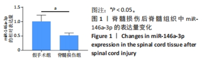
2.1 miR-146a-3p在脊髓损伤后的表达 qPCR结果显示:与假手术组相比,脊髓损伤组miR-146a-3p表达水平显著下降 (P < 0.05),见图1。"


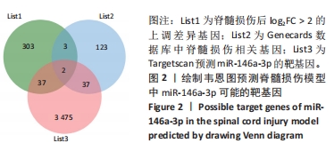
2.2 转录组测序和生物信息学方法预测脊髓损伤后miR-146a-3p的靶基因 利用TargetScan数据库预测miR-146a-3p的潜在靶基因,并与脊髓损伤后345个log2FC > 2的上调差异基因和Genecards数据库中Score > 20的脊髓损伤靶基因取交集得到IGF-1和Serpine1两个基因,见图2。"

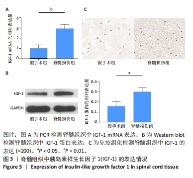
2.3 脊髓损伤后IGF-1的表达 qPCR结果显示:与假手术组相比,脊髓损伤组IGF-1表达水平显著上升(P < 0.05),见图3A。Western blot结果显示:与假手术组相比,脊髓损伤组IGF-1蛋白表达量显著上升(P < 0.05),见图3B。免疫组化结果显示:与假手术组相比,脊髓损伤组IGF-1蛋白表达量上升,见图3C。"
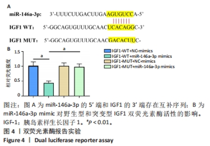
2.4 双荧光素酶报告基因检测结果 查找Starbase数据库发现miR-146a-3p的5’端和IGF-1的3’端存在互补序列,见图4A。双荧光素酶报告基因检测结果表明,与NC组相比,miR-146a-3p mimics显著抑制了IGF1-WT报告分子的荧光素酶活性(P < 0.01),见图4B,对IGF1-MUT报告分子的荧光素酶活性无显著影响。"

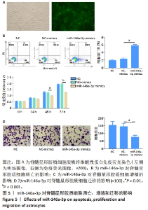
2.5 miR-146a-3p影响脊髓星形胶质细胞增殖、迁移、凋亡 免疫荧光结果显示:脊髓星形胶质细胞呈现出星形和梭形,胶质纤维酸性蛋白阳性细胞数> 95%,见图5A。流式细胞仪检测表明miR-146a-3p会明显促进脊髓星形胶质细胞凋亡(P < 0.001) ,见图5B。同样,CCK-8检测结果表明miR-146a-3p会显著抑制脊髓星形胶质细胞增殖(48 h,P < 0.01;72 h,P < 0.001),见图5C。Transwell结果显示miR-146a-3p会显著抑制脊髓星形胶质细胞的迁移能力(P < 0.01),见图5D。"

| [1] FAN Q, CAVUS O, XIONG L, et al. Spinal Cord Injury: How Could Acupuncture Help? J Acupunct Meridian Stud. 2018;11(4):124-132. [2] FENG H, XU H, ZHANG H, et al. Epidemiological profile of 338 traumatic spinal cord injury cases in Shandong province, China. Spinal Cord. 2022; 60(7):635-640. [3] CHEN J, CHEN Z, ZHANG K, et al. Epidemiological features of traumatic spinal cord injury in Guangdong Province, China. J Spinal Cord Med. 2021; 44(2):276-281. [4] KHADOUR FA, KHADOUR YA, MENG L, et al. Epidemiological features of traumatic spinal cord injury in Wuhan, China. J Orthop Surg Res. 2023;18(1):72. [5] ANJUM A, YAZID MD, FAUZI DAUD M, et al. Spinal Cord Injury: Pathophysiology, Multimolecular Interactions, and Underlying Recovery Mechanisms. Int J Mol Sci. 2020;21(20):7533. [6] SILVER J, MILLER JH. Regeneration beyond the glial scar. Nat Rev Neurosci. 2004;5(2):146-156. [7] ORR MB, GENSEL JC. Spinal Cord Injury Scarring and Inflammation: Therapies Targeting Glial and Inflammatory Responses. Neurotherapeutics. 2018;15(3):541-553. [8] LI X, LI M, TIAN L, et al. Reactive Astrogliosis: Implications in Spinal Cord Injury Progression and Therapy. Oxid Med Cell Longev. 2020;2020:9494352. [9] LI C, WU Z, ZHOU L, et al. Temporal and spatial cellular and molecular pathological alterations with single-cell resolution in the adult spinal cord after injury. Signal Transduct Target Ther. 2022;7(1):65. [10] ZHANG C, WANG MM, ZHANG Y, et al. Downregulation of miRNA-127-5p aggravates spinal cord injury through activating MAPK1. Eur Rev Med Pharmacol Sci. 2019;23(24):10617-10622. [11] PINCHI E, FRATI A, CANTATORE S, et al. Acute Spinal Cord Injury: A Systematic Review Investigating miRNA Families Involved. Int J Mol Sci. 2019;20(8):1841. [12] GUO XD, HE XG, YANG FG, et al. Research progress on the regulatory role of microRNAs in spinal cord injury. Regen Med. 2021;16(5):465-476. [13] YU DS, LV G, MEI XF, et al. MiR-200c regulates ROS-induced apoptosis in murine BV-2 cells by targeting FAP-1. Spinal Cord. 2015;53(3):182-189. [14] 朱琳,郭世武,刘锦波,等.miR-29a调控水通道蛋白4抑制过氧化氢诱导的星形胶质细胞凋亡[J].中国组织工程研究,2023,27(19):2993-2998. [15] DONG J, XIA R, ZHANG Z, et al. lncRNA MEG3 aggravated neuropathic pain and astrocyte overaction through mediating miR-130a-5p/CXCL12/CXCR4 axis. Aging (Albany NY). 2021;13(19):23004-23019. [16] KIERAN NW, SURESH R, DORION MF, et al. MicroRNA-210 regulates the metabolic and inflammatory status of primary human astrocytes. J Neuroinflammation. 2022;19(1):10. [17] NAKANO M, KUBOTA K, KOBAYASHI E, et al. Bone marrow-derived mesenchymal stem cells improve cognitive impairment in an Alzheimer’s disease model by increasing the expression of microRNA-146a in hippocampus. Sci Rep. 2020;10(1):10772. [18] JIANG D, GONG F, GE X, et al. Neuron-derived exosomes-transmitted miR-124-3p protect traumatically injured spinal cord by suppressing the activation of neurotoxic microglia and astrocytes. J Nanobiotechnology. 2020;18(1):105. [19] FAN W, LIANG C, OU M, et al. MicroRNA-146a Is a Wide-Reaching Neuroinflammatory Regulator and Potential Treatment Target in Neurological Diseases. Front Mol Neurosci. 2020;13:90. [20] JOVIČIĆ A, ROSHAN R, MOISOI N, et al. Comprehensive expression analyses of neural cell-type-specific miRNAs identify new determinants of the specification and maintenance of neuronal phenotypes. J Neurosci. 2013;33(12):5127-5137. [21] SISON SL, PATITUCCI TN, SEMINARY ER, et al. Astrocyte-produced miR-146a as a mediator of motor neuron loss in spinal muscular atrophy. Hum Mol Genet. 2017;26(17):3409-3420. [22] ZHANG H, WANG Y, LIAN L, et al. Glycine-Histidine-Lysine (GHK) Alleviates Astrocytes Injury of Intracerebral Hemorrhage via the Akt/miR-146a-3p/AQP4 Pathway. Front Neurosci. 2020;14:576389. [23] JUNG HJ, SUH Y. Regulation of IGF -1 signaling by microRNAs. Front Genet. 2015;5:472. [24] TALIFU Z, QIN C, XIN Z, et al. The Overexpression of Insulin-Like Growth Factor-1 and Neurotrophin-3 Promote Functional Recovery and Alleviate Spasticity After Spinal Cord Injury. Front Neurosci. 2022;16:863793. [25] PAN S, QI Z, LI Q, et al. Graphene oxide-PLGA hybrid nanofibres for the local delivery of IGF-1 and BDNF in spinal cord repair. Artif Cells Nanomed Biotechnol. 2019;47(1):651-664. [26] ZHANG D, YUAN Y, ZHU J, et al. Insulin-like growth factor 1 promotes neurological functional recovery after spinal cord injury through inhibition of autophagy via the PI3K/Akt/mTOR signaling pathway. Exp Ther Med. 2021;22(5):1265. [27] ZHAO Z, ZHANG L, GUO XD, et al. Rosiglitazone Exerts an Anti-depressive Effect in Unpredictable Chronic Mild-Stress-Induced Depressive Mice by Maintaining Essential Neuron Autophagy and Inhibiting Excessive Astrocytic Apoptosis. Front Mol Neurosci. 2017;10:293. [28] BURKHARDT U, WOJCIK B, ZIMMERMANN M, et al. Phospholipase D is a target for inhibition of astroglial proliferation by ethanol. Neuropharmacology. 2014;79:1-9. [29] ZHAO L, ZHANG B, HUANG S, et al. Insulin-Like Growth Factor-1 Enhances Motoneuron Survival and Inhibits Neuroinflammation After Spinal Cord Transection in Zebrafish. Cell Mol Neurobiol. 2022;42(5):1373-1384. [30] YUAN LJ, ZHANG M, CHEN S, et al. Anti-inflammatory effect of IGF-1 is mediated by IGF-1R cross talk with GPER in MPTP/MPP+-induced astrocyte activation. Mol Cell Endocrinol. 2021;519:111053. [31] ACAZ-FONSECA E, ORTIZ-RODRIGUEZ A, AZCOITIA I, et al. Notch signaling in astrocytes mediates their morphological response to an inflammatory challenge. Cell Death Discov. 2019;5:85. [32] OKADA S, HARA M, KOBAYAKAWA K, et al. Astrocyte reactivity and astrogliosis after spinal cord injury. Neurosci Res. 2018;126:39-43. [33] HARA M, KOBAYAKAWA K, OHKAWA Y, et al. Interaction of reactive astrocytes with type I collagen induces astrocytic scar formation through the integrin-N-cadherin pathway after spinal cord injury. Nat Med. 2017;23(7):818-828. [34] ANDERSON MA, BURDA JE, REN Y, et al. Astrocyte scar formation aids central nervous system axon regeneration. Nature. 2016;532(7598):195-200. [35] GU Y, CHENG X, HUANG X, et al. Conditional ablation of reactive astrocytes to dissect their roles in spinal cord injury and repair. Brain Behav Immun. 2019;80:394-405. [36] 杨拓.星形胶质细胞重编程技术治疗脊髓损伤的研究[D].长春:吉林大学,2020. [37] TAMARU T, KOBAYAKAWA K, SAIWAI H, et al. Glial scar survives until the chronic phase by recruiting scar-forming astrocytes after spinal cord injury. Exp Neurol. 2023;359:114264. [38] HOU J, BI H, GE Q, et al. Heterogeneity analysis of astrocytes following spinal cord injury at single-cell resolution. FASEB J. 2022;36(8):e22442. [39] WANG J, CHENG C, LIU Z, et al. Inhibition of A1 Astrocytes and Activation of A2 Astrocytes for the Treatment of Spinal Cord Injury. Neurochem Res. 2023;48(3):767-780. [40] WANG X, ZHANG Z, ZHU Z, et al. Photobiomodulation Promotes Repair Following Spinal Cord Injury by Regulating the Transformation of A1/A2 Reactive Astrocytes. Front Neurosci. 2021;15:768262. [41] CHANG J, QIAN Z, WANG B,et al. Transplantation of A2 type astrocytes promotes neural repair and remyelination after spinal cord injury. Cell Commun Signal. 2023;21(1):37. |
| [1] |
Song Jiating, Chen Jianmin, Wang Kewen, Huang Lanying, Xu Senming, Gui Yuchang, Xu Jianwen.
Metabolomics analysis of serum and urine in patients with traumatic spinal cord injury #br#
#br#
[J]. Chinese Journal of Tissue Engineering Research, 2024, 28(在线): 1-6.
|
| [2] | Zhou Bangyu, Li Jie, Ruan Yushang, Geng Funeng, Li Shaobo. Effects of Periplaneta americana powder on motor function and autophagic protein Beclin-1 in rats undergoing spinal cord hemisection [J]. Chinese Journal of Tissue Engineering Research, 2024, 28(8): 1223-1228. |
| [3] | Liu Kexin, Song Lijuan, Wu Yige, Han Guangyuan, Miao Zhuyue, Wei Ruheng, Xiao Baoguo, Ma Cungen, Huang Jianjun. Hydroxysafflor yellow A intervenes astrocyte lipocalin 2 expression after cerebral ischemia/reperfusion injury [J]. Chinese Journal of Tissue Engineering Research, 2024, 28(7): 1063-1069. |
| [4] | Zeng Fanzhuo, Li Yuxin, Sun Jiachen, Gu Xinyang, Wen Shan, Tian He, Mei Xifan. Efficient strategies for microglia replacement in spinal cord injury models [J]. Chinese Journal of Tissue Engineering Research, 2024, 28(7): 1007-1014. |
| [5] | Liu Tao, Zhang Wenkai, Ma Ziqian, Zhang Yan, Chen Xueming. Riluzole interferes with the activation of NLRP3 inflammasome in microglia of rats with spinal cord injury [J]. Chinese Journal of Tissue Engineering Research, 2024, 28(7): 1036-1042. |
| [6] | Chen Zepeng, Hou Yonghui, Chen Shudong, Hou Yu, Lin Dingkun. Tauroursodeoxycholic acid treats spinal cord injury by reducing apoptosis of spinal cord neurons under glucose and oxygen deprivation [J]. Chinese Journal of Tissue Engineering Research, 2024, 28(4): 528-534. |
| [7] | Tao Jingwei, Zhou Jingya, Zhao Yi, Ren Jingpei, Hu Chuanyu, Xu Lin, Mu Xiaohong, Fan Xiao. Effect and mechanism of tetramethylpyrazine regulating ferroptosis in rats with spinal cord injury [J]. Chinese Journal of Tissue Engineering Research, 2024, 28(26): 4158-4163. |
| [8] | Li Zhen, Sun Xiao, Xie Yongpeng, Rong Wang, Sun Haitao. Neuron-derived extracellular vesicles promote neurogenesis of neural stem cells [J]. Chinese Journal of Tissue Engineering Research, 2024, 28(25): 3994-3999. |
| [9] | Dai Jiafeng, Wang Lizhao, Han Qi, Shen Hongxing. Role of synaptic input remodeling of corticospinal motor neurons after spinal cord injury [J]. Chinese Journal of Tissue Engineering Research, 2024, 28(25): 4054-4059. |
| [10] | Long Zhisheng, Gong Feipeng, Wen Jiabin, Min Huan, Shu Yang, Lai Zhuoxi, Chen Gang. Bone marrow mesenchymal stem cell exosomes combined with epigallocatechin-3-gallate in treatment of spinal cord ischemia/reperfusion injury in rats [J]. Chinese Journal of Tissue Engineering Research, 2024, 28(19): 2953-2959. |
| [11] | Chen Na, Wang Yanlin, Sun Huifang, Fan Feiyan, Li Donghong, Zhang Yunke. Shexiang Huangqi compound dripping pills-containing serum promotes proliferation and differentiation of bone marrow mesenchymal stem cells [J]. Chinese Journal of Tissue Engineering Research, 2024, 28(19): 2960-2966. |
| [12] | He Wanyu, Cheng Leping. Strategies and advance on stem cell transplantation for repair of spinal cord injury [J]. Chinese Journal of Tissue Engineering Research, 2024, 28(19): 3090-3096. |
| [13] | Zhan Lifen, Ai Kun, Zeng Xuejiu, Liang Rouyun, Ding Qiangsheng, Zhang Hong. Application and prospect of reconstructing bladder micturition reflex in neurogenic bladder after spinal cord injury [J]. Chinese Journal of Tissue Engineering Research, 2024, 28(18): 2925-2931. |
| [14] | Xu Zihan, Bi Yunfeng, Li Jiang, Zhang Zongliang, Song Chen, Dong Jie, Liu Dong. Repetitive magnetic stimulation of S3 nerve root and M1 area for treating urinary retention after spinal cord injury [J]. Chinese Journal of Tissue Engineering Research, 2024, 28(11): 1719-1723. |
| [15] | Shang Wenya, Ren Yafeng, Li Bing, Wei Huilin, Zhang Zhilan, Huang Xiaomeng, Huang Jing. Regulatory mechanisms and therapeutic strategies for pyroptosis after spinal cord injury [J]. Chinese Journal of Tissue Engineering Research, 2024, 28(11): 1772-1779. |
| Viewed | ||||||
|
Full text |
|
|||||
|
Abstract |
|
|||||