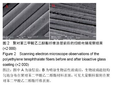
| [1]Zhang Q,Wu X.Biomechanical research progress of non-contact anterior cruciate ligament injury.Zhongguo Zuzhi Gongcheng Yanjiu.2013;17(17):3164-3173.[2]Everhart JS,Flanigan DC,Simon RA,et al.Association of noncontact anterior cruciate ligament injury with presence and thickness of a bony ridge on the anteromedial aspect of the femoral intercondylar notch.Am J Sports Med. 2010;38(8):1667-1673.[3]Hsu SL,Wang CJ.Graft failure versus graft fixation in ACL reconstruction: histological and immunohistochemical studies in rabbits.Arch Orthop Trauma Surg.2013;133(9):1197-1202.[4]Andernord D,Bjornsson H,Petzold M,et al.Surgical predictors of early revision surgery after anterior cruciate ligament reconstruction: results from the swedish national knee ligament register on 13,102 patients. Am J Sports Med. 2014;42(7): 1574-1582[5]张力,靳安民,田京.前交叉韧带重建术后腱-骨和骨-骨界面愈合的比较研究[J].中国修复重建外科杂志,2009,23(4):473-477.[6]Suzuki T,Shino K,Otsubo H,et al.Biomechanical comparison between the rectangular-tunnel and the round-tunnel anterior cruciate ligament reconstruction procedures with a bone-patellar tendon-bone graft.Arthroscopy. 2014;30(10):1294-1302.[7]Herbort M,Tecklenburg K,Zantop T,et al.Single-bundle anterior cruciate ligament reconstruction.A biomechanical cadaveric study of a rectangular quadriceps and bone-patellar tendon: bone graft configuration versus a round hamstring graft.Arthroscopy.2013;29(12):1981-1990.[8]赵耀,翟文亮.富血小板血浆复合小牛脱蛋白骨重建前交叉韧带腱-骨愈合的组织学观察[J].中国修复重建外科杂志, 2010, 24(11): 1323-1329.[9]孙磊,吴波,罗永忠,等.由外向内与经胫骨建立股骨隧道保留残迹重建前交叉韧带的病例对照研究[J].中国骨伤, 2013,26(5): 397-401.[10]Yang JH,Chang M,Kwak DS,et al.Volume and contact surface area analysis of bony tunnels in single and double bundle anterior cruciate ligament reconstruction using autograft tendons: in vivo three-dimensional imaging analysis.Clin Orthop Surg.2014;6(3):290-297.[11]Yang D,Cheon SH,Oh CW,et al.A comparison of the fixation strengths provided by different intraosseous tendon lengths during anterior cruciate ligament reconstruction: a biomechanical study in a porcine tibial model.Clin Orthop Surg.2014;6(2):173-179.[12]Hsu S,Wang C. The use of demineralized bone matrix for anterior cruciate ligament reconstruction: a radiographic,histologic,and immunohistochemical study in rabbits. J Surg Res.2014;187(1):219-224.[13]毕海勇,孙秀江,慕宏杰,等.保留并牵张缝合 Rigidfix 固定胫骨残迹的前交叉韧带重建[J].中华关节外科杂志, 2013,7(1):25-29.[14]Yuan F,Zhou W,Cai J,et al.Optimal graft length for anterior cruciate ligament reconstruction: a biomechanical study in beagles.Orthopedics.2013;36(5):e588-e592.[15]Lessim S,Migonney V,Thoreux P,et al.PolyNaSS bioactivation of LARS artificial ligament promotes human ligament fibroblast colonisation in vitro.Biomed Mater Eng. 2013;23(4):289-297.[16]Silva A,Sampaio R,Fernandes R,et al.Is there a role for adult non-cultivated bone marrow stem cells in ACL reconstruction? Knee Surg Sports Traumatol Arthrosc. 2014;22(1): 66-71.[17]Lu H,Zheng C,Wang Z,et al.Effects of low-intensity pulsed ultrasound on new trabecular bone during bone-tendon junction healing in a rabbit model: a synchrotron radiation micro-CT study.PLoS One.2015;10(4):e0124724.[18]龚熹,余家阔,敖英芳,等.最大化保留残端结合内减张技术单束重建后交叉韧带临床疗效观察[J].中国运动医学杂志, 2013,32(2): 101-103,156.[19]Wolf BR,Ramme AJ,Wright RW,et al.Variability in ACL tunnel placement observational clinical study of surgeon ACL tunnel variability.Am J Sports Med.2013;41(6):1265-1273.[20]Lui PP,Lee YW,Mok TY,et al.Local administration of alendronate reduced peri-tunnel bone loss and promoted graft-bone tunnel healing with minimal systemic effect on bone in contralateral knee.J Orthop Res. 2013;31(12):1897-1906.[21]Kievit AJ,Jonkers FJ,Barentsz JH,et al.A cross-sectional study comparing the rates of osteoarthritis, laxity, and quality of life in primary and revision anterior cruciate ligament reconstructions.Arthroscopy.2013;29(5):898-905.[22]张辉,冯华,洪雷,等.导航辅助下前十字韧带单束与双束重建的比较研究[J].中华骨科杂志,2012,32(6):557-564.[23]吴贵佑,章亚东,汪喜顺,等.保留残端同种异体肌腱重建前交叉韧带:更早恢复患膝关节稳定性及运动功能[J].中国组织工程研究, 2015,19(11):1727-1731. |