Chinese Journal of Tissue Engineering Research ›› 2017, Vol. 21 ›› Issue (17): 2659-2664.doi: 10.3969/j.issn.2095-4344.2017.17.006
Previous Articles Next Articles
Hepatic failure patient’s serum before and after plasmapheresis induces the differentiation of umbilical cord mesenchymal stem cells into hepatic cells
Wang Jing-bo1, Geng Da-ying2, Li Xiao-ying1, Chen Li3, Xu Feng3, Shi Zhao-zhang4
- 1Third Department, 2Central Laboratory, 4Department of Intervention, Jinan Municipal Hospital for Infectious Diseases, Jinan 250021, Shandong Province, China; 3Cell Biology, Jinan 250021, Shandong Province, China
-
Revised:2017-02-13Online:2017-06-18Published:2017-06-29 -
Contact:Shi Zhao-zhang, Associate chief physician, Department of Intervention, Jinan Municipal Hospital for Infectious Diseases, Jinan 250021, Shandong Province, China -
About author:Wang Jing-bo, Master, Associate chief physician, Third Department, Jinan Municipal Hospital for Infectious Diseases, Jinan 250021, Shandong Province, China -
Supported by:the Non-funded Project of Jinan Science and Technology Department in 2012; the Medical and Health Development Plan of Shandong Province, No. 2014WS0006
CLC Number:
Cite this article
Wang Jing-bo, Geng Da-ying, Li Xiao-ying, Chen Li, Xu Feng, Shi Zhao-zhang. Hepatic failure patient’s serum before and after plasmapheresis induces the differentiation of umbilical cord mesenchymal stem cells into hepatic cells[J]. Chinese Journal of Tissue Engineering Research, 2017, 21(17): 2659-2664.
share this article
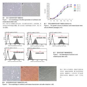
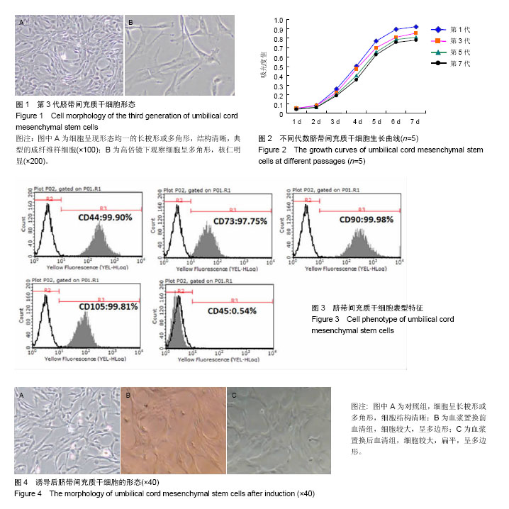
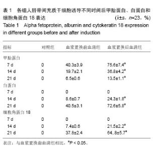
2.1 脐带间充质干细胞形态和增殖能力 接种后6-10 d可见组织块周围有成纤维样细胞爬出,2周左右细胞达到70%-80%融合。传代后,细胞呈现形态均一的长梭形、多角形,而且细胞生长迅速,一般传代两三天后即可达到80%融合,多以平行或漩涡状生长,见图1。 采用CCK-8法检测各代脐带间充质干细胞的增殖活性,可见各代细胞潜伏期均在一两天,细胞生长较缓慢,随即进入对数生长期,持续三四天后,第6天细胞进入平台期,细胞增殖缓慢。第1,3,5代细胞生长迅速,增殖较快,细胞数量也高于同期的第7代细胞,表明细胞代数越早,细胞活力越高,见图2。 2.2 脐带间充质干细胞表型 体外培养的脐带间充质干细胞高表达CD44,CD90,CD73和CD105,纯度在95%以上,不表达造血细胞标记CD45,具备典型的脐带间充质干细胞表型特征,见图3。 2.3 诱导后脐带间充质干细胞向肝细胞分化的形态变化 加入血浆置换前或置换后肝衰竭患者血清培养的脐带间充质干细胞形态发生改变,由原来的长梭形变为多边形,细胞较大而扁平,对照组细胞仍呈典型的长梭形,未发生任何变化,见图4。 2.4 诱导后甲胎蛋白、白蛋白和细胞角蛋白18的表达水平 对照组细胞在培养过程中均未见3种蛋白表达;实验组诱导后甲胎蛋白、白蛋白和细胞角蛋白18的表达均有显著变化,血浆置换后血清组3种蛋白表达水平明显高于血浆置换前血清组,差异有显著性意义(P < 0.05)。甲胎蛋白阳性率在培养第7天达到峰值,培养14,21 d表达变弱;白蛋白和细胞角蛋白在培养14 d时开始表达,培养21 d表达变强,见表1。"
"

| [1] Mendizabal M, Silva MO. Liver transplantation in acute liver failure: A challenging scenario. World J Gastroenterol. 2016; 22(4):1523-1531. [2] Kirnap M, Akdur A, Ozcay F, et al. Liver Transplant for Fulminant Hepatic Failure: A Single-Center Experience. Exp Clin Transplant. 2015;13(4):339-343.[3] McCaughan GW, Crawford M, Sandroussi C, et al. Assessment of adult patients with chronic liver failure for liver transplantation in 2015: who and when. Intern Med J. 2016;46(4):404-412.[4] Nevens F, Laleman W. Artificial liver support devices as treatment option for liver failure. Best Pract Res Clin Gastroenterol. 2012;26(1):17-26.[5] Naruse K, Tang W, Makuuch M. Artificial and bioartificial liver support: a review of perfusion treatment for hepatic failure patients. World J Gastroenterol. 2007;13(10):1516-1521.[6] Kobayashi N. Life support of artificial liver: development of a bioartificial liver to treat liver failure. J Hepatobiliary Pancreat Surg. 2009;16(2):113-117.[7] Kestendjieva S, Kyurkchiev D, Tsvetkova G, et al. Characterization of mesenchymal stem cells isolated from the human umbilical cord. Cell Biol Int. 2008;32(7):724-732.[8] Jin HJ, Bae YK, Kim M,Comparative analysis of human mesenchymal stem cells from bone marrow, adipose tissue, and umbilical cord blood as sources of cell therapy.Int J Mol Sci. 2013;14(9):17986-18001.[9] 张惟,陈松,王继明,等.人脐带间充质干细胞向神经细胞分化的实验研究[J].临床眼科杂志,2015,23(4):365-370.[10] Wang YK, Chen CS. Cell adhesion and mechanical stimulation in the regulation of mesenchymal stem cell differentiation. J Cell Mol Med. 2013;17(7):823-832.[11] Xue HL, Zeng WZ, Wu XL, et al. Clinical therapeutic effects of human umbilical cord-derived mesenchymal stem cells transplantation in the treatment of end-stage liver disease. Transplant Proc. 2015;47(2):412-418.[12] Shi M, Zhang Z, Xu R, et al. Human mesenchymal stem cell transfusion is safe and improves liver function in acute-on-chronic liver failure patients. Stem Cells Transl Med. 2012;1(10):725-731.[13] Pons-Estel GJ, Salerni GE, Serrano RM, et al. Therapeutic plasma exchange for the management of refractory systemic autoimmune diseases: report of 31 cases and review of the literature. Autoimmun Rev. 2011;10(11):679-684.[14] Maiwall R, Moreau R. Plasma exchange for acute on chronic liver failure: is there a light at the end of the tunnel. Hepatol Int. 2016;10(3):387-389.[15] Kribben A, Gerken G, Haag S, et al. Effects of fractionated plasma separation and adsorption on survival in patients with acute-on-chronic liver failure. Gastroenterology. 2012;142(4): 782-789.e3.[16] Romanov YA, Svintsitskaya VA, Smirnov VN. Searching for alternative sources of postnatal human mesenchymal stem cells: candidate MSC-like cells from umbilical cord. Stem Cells. 2003;21(1):105-110.[17] Covas DT, Siufi JL, Silva AR, et al. Isolation and culture of umbilical vein mesenchymal stem cells. Braz J Med Biol Res. 2003;36(9):1179-1183.[18] Wang HS, Hung SC, Peng ST, et al. Mesenchymal stem cells in the Wharton's jelly of the human umbilical cord. Stem Cells. 2004;22(7):1330-1337.[19] Kisiel AH, McDuffee LA, Masaoud E, et al. Isolation, characterization, and in vitro proliferation of canine mesenchymal stem cells derived from bone marrow, adipose tissue, muscle, and periosteum. Am J Vet Res. 2012;73(8): 1305-1317. [20] Roy S, Arora S, Kumari P, et al. A simple and serum-free protocol for cryopreservation of human umbilical cord as source of Wharton's jelly mesenchymal stem cells. Cryobiology. 2014;68(3):467-472.[21] Cao H, Hui Q, Yan Y, et al. Pretreatments with injured microenvironmental signals altered the characteristics of human umbilical cord mesenchymal stem cells. Biotechnol Lett. 2016;38(1):157-165.[22] Nan C, Shi Y, Zhao Z, et al. Monosialoteterahexosyl ganglioside induces the differentiation of human umbilical cord-derived mesenchymal stem cells into neuron-like cells. Int J Mol Med. 2015;36(4):1057-1062.[23] Raoufil A, Aminil A, Azadbakht M, et al. Production of hepatocyte-like cells from human umbilical vein mesenchymal stem cells. Ital J Anat Embryol. 2015;120(3):150-161.[24] Kocaefe C, Balci D, Hayta BB, et al. Reprogramming of human umbilical cord stromal mesenchymal stem cells for myogenic differentiation and muscle repair. Stem Cell Rev. 2010;6(4):512-522.[25] Liu WW, Yu W, Chen JY, et al. Effects of human umbilical cord mesenchymal stem cells in the treatment of paraquat-induced lung injury. Zhonghua Lao Dong Wei Sheng Zhi Ye Bing Za Zhi. 2012;30(11):811-815. [26] Shi M, Zhang Z, Xu R, et al. Human mesenchymal stem cell transfusion is safe and improves liver function in acute-on-chronic liver failure patients. Stem Cells Transl Med. 2012;1(10):725-731.[27] 刘波,董静,张骏飞.人脐带间充质干细胞治疗慢加急性肝衰竭患者近期疗效与安全性分析[J]. 实用肝脏病杂志, 2013, 16(1): 29-31.[28] 王全楚,张凌云,王东琳.人脐带血间充质干细胞输注治疗慢性肝衰竭患者的近期疗效观察[J].胃肠病学和肝病学杂志, 2013, 22(1):22-24. [29] 曲乃方,王者令,闫兆平,等.38例慢性肝衰竭患者经干细胞治疗48周效果观察[J].中华细胞与干细胞杂志:电子版, 2013,3(2): 73-77.[30] 任红英,赵钦军,刘拥军,等.脐带间充质干细胞体外诱导分化为肝细胞样细胞的研究[J].山东医药, 2008,48(30):24-26. [31] An SY, Han J, Lim HJ, et al. Valproic acid promotes differentiation of hepatocyte-like cells from whole human umbilical cord-derived mesenchymal stem cells. Tissue Cell. 2014;46(2):127-135.[32] Xue G, Han X, Ma X, et al. Effect of Microenvironment on Differentiation of Human Umbilical Cord Mesenchymal Stem Cells into Hepatocytes In Vitro and In Vivo. Biomed Res Int. 2016;2016:8916534.[33] Zheng G, Liu Y, Jing Q, et al. Differentiation of human umbilical cord-derived mesenchymal stem cells into hepatocytes in vitro. Biomed Mater Eng. 2015;25(1 Suppl): 145-157.[34] 薛改,韩华,陈泽纱,等.体外诱导人脐带间充质干细胞定向分化为肝细胞的研究[J].肝脏,2016, 21(9):733-737.[35] 闫俊卿,韩涛,朱争艳. 人脐带间充质干细胞生物学特性及向类肝细胞的分化[J].世界华人消化杂志,2008,16(15):1639-1644.[36] 任红英,赵钦军,刘拥军,等.脐带间充质干细胞体外诱导分化为肝细胞样细胞的研究[J].山东医药,2008,48(30):24-26.[37] Borhani-Haghighi M, Talaei-Khozani T, Ayatollahi M, et al. Wharton's Jelly-derived Mesenchymal Stem Cells can Differentiate into Hepatocyte-like Cells by HepG2 Cell Line Extract. Iran J Med Sci. 2015;40(2):143-151.[38] Zhang YN, Lie PC, Wei X. Differentiation of mesenchymal stromal cells derived from umbilical cord Wharton's jelly into hepatocyte-like cells. Cytotherapy. 2009;11(5):548-558.[39] Kang XQ, Zang WJ, Bao LJ, et al. Fibroblast growth factor-4 and hepatocyte growth factor induce differentiation of human umbilical cord blood-derived mesenchymal stem cells into hepatocytes. World J Gastroenterol. 2005;11(47):7461-7465.[40] Talaei-Khozani T, Borhani-Haghighi M, Ayatollahi M, et al. An in vitro model for hepatocyte-like cell differentiation from Wharton's jelly derived-mesenchymal stem cells by cell-base aggregates. Gastroenterol Hepatol Bed Bench. 2015;8(3): 188-199.[41] Vojdani Z, Khodabandeh Z, Jaberipour M, Hosseini A, et al. The influence of fibroblast growth factor 4 on hepatogenic capacity of Wharton's jelly mesenchymal stromal cells. Rom J Morphol Embryol. 2015; 56(3):1043-1050.[42] Lee KD, Kuo TK, Whang-Peng J, et al. In vitro hepatic differentiation of human mesenchymal stem cells. Hepatology. 2004;40(6):1275-1284.[43] Yan C, Xue G, Wu L, et al. Differentiation of human umbilical cord mesenchymal stem cells into hepatocytes induced by rat fibrotic liver tissue extracts. Zhongguo Xiu Fu Chong Jian Wai Ke Za Zhi. 2015;29(7):878-883.[44] Zhang GQ, Fang CH, Chi DZ. A study of rat mesenchymal stem cells (MSCs) differentiating into liver cells when co-cultured with rat hepatocytes. Zhonghua Gan Zang Bing Za Zhi. 2005;13(9):648-651.[45] 毛德文,邱华.甲胎蛋白对肝衰竭肝细胞再生及预后判定价值的研究[J].实用肝脏病杂志, 2006,9(6):378-380.[46] Lange C, Bassler P, Lioznov MV, et al. Liver-specific gene expression in mesenchymal stem cells is induced by liver cells. World J Gastroenterol. 2005;11(29):4497-4504. |
| [1] | Wang Jing, Xiong Shan, Cao Jin, Feng Linwei, Wang Xin. Role and mechanism of interleukin-3 in bone metabolism [J]. Chinese Journal of Tissue Engineering Research, 2022, 26(8): 1260-1265. |
| [2] | Xiao Hao, Liu Jing, Zhou Jun. Research progress of pulsed electromagnetic field in the treatment of postmenopausal osteoporosis [J]. Chinese Journal of Tissue Engineering Research, 2022, 26(8): 1266-1271. |
| [3] | Tian Chuan, Zhu Xiangqing, Yang Zailing, Yan Donghai, Li Ye, Wang Yanying, Yang Yukun, He Jie, Lü Guanke, Cai Xuemin, Shu Liping, He Zhixu, Pan Xinghua. Bone marrow mesenchymal stem cells regulate ovarian aging in macaques [J]. Chinese Journal of Tissue Engineering Research, 2022, 26(7): 985-991. |
| [4] | Hou Jingying, Guo Tianzhu, Yu Menglei, Long Huibao, Wu Hao. Hypoxia preconditioning targets and downregulates miR-195 and promotes bone marrow mesenchymal stem cell survival and pro-angiogenic potential by activating MALAT1 [J]. Chinese Journal of Tissue Engineering Research, 2022, 26(7): 1005-1011. |
| [5] | Liang Xuezhen, Yang Xi, Li Jiacheng, Luo Di, Xu Bo, Li Gang. Bushen Huoxue capsule regulates osteogenic and adipogenic differentiation of rat bone marrow mesenchymal stem cells via Hedgehog signaling pathway [J]. Chinese Journal of Tissue Engineering Research, 2022, 26(7): 1020-1026. |
| [6] | Wen Dandan, Li Qiang, Shen Caiqi, Ji Zhe, Jin Peisheng. Nocardia rubra cell wall skeleton for extemal use improves the viability of adipogenic mesenchymal stem cells and promotes diabetes wound repair [J]. Chinese Journal of Tissue Engineering Research, 2022, 26(7): 1038-1044. |
| [7] | Zhu Bingbing, Deng Jianghua, Chen Jingjing, Mu Xiaoling. Interleukin-8 receptor enhances the migration and adhesion of umbilical cord mesenchymal stem cells to injured endothelium [J]. Chinese Journal of Tissue Engineering Research, 2022, 26(7): 1045-1050. |
| [8] | Fang Xiaolei, Leng Jun, Zhang Chen, Liu Huimin, Guo Wen. Systematic evaluation of different therapeutic effects of mesenchymal stem cell transplantation in the treatment of ischemic stroke [J]. Chinese Journal of Tissue Engineering Research, 2022, 26(7): 1085-1092. |
| [9] | Guo Jia, Ding Qionghua, Liu Ze, Lü Siyi, Zhou Quancheng, Gao Yuhua, Bai Chunyu. Biological characteristics and immunoregulation of exosomes derived from mesenchymal stem cells [J]. Chinese Journal of Tissue Engineering Research, 2022, 26(7): 1093-1101. |
| [10] | Huang Chenwei, Fei Yankang, Zhu Mengmei, Li Penghao, Yu Bing. Important role of glutathione in stemness and regulation of stem cells [J]. Chinese Journal of Tissue Engineering Research, 2022, 26(7): 1119-1124. |
| [11] | Huang Chuanjun, Zou Yu, Zhou Xiaoting, Zhu Yangqing, Qian Wei, Zhang Wei, Liu Xing. Transplantation of umbilical cord mesenchymal stem cells encapsulated in RADA16-BDNF hydrogel promotes neurological recovery in an intracerebral hemorrhage rat model [J]. Chinese Journal of Tissue Engineering Research, 2022, 26(4): 510-515. |
| [12] | Yang Sidi, Wang Qian, Xu Nuo, Wang Ronghan, Jin Chuanqi, Lu Ying, Dong Ming. Biodentine enhances the proliferation and differentiation of osteoblasts through upregulating bone morphogenetic protein-2 [J]. Chinese Journal of Tissue Engineering Research, 2022, 26(4): 516-520. |
| [13] | Kang Kunlong, Wang Xintao. Research hotspot of biological scaffold materials promoting osteogenic differentiation of bone marrow mesenchymal stem cells [J]. Chinese Journal of Tissue Engineering Research, 2022, 26(4): 597-603. |
| [14] | Cao Wei, Mao Furong, Hu Xiaohua, Yang Xiaohong. N-6 methyladenosine RNA methylation regulates osteogenic and adipogenic differentiation of bone marrow mesenchymal stem cells [J]. Chinese Journal of Tissue Engineering Research, 2022, 26(2): 266-270. |
| [15] | Fan Danyang, Fu Runze, Mi Jiajing, Liu Chunyan. Expression and role of cannabinoid receptors during bone remodeling [J]. Chinese Journal of Tissue Engineering Research, 2022, 26(2): 283-288. |
| Viewed | ||||||
|
Full text |
|
|||||
|
Abstract |
|
|||||