| [1] Gámez Escalona JA. The embryonic stem cells research. Example of biotechnology progress under extra-scientific pressure. Cuad Bioet. 2013;24(82):443-462.
[2] Rigby B. Revocation of European patent for neural progenitors highlights patent challenges for inventions relating to human embryonic stem cells. Expert Opin Ther Pat. 2013; 23(11):1395-1398.
[3] Zhang YN, Lie PC, Wei X. Differentiation of mesenchymal stromal cells derived from umbilical cord Wharton's jelly into hepatocyte-like cells. Cytotherapy. 2009;11(5):548-558.
[4] Woodbury D, Schwarz EJ, Prockop DJ, et al. Adult rat and human bone marrow stromal cells differentiate into neurons. J Neurosci Res. 2000;61(4):364-370.
[5] Paldino E, Cenciarelli C, Giampaolo A, et al. Induction of dopaminergic neurons from human Wharton's jelly mesenchymal stem cell by forskolin. J Cell Physiol. 2014; 229(2):232-244.
[6] Alves da Silva ML, Costa-Pinto AR, Martins A, et al. Conditioned medium as a strategy for human stem cells chondrogenic differentiation. J Tissue Eng Regen Med. 2015; 9(6):714-723.
[7] Song X, Xie Y, Liu Y, et al. Beneficial effects of coculturing synovial derived mesenchymal stem cells with meniscus fibrochondrocytes are mediated by fibroblast growth factor 1: increased proliferation and collagen synthesis. Stem Cells Int. 2015;2015:926325.
[8] Yu FX, Hu WJ, He B, et al. Bone marrow mesenchymal stem cells promote osteosarcoma cell proliferation and invasion. World J Surg Oncol. 2015;13:52.
[9] Jang KM, Lim HC, Jung WY, et al. Efficacy and Safety of Human Umbilical Cord Blood-Derived Mesenchymal Stem Cells in Anterior Cruciate Ligament Reconstruction of a Rabbit Model: New Strategy to Enhance Tendon Graft Healing. Arthroscopy. 2015 Apr 13. [Epub ahead of print]
[10] Banerjee A, Bizzaro D, Burra P, et al. Umbilical cord mesenchymal stem cells modulate dextran sulfate sodium induced acute colitis in immunodeficient mice. Stem Cell Res Ther. 2015;6:79.
[11] Sela M, Tirza G, Ravid O, et al. NOX1-induced accumulation of reactive oxygen species in abdominal fat-derived mesenchymal stromal cells impinges on long-term proliferation. Cell Death Dis. 2015;6:e1728.
[12] Ulrich C, Abruzzese T, Maerz JK, et al. Human Placenta-Derived CD146-Positive Mesenchymal Stromal Cells Display a Distinct Osteogenic Differentiation Potential. Stem Cells Dev. 2015;24(13):1558-1569.
[13] Cameron SH, Alwakeel AJ, Goddard L, et al. Delayed post-treatment with bone marrow-derived mesenchymal stem cells is neurorestorative of striatal medium-spiny projection neurons and improves motor function after neonatal rat hypoxia-ischemia. Mol Cell Neurosci. 2015;68:56-72.
[14] Zhang Y, Chopp M, Meng Y, et al. Effect of exosomes derived from multipluripotent mesenchymal stromal cells on functional recovery and neurovascular plasticity in rats after traumatic brain injury. J Neurosurg. 2015;122(4):856-867.
[15] Salic A, Mitchison TJ. A chemical method for fast and sensitive detection of DNA synthesis in vivo. Proc Natl Acad Sci U S A. 2008;105(7):2415-2420.
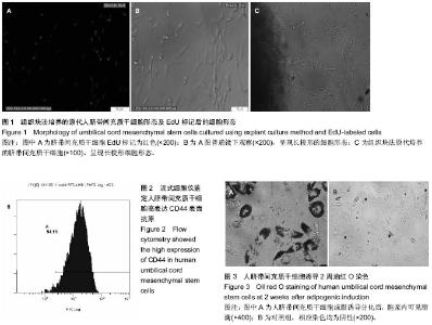
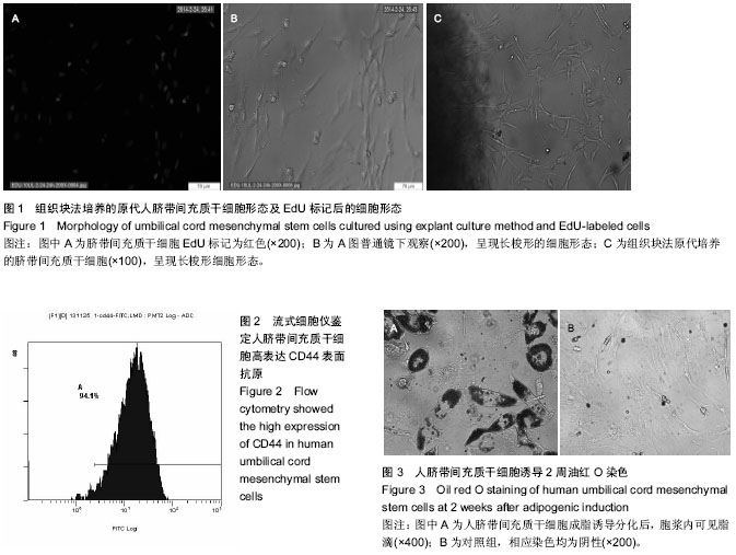
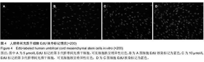
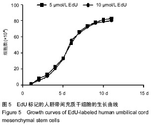
[16] 张飞,王一雄,武忠炎,等.人脐带间充质干细胞生物特性比较:胰酶冷消化和组织块法体外培养[J].中国组织工程研究,2014, 18(41):6614-6619.
[17] Tomar GB, Srivastava RK, Gupta N, et al. Human gingiva-derived mesenchymal stem cells are superior to bone marrow-derived mesenchymal stem cells for cell therapy in regenerative medicine. Biochem Biophys Res Commun. 2010;393(3):377-383.
[18] Sun L, Fan X, Zhang L, et al. Bone mesenchymal stem cell transplantation via four routes for the treatment of acute liver failure in rats. Int J Mol Med. 2014;34(4):987-996.
[19] Qiu X, Villalta J, Ferretti L, et al. Effects of intravenous injection of adipose-derived stem cells in a rat model of radiation therapy-induced erectile dysfunction. J Sex Med. 2012;9(7):1834-1841.
[20] Orabi H, Lin G, Ferretti L, et al. Scaffoldless tissue engineering of stem cell derived cavernous tissue for treatment of erectile function. J Sex Med. 2012;9(6): 1522-1534.
[21] Lin CS, Xin ZC, Wang Z, et al. Stem cell therapy for erectile dysfunction: a critical review. Stem Cells Dev. 2012;21(3): 343-351.
[22] Fandel TM, Albersen M, Lin G, et al. Recruitment of intracavernously injected adipose-derived stem cells to the major pelvic ganglion improves erectile function in a rat model of cavernous nerve injury. Eur Urol. 2012;61(1):201-210. |