Chinese Journal of Tissue Engineering Research ›› 2014, Vol. 18 ›› Issue (47): 7579-7584.doi: 10.3969/j.issn.2095-4344.2014.47.007
Previous Articles Next Articles
Polylactic-co-glycolic acid complex with different concentrations of Schwann cells for peripheral nerve regeneration
Sun Huan-wei, Zhang Tie-hui, You Xin-yan, Ren Yuan-fei, Zhong Sheng
- Department of Hands and Feet Microsurgery, Dalian Municipal Central Hospital, Dalian 116033, Liaoning Province, China
-
Revised:2014-10-12Online:2014-11-19Published:2014-11-19 -
Contact:Zhong Sheng, Master, Department of Hands and Feet Microsurgery, Dalian Municipal Central Hospital, Dalian 116033, Liaoning Province, China -
About author:Sun Huan-wei, Master, Chief physician, Department of Hands and Feet Microsurgery, Dalian Municipal Central Hospital, Dalian 116033, Liaoning Province, China -
Supported by:the Project of Dalian Health Bureau, No. 201410040
CLC Number:
Cite this article
Sun Huan-wei, Zhang Tie-hui, You Xin-yan, Ren Yuan-fei, Zhong Sheng. Polylactic-co-glycolic acid complex with different concentrations of Schwann cells for peripheral nerve regeneration[J]. Chinese Journal of Tissue Engineering Research, 2014, 18(47): 7579-7584.
share this article
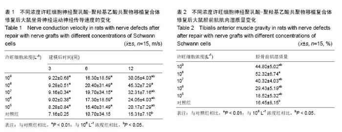
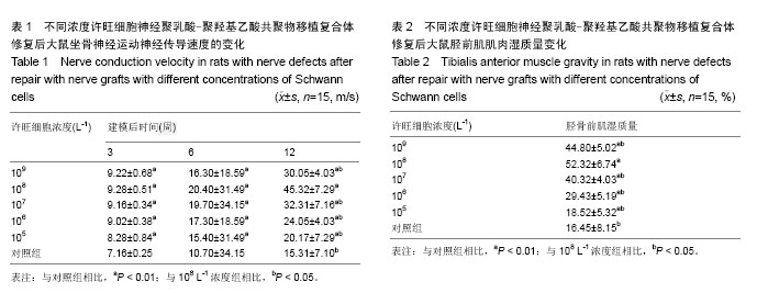
2.1 实验动物数量分析 大鼠90只均造模成功,其中3只在实验过程中死于建模后感染,予以剔除,并补充重新制作动物模型,最终90只大鼠进入结果分析。 2.2 许旺细胞鉴定结果 培养24 h后可见部分贴壁许旺细胞,5 d后形态清晰、立体感强。胞核呈不规则形,胞体呈长梭形,突起细长,多为双极或多极,偶见扁平椭圆形细胞。 2.3 不同浓度许旺细胞神经聚乳酸-聚羟基乙酸共聚物移植复合体修复后大鼠坐骨神经运动神经传导速度的变化 电生理实验结果显示,含许旺细胞神经移植复合体各时间点神经传导速率均高于对照组(P < 0.01)。且在105 L-1至108 L-1浓度组中,许旺细胞浓度与神经传导速率成正相关剂量依存关系。109 L-1至108 L-1浓度组,旺细胞浓度与运动神经传导速率成负相关。随时间变化,至建模后12周时,107 L-1至109 L-1浓度组之间运动神经传导速率差异有显著性意义(P < 0.05)。其中108 L-1组运动神经传导速度优于其他各组。见表1。 2.4 不同浓度许旺细胞神经聚乳酸-聚羟基乙酸共聚物移植复合体修复后大鼠胫前肌肌肉湿质量变化 建模后12周,6组大鼠的胫前肌肌萎缩有不同程度的恢复,含许旺细胞神经移植复合体胫前肌湿质量高于对照组(P < 0.01)。且在105 L-1至108 L-1浓度组中,许旺细胞浓度与胫前肌湿质量成正相关剂量依存关系。109 L-1至108 L-1浓度组,许旺细胞浓度与胫前肌湿质量成负相关。107 L-1至109 L-1浓度组之间胫前肌湿质量差异有显著性意义(P < 0.05)。其中108 L-1浓度组胫前肌湿质量优于其他各组(P < 0.05)。见表2。"
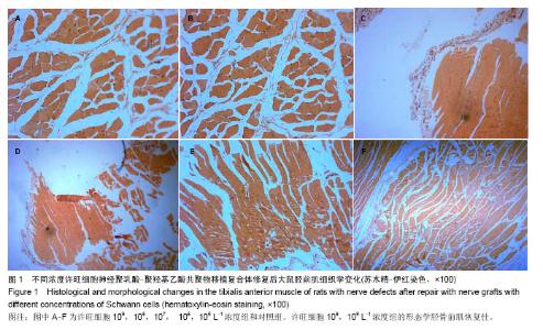
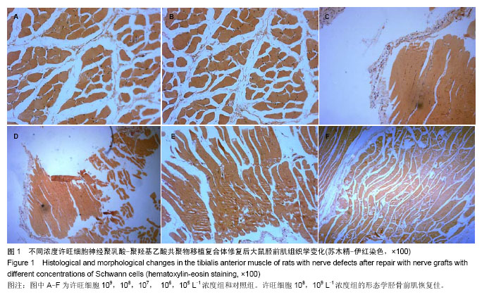
2.5 不同浓度许旺细胞神经聚乳酸-聚羟基乙酸共聚物移植复合体修复后大鼠胫前肌组织学变化 苏木精-伊红染色显示,含许旺细胞神经移植复合体各组正常肌纤维数均多于对照组,其中许旺细胞108,109 L-1浓度组的形态学胫骨前肌恢复佳,肌纤维细条样、波浪状,同向而行,长短、粗细及疏密大致一致,而对照组排列紊乱,神经纤维长短、粗细及疏密不一致,混杂。见图1。"
| [1] Chung KF, J Thorac Dis. Approach to chronic cough: the neuropathic basis for cough hypersensitivity syndrome. 2014;6(Suppl 7):S699-S707. [2] Sumalatha S, D Souza AS, Yadav JS, et al. An unorthodox innervation of the gluteus maximus muscle and other associated variations: a case report. Australas Med J. 2014; 7(10):419-422. [3] Piao C, Li P, Liu G, et al. Viscoelasticity of repaired sciatic nerve by poly (lactic-co-glycolic acid) tubes. Neural Regen Res. 2013;8(33):3131-3138. [4] Zhu S, Li J, Zhu Q, et al.Differentiation of human amniotic epithelial cells into Schwann like cells via indirect co culture withSchwann cells in vitro. Mol Med Rep. 2014 [Epub ahead of print]. [5] Gordon A, Adamsky K, Vainshtein A, et al. Caspr and caspr2 are required for both radial and longitudinal organization of myelinated axons. J Neurosci. 2014;34(45): 14820-14826. [6] Thoma EC, Merkl C, Heckel T, et al. Chemical conversion of human fibroblasts into functional schwann cells. Stem Cell Reports. 2014;3(4):539-547. [7] Gao R, Wang L, Sun J, et al. MiR-204 promotes apoptosis in oxidative stress-induced rat Schwann cells by suppressing neuritin expression. FEBS Lett. 2014;588(17):3225-3232. [8] Sulaiman W, Dreesen TD. Effect of local application of transforming growth factor-β at the nerve repair site following chronic axotomy and denervation on the expression of regeneration-associated genes.J Neurosurg. 2014;121(4): 859-874. [9] Zong HB, Zhao HX, Zhao YL, et al. Nanoparticles carrying neurotrophin-3-modified Schwann cells promote repair of sciatic nerve defects.Neural Regen Res. 2013;8(14): 1262-1268. [10] Ye FG, Li HY, Qiao GG,et al. Platelet-rich plasma gel in combination with Schwann cells for repair of sciatic nerve injury. Neural Regen Res.2012;7(29): 2286-2292. [11] He B, Tao HY, Liu SQ. Neuroprotective effects of carboxymethylated chitosan on hydrogen peroxide induced apoptosis inSchwann cells. Eur J Pharmacol. 2014;740: 127-134. [12] Painter MW, Brosius Lutz A, Cheng YC, et al. Diminished Schwann cell repair responses underlie age-associated impaired axonal regeneration. Neuron. 2014;83(2): 331-343. [13] Chen C, Tang P, Zhang X. Reconstruction of proper digital nerve defects in the thumb using a pedicle nerve graft. Plast Reconstr Surg. 2012;130(5):1089-1097. [14] Yoon WY, Lee BI. Fingertip reconstruction using free toe tissue transfer without venous anastomosis. Arch Plast Surg. 2012;39(5):546-550. [15] Park SY, Ki CS, Park YH, et al. Functional recovery guided by an electrospun silk fibroin conduit after sciatic nerve injury in rats. J Tissue Eng Regen Med. 2012 [Epub ahead of print]. [16] Mohammadi R, Amini K, Eskafian H. Betamethasone- enhanced vein graft conduit accelerates functional recovery in the rat sciatic nervegap. J Oral Maxillofac Surg. 2013;71(4): 786-792. [17] Paun CC, Pijl BJ, Siemiatkowska AM, et al. A novel crumbs homolog 1 mutation in a family with retinitis pigmentosa, nanophthalmos, and optic disc drusen. Mol Vis. 2012;18: 2447-2453. [18] Carr AL, Sun L, Lee E, et al. The human oncogene SCL/TAL1 interrupting locus is required for mammalian dopaminergic cell proliferation through the Sonic hedgehog pathway. Cell Signal. 2014;26(2):306-312. [19] Mohammadi R, Sanaei N, Ahsan S, et al. Repair of nerve defect with chitosan graft supplemented by uncultured characterized stromal vascular fraction in streptozotocin induced diabetic rats. Int J Surg. 2014;12(1):33-40. [20] Mohammadi R, Masoumi-Verki M, Ahsan S, et al. Improvement of peripheral nerve defects using a silicone conduit filled with hepatocyte growth factor. Oral Surg Oral Med Oral Pathol Oral Radiol. 2013;116(6):673-679. [21] Marlin S, Chantot-Bastaraud S, David A, et al.Discovery of a large deletion of KAL1 in 2 deaf brothers. Otol Neurotol. 2013; 34(9):1590-1594. [22] Guo L, Lee AA, Rizvi TA, et al. The protein kinase A regulatory subunit R1A (Prkar1a) plays critical roles in peripheral nervedevelopment. J Neurosci. 2013;33(46):17967-17975. [23] Liu K, Zhu W, Shi J, et al. Foot drop caused by lumbar degenerative disease: clinical features, prognostic factors of surgical outcome and clinical stage. PLoS One. 2013;8(11): e80375. [24] Burke RM, Norman TA, Haydar TF, et al. BMP9 ameliorates amyloidosis and the cholinergic defect in a mouse model of Alzheimer's disease. Proc Natl Acad Sci U S A. 2013;110 (48):19567-19572. [25] Kim DW, Kim US. Isolated complete bitemporal hemianopia in traumatic chiasmal syndrome.Indian J Ophthalmol. 2013; 61(12):759-760. [26] Lv L, Wang Y, Zhang J, et al. Healing of periodontal defects and calcitonin gene related peptide expression following inferior alveolar nerve transection in rats. J Mol Histol. 2014; 45(3):311-320. [27] Sahakyants T, Lee JY, Friedrich PF, et al. Return of motor function after repair of a 3-cm gap in a rabbit peroneal nerve: a comparison of autograft, collagen conduit, and conduit filled with collagen-GAG matrix.J Bone Joint Surg Am. 2013;95(21): 1952-1958. [28] Van Dine SE, Salem E, George E, et al. Cellular and axonal diversity in molecular layer heterotopia of the rat cerebellar vermis.Biomed Res Int. 2013;2013:805467. [29] Hazer DB, Bal E, Nurlu G, et al.In vivo application of poly-3-hydroxyoctanoate as peripheral nerve graft. J Zhejiang Univ Sci B. 2013;14(11):993-1003. [30] Kim JM, Park KH, Kim SJ, et al. Comparison of localized retinal nerve fiber layer defects in highly myopic, myopic, and non-myopic patients with normal-tension glaucoma: a retrospective cross-sectional study. BMC Ophthalmol. 2013; 13:67. [31] Chowdhury S, Wang S, Patterson BW, et al.The combination of GIP plus xenin-25 indirectly increases pancreatic polypeptide release in humans with and without type 2 diabetes mellitus. Regul Pept. 2013;187:42-50. [32] Darcy MJ, Trouche S, Jin SX, et al. Age-dependent role for Ras-GRF1 in the late stages of adult neurogenesis in the dentate gyrus.Hippocampus. 2014;24(3):315-325. [33] Marinescu SA, Z?rnescu O, Mihai IR, et al. An animal model of peripheral nerve regeneration after the application of a collagen-polyvinyl alcohol scaffold and mesenchymal stem cells. Rom J Morphol Embryol. 2014;55(3):891-903. [34] Huang L, Li R, Liu W, et al. Dynamic culture of a thermosensitive collagen hydrogel as an extracellular matrix improves the construction of tissue-engineered peripheral nerve. Neural Regen Res. 2014;9(14):1371-1378. [35] Liu H, Wen W, Hu M, et al. Chitosan conduits combined with nerve growth factor microspheres repair facial nerve defects. Neural Regen Res. 2013;8(33):3139-3147. [36] Biazar E, Keshel SH, Pouya M. Efficacy of nanofibrous conduits in repair of long-segment sciatic nerve defects. Neural Regen Res. 2013;8(27):2501-2509. [37] Biazar E, Keshel SH, Pouya M, et al. Nanofibrous nerve conduits for repair of 30-mm-long sciatic nerve defects. Neural Regen Res. 2013;8(24):2266-2274. [38] Zhang L, Zhang J, Li B, et al. Transplanted epidermal neural crest stem cell in a peripheral nerve gap. Sheng Wu Gong Cheng Xue Bao. 2014;30(4):605-614. [39] Riccio M, Pangrazi PP, Parodi PC, et al. The amnion muscle combined graft (AMCG) conduits: a new alternative in the repair of wide substance loss of peripheral nerves. Microsurgery. 2014;34(8):616-622. [40] Wu MC, Yuan H, Li KJ, et al. Cellular Transplantation-Based Evolving Treatment Options in Spinal Cord Injury. Cell Biochem Biophys. 2014 [Epub ahead of print]. [41] Rochkind S, Nevo Z. Recovery of peripheral nerve with massive loss defect by tissue engineered guiding regenerative gel. Biomed Res Int. 2014;2014:327578. [42] Cirillo V, Clements BA, Guarino V, et al. A comparison of the performance of mono- and bi-component electrospun conduits in a rat sciatic model. Biomaterials. 2014;35(32): 8970-8982. |
| [1] | Zeng Yanhua, Hao Yanlei. In vitro culture and purification of Schwann cells: a systematic review [J]. Chinese Journal of Tissue Engineering Research, 2021, 25(7): 1135-1141. |
| [2] | Wu Zhifeng, Luo Min. Biomechanical analysis of chemical acellular nerve allograft combined with bone marrow mesenchymal stem cell transplantation for repairing sciatic nerve injury [J]. Chinese Journal of Tissue Engineering Research, 2020, 24(7): 991-995. |
| [3] | Wang Jing, Lu Changfeng, Peng Jiang, Zhu Chen, Xu Wenjing, Cheng Xiaoqing, Fang Jie, Zhu Yaqiong, Zhao Yanxu, Jiang Wen, Xu Hongguang, Wang Yu. Establishment and evaluation of traumatic neuroma model [J]. Chinese Journal of Tissue Engineering Research, 2020, 24(5): 716-719. |
| [4] | Li Qinwen, Liang Jie, Wang Dongmei, Shang Zhenghui. Fibrotic changes in rat dorsal root ganglion following chronic sciatic nerve compression [J]. Chinese Journal of Tissue Engineering Research, 2020, 24(29): 4686-4691. |
| [5] | Gao Jianbo, Xia Bing, Li Shengyou, Yang Yujie, Ma Teng, Yu Peng, Luo Zhuojing, Huang Jinghui. Effect of nanoparticles carrying chondroitin sulfate ABC on the migration of Schwann cells in a magnetic field [J]. Chinese Journal of Tissue Engineering Research, 2020, 24(28): 4526-4532. |
| [6] | Zhu Yaqiong, Jin Zhuang, Wang Jing, Fang Jie, Li Chaochao, Hu Yongqiang, Tian Xiaoqi, Zhang Ying, Song Qing, Wang Yuexiang, Luo Yukun. Ultrasound-guided platelet-rich plasma injection for repair of sciatic nerve crush injury [J]. Chinese Journal of Tissue Engineering Research, 2020, 24(20): 3196-3201. |
| [7] | Luo Xuanxiang, Feng Hu, Jing Li, Pan Bin. Important roles of non-coding RNA in peripheral nerve repair [J]. Chinese Journal of Tissue Engineering Research, 2020, 24(14): 2271-2276. |
| [8] | Liu Jianhang, Liu Hao, Chen Daoyun, Xu Zhiwei, Xie Guixin, Li Jinwei. Advantages and values of magnetic resonance diffusion tensor imaging in clinical diagnosis and treatment [J]. Chinese Journal of Tissue Engineering Research, 2019, 23(8): 1241-1247. |
| [9] | Cao Peng, Wang Haonan, Tian Weifeng, Sun Naichao, Bai Jiangbo, Yu Kunlun, Tian Dehu. Human amniotic membrane repairs acute sciatic nerve injury in rat models [J]. Chinese Journal of Tissue Engineering Research, 2019, 23(7): 1046-1051. |
| [10] | Xia Ling, Wang Pan, Wu Chunfang, Zhang Zhaobo. Application value of surface electromyography in the repair of peripheral nerve injury [J]. Chinese Journal of Tissue Engineering Research, 2019, 23(7): 1142-1148. |
| [11] | Wu Bo, Li Xiangkui, Wang Hua, Wen Zhiyuan. Sustained-release properties and histocompatibility of ropivacaine-coated polyethylene glycol/polylactic acid microspheres implanted around the sciatic nerve [J]. Chinese Journal of Tissue Engineering Research, 2019, 23(34): 5486-5491. |
| [12] | Zong Qiang, Xu Yanan, Qu Tianyi, Li Lijun, Hai Miti·Abuduaini, Ni Dongkui. Magnetic verapamil nanoparticles promote peripheral nerve regeneration [J]. Chinese Journal of Tissue Engineering Research, 2019, 23(34): 5425-5429. |
| [13] | Zhou Xiaobin1, Wang Dong2, Lin Pengzhao1, Gao Weijing1, Ji Yanlin1, Qi Huan3, Li Jing4, Zhou Junlin2 . Repeated transient ischemia inhibits denervated gastrocnemius atrophy in rat models of sciatic nerve injury [J]. Chinese Journal of Tissue Engineering Research, 2019, 23(23): 3667-3672. |
| [14] | Zhang Liqian1, Xu Chungui1, Li Ziyu1, Yao Fei1, Zha Xiaowei1, Qi Lei2, Jing Juehua1 . Low-frequency pulsed electromagnetic field promotes neurologic function recovery after delayed repair of perioheral nerve injury in rats [J]. Chinese Journal of Tissue Engineering Research, 2019, 23(11): 1711-1716. |
| [15] | Yang Kai-yun, Sun Pei-pei, Wu Wen-liang, Liu Hai-chun. Interactions of Notch signaling pathway and miR-21 in peripheral nerve repair [J]. Chinese Journal of Tissue Engineering Research, 2018, 22(32): 5139-5144. |
| Viewed | ||||||
|
Full text |
|
|||||
|
Abstract |
|
|||||