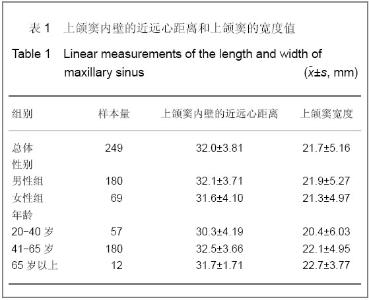
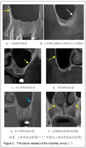
| [1] Raja SV. Management of the posterior maxilla with sinus lift: review of techniques. J Oral Maxillofac Surg. 2009;67(8): 1730-1734.[2] Lin Y, Wang X, Qiu LX, et al. Zhonghua Kouqiang Yixue Zazhi. 1998;33(6):326-328.林野,王兴,邱立新,等.上颌窦提升植骨及同期种植体植入术[J].中华口腔医学杂志,1998,33(6):326-328.[3] Tatum H Jr. Maxillary and sinus implant reconstructions. Dent Clin North Am. 1986;30(2):207-229.[4] Sharan A, Madjar D. Maxillary sinus pneumatization following extractions: a radiographic study. Int J Oral Maxillofac Implants. 2008;23(1):48-56.[5] Kwak HH, Park HD, Yoon HR, et al. Topographic anatomy of the inferior wall of the maxillary sinus in Koreans. Int J Oral Maxillofac Surg. 2004;33(4):382-388.[6] van den Bergh JP, ten Bruggenkate CM, Disch FJ, et al. Anatomical aspects of sinus floor elevations. Clin Oral Implants Res. 2000;11(3):256-265.[7] Xu F, Yang F, Wang RF. Shanghai Kouqiang Yixue. 2011; 20(2):187-190.徐梅,杨帆,王仁飞.正常人上颌窦CBCT影像学特征分析[J].上海口腔医学,2011,20(2):187-190.[8] Uchida Y, Goto M, Katsuki T, et al. A cadaveric study of maxillary sinus size as an aid in bone grafting of the maxillary sinus floor. J Oral Maxillofac Surg. 1998;56(10):1158-1163. [9] Underwood AS. An Inquiry into the Anatomy and Pathology of the Maxillary Sinus. J Anat Physiol. 1910;44(Pt 4):354-369. [10] van Zyl AW, van Heerden WF. A retrospective analysis of maxillary sinus septa on reformatted computerised tomography scans. Clin Oral Implants Res. 2009;20(12): 1398-1401.[11] Naitoh M, Suenaga Y, Kondo S, et al. sessment of maxillary sinus septa using cone-beam computed tomography: etiological consideration. Clin Implant Dent Relat Res. 2009; 11 Suppl 1:e52-58.[12] Kim MJ, Jung UW, Kim CS, et al. Maxillary sinus septa: prevalence, height, location, and morphology. A reformatted computed tomography scan analysis. J Periodontol. 2006; 77(5):903-908. [13] Betts NJ, Miloro M. Modification of the sinus lift procedure for septa in the maxillary antrum. J Oral Maxillofac Surg. 1994; 52(3): 332-333.[14] Boyne PJ, James RA. Grafting of the maxillary sinus floor with autogenous marrow and bone. J Oral Surg. 1980;38(8): 613-616. [15] González-Santana H, Peñarrocha-Diago M, Guarinos-Carbó J, et al. A study of the septa in the maxillary sinuses and the subantral alveolar processes in 30 patients. J Oral Implantol. 2007;33(6):340-343.[16] Kasabah S,Slezak R,Sim nek A,et al.Evaluation of the accuracy of panoramic radiograph in the definition of maxillary sinus septa.Acta Medica (Hradec Kralove).2002;45(4): 173-175. [17] Staudt J, Breustedt A, Kunz G, et al. Untersuchungen uber die Besonderheiten der arteriellen Versorgung im Kopfbereich beim alten Menschen. Verh Anat Ges. 1977;71(Pt1):725-729. [18] McGregor AD, MacDonald DG. Age changes in the human inferior alveolar artery-a histological study. Br J Oral Maxillofac Surg. 1989;27(5):371-374. [19] Traxler H, Windisch A, Geyerhofer U, et al. Arterial blood supply of the maxillary sinus. Clin Anat. 1999;12(6):417-421. [20] Solar P, Geyerhofer U, Traxler H, et al. Blood supply to the maxillary sinus relevant to sinus floor elevation procedures. Clin Oral Implants Res.1999;10(1):34-44. |