|
[1] TAUROG JD, CHHABRA A, COLBERT RA. Ankylosing spondylitis and axial spondyloarthritis.N Engl J Med.2016;374(26): 2563-2574.
[2] FENG ZX, QIAN BP, MAO SH, et al. Anatomical feature of lumbar and S₁ pedicle in patients with thoracolumbar kyphosis secondary to ankylosing spondylitis.Zhongguo Gu Shang.2017;30(2): 132-136.
[3] ZHANG N, LI H, XU ZK, et al. Computer Simulation of Two-level Pedicle Subtraction Osteotomy for Severe Thoracolumbar Kyphosis in Ankylosing Spondylitis.Indian J Orthop.2017;51(6): 666-671.
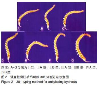
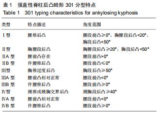
[4] 郑国权,张永刚,王岩,等.强直性脊柱炎后凸畸形的301分型[J].中国脊柱脊髓杂志,2015,25(9):8-13.
[5] 金海明,王向阳.脊柱矢状面畸形截骨角度计算方法的研究进展[J].中华骨科杂志,2016,36(5):298-306.
[6] BEHARI S, TUNGERIA A, JAISWAL AK, et al. The "moustache" sign: localized intervertebral disc fibrosis and panligamentous ossification in ankylosing spondylitis with kyphosis.Neurol India. 2010;58(5):764-767.
[7] BURSTEIN AH, REILLY DT, MARTENS M. Aging of bone tissue: mechanical properties. J Bone Joint Surg Am.1976;58(1):82-86.
[8] SHIRAZI-ADL SA, SHRIVASTAVA SC, AHMED AM. Stress Analysis of the Lumbar Disc-Body Unit in Compression A Three-Dimensional Nonlinear Finite Element Study.Spine(Phila Pa 1976). 1984;9(2): 120-134.
[9] DANISH SF, WILDEN JA, SCHUSTER J. Iatrogenic paraplegia in 2 morbidly obese patients with ankylosing spondylitis undergoing total hip arthroplasty.J Neurosurg Spine.2008;8(1):80-83.
[10] WANG Y, ZHANG YG, ZHENG GQ, et al. Vertebral column decancellation for the management of rigid scoliosis: the effectiveness and safety analysis.Zhonghua Wai Ke Za Zhi. 2010;48(22):1701-1704.
[11] 刘新宇,原所茂,田永昊,等.扩大“蛋壳”结合闭合-张开技术治疗胸腰椎角状后凸畸形[J].中国脊柱脊髓杂志,2014,24(9):779-783.
[12] 张永刚,王岩,张雪松,等.扩大蛋壳技术单纯后路切除青少年胸腰段半脊椎[J].脊柱外科杂志,2007,5(2):69-72.
[13] 林斌,张毕,许洋,等.脊柱去松质骨化截骨术治疗强直性脊柱炎并脊柱后凸畸形[J].临床骨科杂志,2014,17(3):241-244.
[14] 汪飞,邱勇,钱邦平,等.后路全脊椎截骨治疗严重脊柱畸形内固定棒断裂危险因素分析[J].中华骨科杂志,2012,32(10):946-950.
[15] 朱明喜.脊柱后路去松质骨截骨术在脊柱畸形翻修手术中的临床应用[J].颈腰痛杂志,2017,38(5): 455-459.
[16] 钱邦平,黄季晨,邱勇,等.截骨矫形术治疗强直性脊柱炎颈胸段畸形的疗效分析[J].中华骨科杂志,2018,38(4):204-211.
[17] 陈遥,洪正华,洪盾,等.保留中柱经椎弓根开合式截骨术治疗强直性脊柱炎后凸畸形[J].中华骨科杂志,2018,38(22):1349-1356.
[18] 李长明,赵士杰,许建柱,等.经椎弓根楔形截骨联合长节段椎弓根螺钉固定治疗强直性脊柱炎后凸畸形合并胸腰段骨折的短期疗效[J].中华创伤杂志,2019,35(6):501-507.
[19] 齐鹏,宋凯,张永刚,等.单节段脊柱去松质骨截骨与双节段经椎弓根截骨矫正强直性脊柱炎后凸畸形的临床效果比较[J].中国脊柱脊髓杂志, 2015, 25(9): 775-780.
[20] 张智发,杨全中,杨晓清,等.脊柱后路去骨松质截骨术在强直性脊柱炎胸腰段后凸畸形矫正手术中的应用[J].解放军医学院学报, 2016, 37(6): 586-590.
|