Chinese Journal of Tissue Engineering Research ›› 2020, Vol. 24 ›› Issue (23): 3708-3715.doi: 10.3969/j.issn.2095-4344.2692
Previous Articles Next Articles
High-intensity interval training for treating pathological cardiac hypertrophy in spontaneously hypertensive rats: effects and mechanisms
Yuan Guoqiang1, Qin Yongsheng2, Peng Peng2
- 1Department of Physical Education, Zhengzhou University of Aeronautics, Zhengzhou 450015, Henan Province, China; 2Department of Health Service, Logistics University of Chinese People’s Armed Police Forces, Tianjin 300309, China
-
Received:2019-09-04Revised:2019-09-09Accepted:2019-10-19Online:2020-08-18Published:2020-07-30 -
Contact:Peng Peng, MD, Lecturer, Department of Health Service, Logistics University of Chinese People’s Armed Police Forces, Tianjin 300309, China -
About author:Yuan Guoqiang, Master, Lecturer, Department of Physical Education, Zhengzhou University of Aeronautics, Zhengzhou 450015, Henan Province, China -
Supported by:the Natural Science Foundation of Tianjin, No. 17JCYBJC274002
CLC Number:
Cite this article
Yuan Guoqiang, Qin Yongsheng, Peng Peng. High-intensity interval training for treating pathological cardiac hypertrophy in spontaneously hypertensive rats: effects and mechanisms[J]. Chinese Journal of Tissue Engineering Research, 2020, 24(23): 3708-3715.
share this article
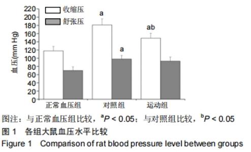
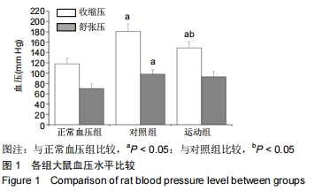
2.1 实验动物数量分析 在实验过程中,对照组死亡1只,运动组死亡1只、拒跑2只,统计时予以剔除,因此最终样本量为41只,其中正常血压组15只,对照组14只和运动组12只。 2.2 血压的变化 与正常血压组[收缩压:(118±11) mm Hg,舒张压:(90±9) mm Hg]比较,对照组、运动组收缩压和舒张压增加[对照组收缩压:(181±15) mm Hg,舒张压:(98± 9) mm Hg;运动组收缩压:(149±12) mm Hg,舒张压:(93± 10) mm Hg],差异有显著性意义(P < 0.05);与对照组比较,运动组收缩压降低(P < 0.05),舒张压的变化差异无显著性意义(P > 0.05),提示8周高强度间歇运动能够降低高血压大鼠收缩压,但对舒张压无影响,见图1。 "
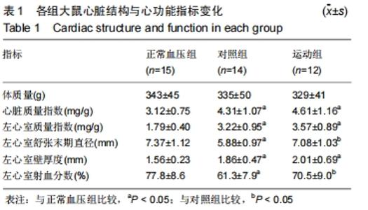
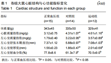
2.3 心脏结构与功能的变化 与正常血压组比较,对照组心脏质量指数、左心室质量指数和左心室壁厚度升高(均P < 0.05),左心室舒张末期直径和左心室射血分数降低(均P < 0.05),说明高血压大鼠左室出现心腔缩窄,室壁增厚,即向心性肥大,同时心功能下降。与对照组比较,运动组左心室舒张末期直径和左心室射血分数升高(均 P < 0.05),体质量、心脏质量指数、左心室质量指数和左心室壁厚度的变化差异无显著性意义(均P > 0.05);与正常血压组比较,运动组心脏质量指数、左心室质量指数和左心室壁厚度升高(均P < 0.05),上述结果提示8周高强度间歇运动诱导左室心腔扩张,即离心性肥大,伴随心功能升高,见表1。 "
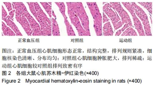
2.4 心肌组织病理学观察与心肌细胞横截面积的变化 各组大鼠心肌苏木精-伊红染色见图2。正常血压组心肌细胞形态正常,结构完整,排列规则紧凑,细胞核染色清晰、分布均匀;对照组心肌细胞肿胀肥大,排列稀疏;运动组心肌细胞较对照组排列致密有序。 "

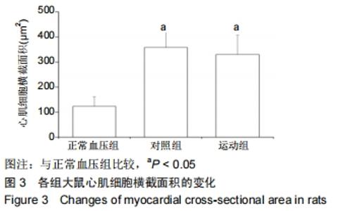
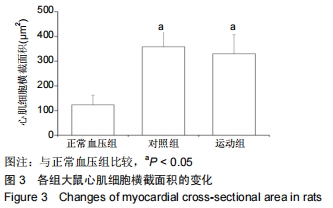
心肌细胞横截面积的变化见图3。与正常血压组比较,对照组和运动组心肌细胞横截面积均升高(均P < 0.05),说明高血压大鼠心肌细胞体积增大;与对照组比较,运动组心肌细胞横截面积的变化差异无显著性意义(P > 0.05),提示8周高强度间歇运动并未改善高血压大鼠心肌细胞肥大。 "
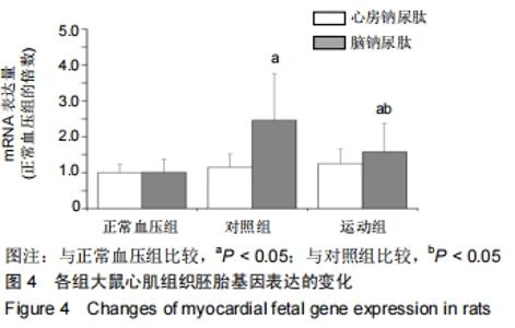
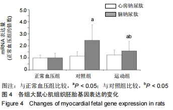
2.5 胚胎基因mRNA表达量的变化 与正常血压组比较,对照组和运动组脑钠尿肽mRNA表达量上调(P < 0.05),说明高血压大鼠心脏胚胎基因重新激活;与对照组比较,运动组脑钠尿肽mRNA表达量降低(P < 0.05),提示8周高强度间歇运动诱导高血压大鼠心脏胚胎基因下调。心房钠尿肽mRNA在各组间比较差异无显著性意义(P > 0.05),说明高血压和运动并未对心房钠尿肽造成影响,见图4。 "
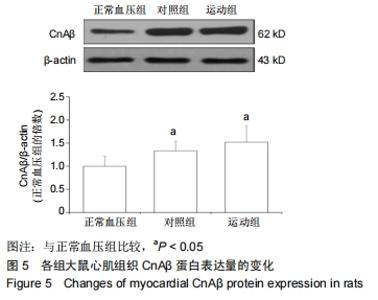
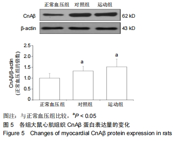
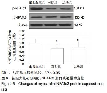
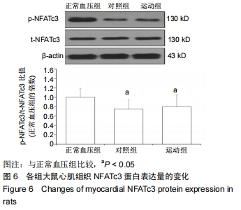
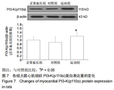
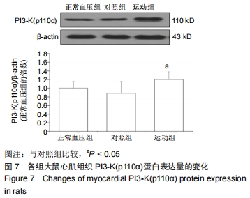
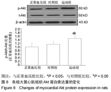
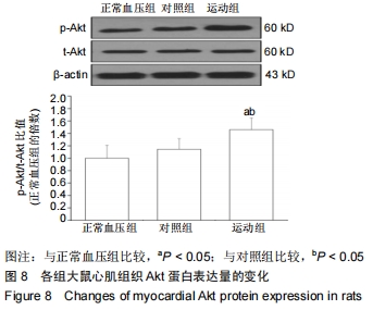
2.6 心脏肥大信号分子蛋白表达量的变化 与正常血压组比较,对照组CnAβ表达升高(P < 0.05),p-NFATc3/t-NFATc3比值下降(P < 0.05)、PI3-K (p110α)无显著变化(P > 0.05),说明高血压诱导Cn/NFAT信号途径激活,但对PI3-K/Akt通路并无影响。与对照组比较,运动组CnAβ和p-NFATc3/ t-NFATc3比值无显著变化(均P > 0.05),PI3-K(p110α)和p-Akt/t-Akt比值升高(均P < 0.05);与正常血压组比较,运动组CnAβ和p-Akt/t-Akt比值升高(P < 0.05),p-NFATc3/ t-NFATc3比值下降(P < 0.05)、PI3-K(p110α)无显著变化(P > 0.05);结果提示8周高强度间歇运动激活PI3-K/Akt途径,但未对Cn/NFAT通路产生抑制作用,见图5-8。 "
"
"
"
| [1]SHENASA M, SHENASA H. Hypertension, left ventricular hypertrophy, and sudden cardiac death. Int J Cardiol. 2017; 237:60-63. [2]BESSEM B, DE BRUIJN MC, NIEUWLAND W, et al. The electrocardiographic manifestations of athlete's heart and their association with exercise exposure. Eur J Sport Sci. 2018;18(4):587-593. [3]NAKAMURA M, SADOSHIMA J. Mechanisms of physiological and pathological cardiac hypertrophy. Nat Rev Cardiol. 2018; 15(7):387-407. [4]SHARMAN JE, LA GERCHE A, COOMBES JS. Exercise and cardiovascular risk in patients with hypertension. Am J Hypertens. 2015;28(2):147-158. [5]JACOBS RA, FLÜCK D, BONNE TC, et al. Improvements in exercise performance with high-intensity interval training coincide with an increase in skeletal muscle mitochondrial content and function. J Appl Physiol (1985). 2013;115(6): 785-793. [6]CHRISTENSEN PM, KRUSTRUP P, GUNNARSSON TP, et al. VO2 kinetics and performance in soccer players after intense training and inactivity. Med Sci Sports Exerc. 2011;43(9): 1716-1724. [7]LITTLE JP, GILLEN JB, PERCIVAL ME, et al. Low-volume high-intensity interval training reduces hyperglycemia and increases muscle mitochondrial capacity in patients with type 2 diabetes. J Appl Physiol (1985). 2011;111(6):1554-1560. [8]CIOLAC EG. High-intensity interval training and hypertension: maximizing the benefits of exercise? Am J Cardiovasc Dis. 2012;2(2):102-110. [9]HUSSAIN SR, MACALUSO A, PEARSON SJ. High-intensity interval training versus moderate-intensity continuous training in the prevention/management of cardiovascular disease. Cardiol Rev. 2016;24(6):273-281. [10]MOUSAVI N, CZARNECKI A, KUMAR K, et al. Relation of biomarkers and cardiac magnetic resonance imaging after marathon running. Am J Cardiol. 2009;103(10):1467-1472. [11]BENITO B, GAY-JORDI G, SERRANO-MOLLAR A, et al. Cardiac arrhythmogenic remodeling in a rat model of long-term intensive exercise training. Circulation. 2011;123(1): 13-22. [12]DA COSTA REBELO RM, SCHRECKENBERG R, SCHLÜTER KD. Adverse cardiac remodelling in spontaneously hypertensive rats: acceleration by high aerobic exercise intensity. J Physiol. 2012;590(21):5389-5400. [13]范朋琦,秦永生,彭朋.不同运动方式对自发性高血压大鼠心脏重塑和运动能力的影响[J].现代预防医学,2018,45(23): 4341-4345. [14]孟宪欣,管泽毅,葛吉生,等.间歇运动干预自发性高血压大鼠病理性心脏肥大:运动强度与健康效应的关系[J].体育科学,2019, 39(6): 73-82. [15]黄凯,彭朋,李晓霞.长期自主跑轮运动对心肌梗死大鼠心肌重塑的影响[J].武警后勤学院学报(医学版),2015,24(5): 337-341. [16]ROSSONI LV, OLIVEIRA RA, CAFFARO RR, et al. Cardiac benefits of exercise training in aging spontaneously hypertensive rats. J Hypertens. 2011;29(12):2349-2358. [17]甄洁,李晓霞. TGF-β/TIMP-1/MMP-1信号通路在有氧运动改善心衰大鼠心脏重塑中的作用[J].中国康复医学杂志,2015, 30(12): 1212-1216. [18]施曼莉,李晓霞.有氧运动对慢性心力衰竭大鼠病理性心脏肥大的影响[J].体育学刊,2015,22(3):127-134. [19]LOVIC D, NARAYAN P, PITTARAS A, et al. Left ventricular hypertrophy in athletes and hypertensive patients. J Clin Hypertens (Greenwich). 2017;19(4):413-417. [20]DZUDIE A, DZEKEM BS, KENGNE AP. NT-pro BNP and plasma-soluble ST2 as promising biomarkers for hypertension, hypertensive heart disease and heart failure in sub-Saharan Africa. Cardiovasc J Afr. 2017;28(6):406-407. [21]DIRKX E, DA COSTA MARTINS PA, DE WINDT LJ. Regulation of fetal gene expression in heart failure. Biochim Biophys Acta. 2013;1832(12):2414-2424. [22]YASUE H, YOSHIMURA M, SUMIDA H, et al. Localization and mechanism of secretion of B-type natriuretic peptide in comparison with those of A-type natriuretic peptide in normal subjects and patients with heart failure. Circulation.1994; 90(1):195-203. [23]YEVES AM, BURGOS JI, MEDINA AJ, et al. Cardioprotective role of IGF-1 in the hypertrophied myocardium of the spontaneously hypertensive rats: A key effect on NHE-1 activity. Acta Physiol (Oxf). 2018;224(2):e13092. [24]MCMULLEN JR, AMIRAHMADI F, WOODCOCK EA, et al. Protective effects of exercise and phosphoinositide 3-kinase(p110alpha) signaling in dilated and hypertrophic cardiomyopathy. Proc Natl Acad Sci U S A. 2007;104(2): 612-617. [25]SAGARA S, OSANAI T, ITOH T, et al. Overexpression of coupling factor 6 attenuates exercise-induced physiological cardiac hypertrophy by inhibiting PI3K/Akt signaling in mice. J Hypertens. 2012;30(4):778-786. [26]MIYACHI M, YAZAWA H, FURUKAWA M, et al. Exercise training alters left ventricular geometry and attenuates heart failure in dahl salt-sensitive hypertensive rats. Hypertension. 2009;53(4):701-707. [27]GARCIARENA CD, PINILLA OA, NOLLY MB, et al. Endurance training in the spontaneously hypertensive rat: conversion of pathological into physiological cardiac hypertrophy. Hypertension. 2009;53(4):708-714. [28]贾祁,李丼,石丼君,等. CaN/NFAT信号通路在有氧运动改善高血压心肌肥大中的作用[J].体育科学,2018,38(12):45-52. [29]NICHOLSON CK, LAMBERT JP, CHOW CW, et al. Chronic exercise downregulates myocardial myoglobin and attenuates nitrite reductase capacity during ischemia-reperfusion. J Mol Cell Cardiol. 2013;64:1-10. [30]KONHILAS JP, WATSON PA, MAASS A, et al. Exercise can prevent and reverse the severity of hypertrophic cardiomyopathy. Circ Res. 2006;98(4):540-548. [31]OLIVEIRA RS, FERREIRA JC, GOMES ER, et al. Cardiac anti-remodelling effect of aerobic training is associated with a reduction in the calcineurin/NFAT signalling pathway in heart failure mice. J Physiol. 2009;587(Pt 15):3899-3910. [32]廖兴林,常芸,高晓嶙,等.力竭运动后不同时相大鼠心肌CnAβ的变化[J].中国运动医学杂志,2009,28(4):388-390. [33]王增喜,李洁,王悦.高强度间歇训练对大鼠心肌线粒体呼吸链复合体活性的影响[J].中国运动医学杂志,2018,37(4):315-322. [34]HOLLOWAY TM, BLOEMBERG D, DA SILVA ML, et al. High intensity interval and endurance training have opposing effects on markers of heart failure and cardiac remodeling in hypertensive rats. PLoS One. 2015;10(3):e0121138. [35]BOUTCHER YN, BOUTCHER SH. Exercise intensity and hypertension: what's new? J Hum Hypertens. 2017;31(3): 157-164. [36]THOMPSON PD, FRANKLIN BA, BALADY GJ, et al. Exercise and acute cardiovascular events placing the risks into perspective: a scientific statement from the American Heart Association Council on Nutrition, Physical Activity, and Metabolism and the Council on Clinical Cardiology. Circulation. 2007;115(17):2358-2368. [37]GILLEN JB, GIBALA MJ. Is high-intensity interval training a time-efficient exercise strategy to improve health and fitness? Appl Physiol Nutr Metab. 2014;39(3):409-412. |
| [1] | Deng Shuang, Pu Rui, Chen Ziyang, Zhang Jianchao, Yuan Lingyan . Effects of exercise preconditioning on myocardial protection and apoptosis in a mouse model of myocardial remodeling due to early stress overload [J]. Chinese Journal of Tissue Engineering Research, 2022, 26(5): 717-723. |
| [2] | Feng Jianbo, Li Chencheng, Liu Jinyue, Wang Xiaomin, Peng Jiachen. Implantation of Kirschner wire with Staphylococcus aureus biofilm establishes a traumatic osteomyelitis model in rats [J]. Chinese Journal of Tissue Engineering Research, 2022, 26(5): 700-705. |
| [3] | Zhang Wanxia, Nijiati·Nuermuhanmode, Maisituremu·Heilili, Liu Jianglong, Liu Di, Maimaitituxun·Tuerdi. An appropriate dose of intra-articular medical ozone injection in a rat model of temporomandibular joint osteoarthritis [J]. Chinese Journal of Tissue Engineering Research, 2022, 26(23): 3700-3705. |
| [4] | Wang Wenhua, Liu Yang, Wang Qiaomin, Lü Yang, Zhao Yaru, Wang Haiping. 5-Azacytidine combined with bone morphogenetic protein 2 induces differentiation of bone marrow mesenchymal stem cells into cardiomyocyte-like cells [J]. Chinese Journal of Tissue Engineering Research, 2022, 26(19): 2958-2963. |
| [5] | Zhu Yaping, Li Xiang, Chen Zhiwen, Liu Ying, Zhao Wenzhuo, Ma Ketao, Gu Qiang. Calcium-sensitive receptors influence pulmonary vascular remodeling in neonatal mice with persistent pulmonary hypertension [J]. Chinese Journal of Tissue Engineering Research, 2022, 26(17): 2708-2712. |
| [6] | Zhu Zheng, Fu Changxi, Ma Wenchao, Ma Gang, Peng peng. Regulatory mechanism of aerobic exercise on cardiac remodeling in spontaneously hypertensive rats [J]. Chinese Journal of Tissue Engineering Research, 2022, 26(14): 2231-2237. |
| [7] | Wang Dong, Ding Hai, Chang Wenju, Wang Jinzi. Biological changes and mechanism of salvianolic acid B in ovariectomized osteoporosis rat models [J]. Chinese Journal of Tissue Engineering Research, 2022, 26(12): 1927-1930. |
| [8] | Lun Zhigang, Jin Jing, Wang Tianyan, Li Aimin. Effect of peroxiredoxin 6 on proliferation and differentiation of bone marrow mesenchymal stem cells into neural lineage in vitro [J]. Chinese Journal of Tissue Engineering Research, 2021, 25(7): 1014-1018. |
| [9] | Chen Junyi, Wang Ning, Peng Chengfei, Zhu Lunjing, Duan Jiangtao, Wang Ye, Bei Chaoyong. Decalcified bone matrix and lentivirus-mediated silencing of P75 neurotrophin receptor transfected bone marrow mesenchymal stem cells to construct tissue-engineered bone [J]. Chinese Journal of Tissue Engineering Research, 2021, 25(4): 510-515. |
| [10] | Yuan Lifen, Cao Shuang, Sun Ting. Effect of angiotensinogen-targeted RNA interference in spontaneously hypertensive rats [J]. Chinese Journal of Tissue Engineering Research, 2021, 25(35): 5626-5631. |
| [11] | Zhang Fucai, Zheng Feng, Wang Furong, Chen Gang . Effects of paeonol on forkhead box O3a/Wnt signal pathway and vertebral bone mineral density in ovariectomized osteoporosis rats [J]. Chinese Journal of Tissue Engineering Research, 2021, 25(32): 5091-5096. |
| [12] | Li Jie, Ma Yuewen, Kang Nan, Zhang Jing, Zhang Yu. Radial extracorporeal shock wave therapy promotes the proliferation of neural stem cells in hippocampus of cerebral infarction rats and inhibits miR-124 expression [J]. Chinese Journal of Tissue Engineering Research, 2021, 25(31): 4981-4987. |
| [13] | Hao Xiaona, Zhang Yingjie, Li Yuyun, Xu Tao. Bone marrow mesenchymal stem cells overexpressing prolyl oligopeptidase on the repair of liver fibrosis in rat models [J]. Chinese Journal of Tissue Engineering Research, 2021, 25(25): 3988-3993. |
| [14] | Huang Zhusong, Lin Yu, Chen Xiang, Lan Jinfu, Guan Yong, Gao Xi. Alcohol extract of Morinda officinalis improves lipid metabolism and bone metabolism in ovariectomized obese rats [J]. Chinese Journal of Tissue Engineering Research, 2021, 25(2): 205-210. |
| [15] | Chen Yutong, Li Chenchen, Liu Yang, Zheng Yaqin, Yang Xihua, An Meiwen. Establishment of an acute radioactive skin injury model in Wistar rats [J]. Chinese Journal of Tissue Engineering Research, 2021, 25(2): 237-241. |
| Viewed | ||||||
|
Full text |
|
|||||
|
Abstract |
|
|||||