Chinese Journal of Tissue Engineering Research ›› 2020, Vol. 24 ›› Issue (14): 2197-2204.doi: 10.3969/j.issn.2095-4344.2582
Previous Articles Next Articles
Establishment and application of paravertebral muscle endurance and left-right symmetry measurement indexes and methods in teenagers
Qiu Dan1, 2, He Hui1, Xiong Kaiyu1, Li Hongjuan1
- 1Beijing Sport University, Beijing 100084, China; 2Baotou Teachers’ College, Inner Mongolia University of Science & Technology, Baotou 014030, Inner Mongolia Autonomous Region, China
-
Received:2019-05-10Revised:2019-05-22Accepted:2019-10-09Online:2020-05-18Published:2020-03-15 -
Contact:He Hui, Associate researcher, Beijing Sport University, Beijing 100084, China -
About author:Qiu Dan, Master, Lecturer, Beijing Sport University, Beijing 100084, China; Baotou Teachers’ College, Inner Mongolia University of Science & Technology, Baotou 014030, Inner Mongolia Autonomous Region, China -
Supported by:the Fundamental Research Fund for the Central Universities, No. 2016YB033; the National Key Research and Development Project, No. 2018YFC2000603
CLC Number:
Cite this article
Qiu Dan, He Hui, Xiong Kaiyu, Li Hongjuan. Establishment and application of paravertebral muscle endurance and left-right symmetry measurement indexes and methods in teenagers[J]. Chinese Journal of Tissue Engineering Research, 2020, 24(14): 2197-2204.
share this article

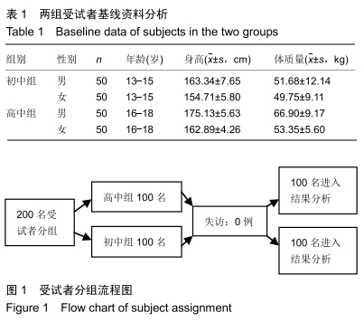
2.1 参与者数量分析 纳入受试者200名,分为2组,试验过程无脱落,全部进入结果分析。 2.2 两组受试者基线资料分析 见表1。受试者分组流程图见图1。 "
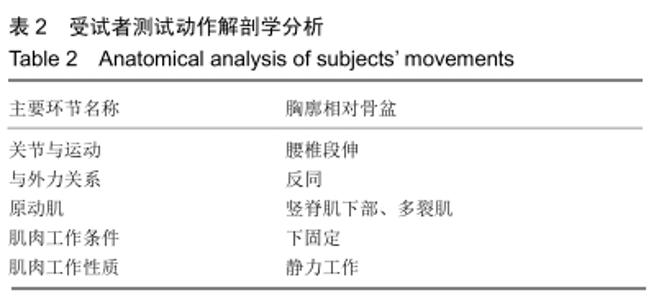
2.3 Biering-Sorensen(B-S)试验姿势动作分析 根据生物力学分析,骨盆及以下部位固定,上身悬空,身体处于水平位,椎旁肌对抗受试者自身髂前上棘以上体质量。测试动作解剖学分析见表2。 "


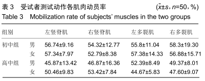
将每个受试者测试开始后第5秒L3-L4双侧竖脊肌和L5-S1双侧多裂肌的积分肌电值除以该块肌肉最大等长随意收缩时的积分肌电值,得到受试者做B-S测试中每块肌肉的动员率,见表3。可以看出,在做该动作时左竖脊肌、右竖脊肌、左多裂肌、右多裂肌动员率在40%-60%最大等长随意收缩之间,为中等负荷强度,受试者负自身髂前上棘以上体质量,负重具有自身特异性,具有实际价值,可以作为评定椎旁肌耐力的动作姿势。 "

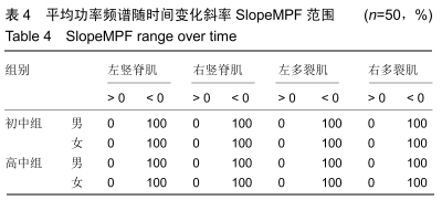
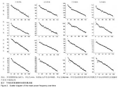
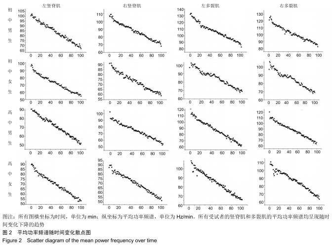
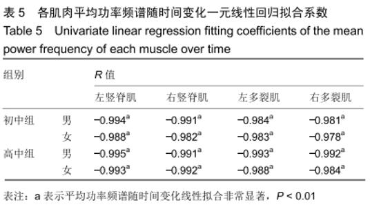
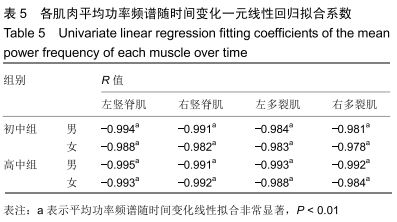
2.4 肌电指标筛选 选取肌电频域指标平均功率频谱随时间变化斜率SlopeMPF,时域指标积分肌电值随时间变化斜率SlopeIEMG。进行一致性、可靠性检验,筛选适合青少年椎旁肌耐力测评的指标。 2.4.1 肌电指标变化一致性研究 (1)频域指标平均功率频谱随时间变化斜率SlopeMPF:①SlopeMPF范围:将所有受试者在进行整个B-S实验测试中各块肌肉平均功率频谱值做一元线性回归,得到平均功率频谱随时间变化斜率SlopeMPF,比较SlopeMPF>0和SlopeMPF<0占该组总数的百分比,结果见表4。可以看出,各组学生平均功率频谱随时间变化斜率SlopeMPF<0的人数占该组人数的100%,即所有受试者在做该测试时,频域指标平均功率频谱均呈现随时间变化下降的趋势。②平均功率频谱随时间变化一元线性回归检验:将所有受试者的测量时间(t)分为100份,平均功率频谱随时间变化的散点图见图2。③各组一元线性回归拟合系数:见表5。可以看出左竖脊肌、右竖脊肌、左多裂肌、右多裂肌的平均功率频谱变化与时间变化成高度的线性负相关,即在整个测试过程中,所有受试者平均功率频谱随时间的延长出现持续下降。 "
"
"
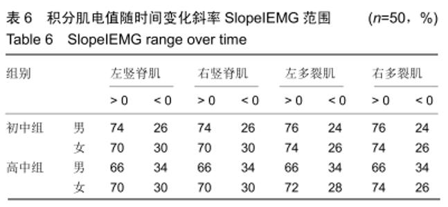
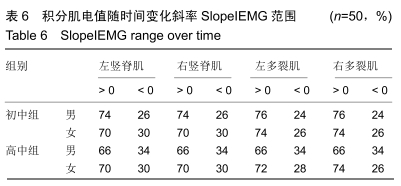
(2)时域指标积分肌电值随时间变化斜率SlopeIEMG: 将所有受试者在进行整个B-S实验测试中各块肌肉积分肌电值做一元线性回归,得到积分肌电值随时间变化斜率SlopeIEMG,比较SlopeIEMG>0和SlopeIEMG<0占该组学生的百分比。结果见表6。可以看出,在测试过程中,各年龄组积分肌电值的变化增加和减少都有出现,无规律性。 "
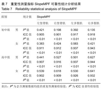
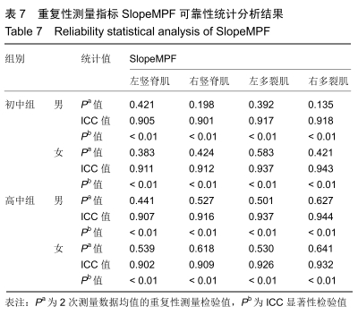
2.4.2 肌电指标变化可靠性研究 对受试者进行重复2次椎旁肌耐力测试。 (1)频域指标SlopeMPF的可靠性研究:比较分析2次测试结果,SlopeMPF可靠性统计分析结果见表7。可以看出所有受试者各肌肉SlopeMPF指标重复性检验均P > 0.05,差异无显著性意义,各组右竖脊肌、左多裂肌、右多裂肌组内的相关系数均为0.9以上,且具有非常显著性,可靠性佳。 "
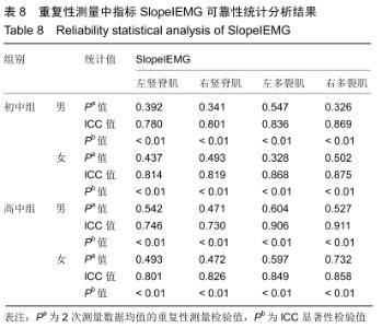
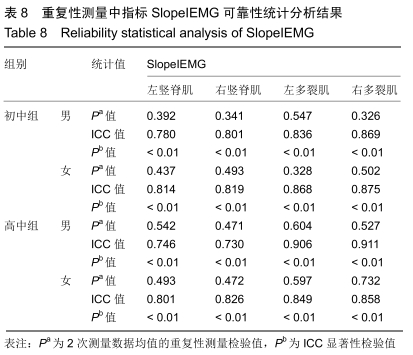
(2)时域指标SlopeIEMG的可靠性研究:比较分析2次测试结果,时域指标SlopeIEMG可靠性统计分析结果见表8。可以看出各组学生在2次重复性测量中,左竖脊肌、右竖脊肌、左多裂肌、右多裂肌4块肌肉SlopeIEMG指标均值的重复性检验均P > 0.05,差异无显著性意义。初中男生组和高中男生组部分ESSlopeIEMG组内相关系数小于0.8,可靠性欠佳,其余各肌肉SlopeIEMG组内相关系数均在0.8以上,可靠性可以接受。 "
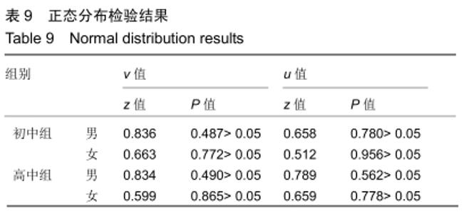
通过以上的对比研究,确定以MPFslope作为评价青少年椎旁肌耐力的评价指标,其具有一致性和可靠性的优点。 2.5 青少年椎旁肌耐力及左右侧对称性的评价与分析 2.5.1 正态分布检验 对初中组、高中组学生椎旁肌耐力及左右侧对称性测试结果采用非参数K-S正态分布检验,结果见表9。可以看出,初中、高中组学生椎旁肌耐力和左右侧对称性测试结果均服从正态分布。 "


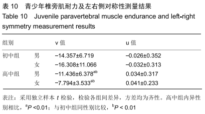
2.5.2 拟合评价指标 研究表明从几块肌肉得出的肌电图指数,如平均值和极值,通常能够反映更有效和可靠的结果[29-30]。左竖脊肌、右竖脊肌、左多裂肌、右多裂肌在维持脊柱稳定性和保持躯干直立状态起重要作用,其中任何一块肌肉耐力不足都会影响整个椎旁肌的功能状况,继而引起脊柱的不稳定。因此,研究将此4块椎旁肌作为共同研究对象,采用拟合计算,得到指数v和指数u,分别用来评价椎旁肌耐力及左右侧对称情况。具体计算如下公式: 复合性指标v作为椎旁肌耐力指数来评价椎旁肌耐力,以整个测试过程左竖脊肌、右竖脊肌、左多裂肌、右多裂肌四块肌肉slopeMPF的平均值来表示;复合性指标u作为椎旁肌左右侧对称性指数,综合考虑竖脊肌和多裂肌双侧肌肉,以左右竖脊肌slopeMPF差值相对值和左右多裂肌slopeMPF差值相对值之和来表示,u > 0反映椎旁肌左侧肌肉力量耐力较右侧强,u < 0反映椎旁肌右侧肌肉力量耐力较左侧强。 2.5.3 评价测试结果 将初中组、高中组所有受试者测试结果按上述计算方法处理,得到结果见表10。可以看出,高中组和初中组同性别学生相比较,椎旁肌耐力增长并有非常显著性。初中组男、女生椎旁肌耐力差异不显著,高中组男生椎旁肌耐力低于女性且具有非常显著性。初中组男、女生椎旁肌对称性均为右侧肌肉耐力强于左侧,高中组均为左侧肌肉耐力强于右侧,但是差异均无显著性意义。 "
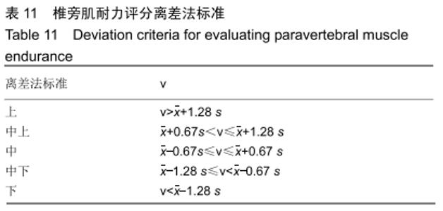
2.5.4 建立评价标准 (1)椎旁肌耐力评分标准:利用离差法对青少年椎旁肌耐力进行评估,制定上、中上、中、中下、下5个等级,评估标准见表11。 "


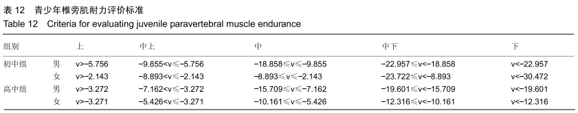
根据初中组、高中组学生测试结果制定评价标准,将各年龄组学生的椎旁肌耐力分为5个等级,如表12。 "
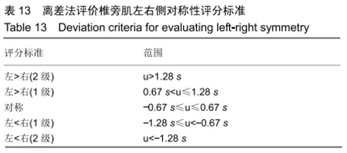
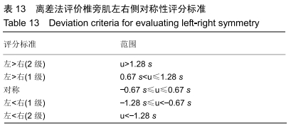
(2)椎旁肌耐力左右侧对称性评分标准:利用离差法来对青少年椎旁肌耐力左右侧对称性进行评估。u=0表示椎旁肌耐力完全对称,以u=0为基准,即离差法中平均数所处的位置,按照不同组别的标准差,将各组学生椎旁肌耐力对称性分为5个等级进行评估,左>右(2级)表示左侧强于右侧,左>右(1级)表示左侧微强于右侧,对称表示左右侧基本对称;左<右(1级)表示右侧微强于左侧,左<右(2级)表示右侧强于左侧。评估标准见表13。 "
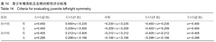
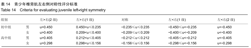
根据初中组、高中组学生测试结果制定评价标准,将各年龄组学生的椎旁肌左右侧对称性结果分为5个等级,见表14。 "
| [1] RUSSO M, DECKERS K, ELDABE S, et al.Muscle control and non-specific chronic low back pain.Neuromodulation. 2018;21(1):1-9. [2] LIONEL GOTTSCHALK IV, CAROLYN ENG BS, SAMUEL WARD PT, et al.The multifidus muscle is the strongest stabilizer of the lumbar spine.The Spine Journal.2007;7(5):76S-76S. [3] 王晨,田野,沈晓龙,等.初发腰腿痛年轻人群中腰椎间盘退变与椎旁肌群改变的相关性[J].中国矫形外科杂志, 2018,,441(7):589-593. [4] KALICHMAN L, CARMELI E, BEEN E. The Association between Imaging Parameters of the Paraspinal Muscles, Spinal Degeneration, and Low Back Pain.Biomed Res Int. 2017;2017:2562957. [5] MAHAUDENS P, BANSE X, MOUSNY M, et al.Gait in adolescent idiopathic scoliosis:kinematics and electromyographic analysis.Eur Spine J. 2009;18(4):512-521. [6] TAKAHASHI Y, KOU I, TAKAHASHI A, et al.A genome-wide association study identifes common variants near LBX1 associated with adolescent idiopathic scoliosis.Nat Genet.2011;43(12): 1237–1240. [7] WAJCHENBERG M, ASTUR N, KANAS M, et al.Adolescent idiopathic scoliosis: Current concepts on neurological and muscular etiologies. Scoliosis Spinal Disord. 2016;11:4 [8] BURWELL RG, DANGERFELD PH, MOULTON A, et al.Adolescent idiopathic scoliosis (AIS), environment, exposome and epigenetics:A molecular perspective of postnatal normal spinal growth and the etiopathogenesis of AIS with consideration of a network approach and possible implications for medical therapy.Scoliosis.2011;6(1): 26. [9] NEWTON EDE MM, JONES SW.Adolescent idiopathic scoliosis: evidence for intrinsic factors driving aetiology and progression.Int Orthop. 2016;40(10):2075-2080. [10] MODI HN, SUH SW, YANG JH, et al.Spontaneous regression of curve in immature idiopathic scoliosis – does spinal column play a role to balance? An observation with literature review.J Orthop Surg Res. 2010; 5: 80. [11] JENG J, YICHEN M, XINMENG J, et al.Volumetric and Fatty Infiltration Imbalance of Deep Paravertebral Muscles in Adolescent Idiopathic Scoliosis.Medical Science Monitor.2017;23:2089-2095. [12] WONG C.Mechanism of right thoracic adolescent idiopathic scoliosis at risk for progression; A unifying pathway of development by normal growth and imbalance.Scoliosis.2015; 10: 2. [13] SHARMA S, LONDONO D, ECKALBAR WL, et al.A PAX1 enhancer locus is associated with susceptibility to idiopathic scoliosis in females. Nat Commun.2015; 6: 6452. [14] ZHU Z, TANG NL, XU L, et al.Genome-wide association study identifes new susceptibility loci for adolescent idiopathic scoliosis in Chinese girls. Nat Commun.2015; 6: 8355. [15] 袁望舒,陈丽霞,沈建雄,等,青少年特发性脊柱侧凸患者顶椎椎旁肌表面肌电信号与Cobb角及轴向躯干旋转角的相关性[J].中国组织工程研究, 2019,23(24):3824-3828. [16] 邱丹,何辉,熊开宇.不同强度抗阻训练对青年男性腰部肌肉形态及机能的影响[J].中国运动医学杂志, 2019, 38(2):32-39. [17] HÄGG GM.Interpretation of EMG spectral alternations and alternation indexes at sustained contraction.Appl Physiol.1992;73(4): 1211-1217 [18] 李扬政,李建华,吴方超.基于不同角度的桥式运动表面肌电特征分析[J].中国运动医学杂志, 2019(4).267-270 [19] SEBASTIAN B, FERDINAND B, SCHNAKE KJ, et al.The relationship between functionality and erector spinae activity in patients with specific low back pain during dynamic and static movements.Gait Posture. 2018;66:208-213. [20] HIDES J, GILMORE C, STANTON W, et al.Multifidus size and symmetry among chronic LBP and healthy asymptomatic subjects. Man Ther.2008;13(1):43-49. [21] MACDONALD D, MOSELEY GL, HODGES PW, et al.Why do some patients keep hurting their back? Evidence of ongoing back muscle dysfunction during remission from recurrent back pain.Pain. 2009; 142(3):183-188. [22] BARKER KL, SHAMLEY DR, JACKSON D.Changes in the cross-sectional area of multifidus and psoas in patients with unilateral back pain: the relationship to pain and disability.Spine.2004; 29(22): 515-519. [23] FARAHPOUR N, GHASEMI S, ALLARD P, et al.Electromyographic responses of erector spinae and lower limb’s muscles to dynamic postural perturbations in patients with adolescent idiopathic scoliosis.J Electromyogr Kinesiol. 2014;24(5):645-651. [24] CHEUNG J, HALBERTSMA JP, VELDHUIZEN AG, et al.A preliminary study on electromyographic analysis of the paraspinal musculature in idiopathic scoliosis.Eur Spine J. 2005;14(2):130-137. [25] DE OLIVEIRA AS, GIANINI PE, CAMARINI PM, et al. Electromyographic Analysis of Paravertebral Muscles in Patients with Idiopathic Scoliosis.Spine (Phila Pa 1976). 2011;36(5):E334-339. [26] FARAHPOUR N, YOUNESIAN H, BAHRPEYMA F. Electromyographic activity of erector spinae and external oblique muscles during trunk lateral bending and axial rotation in patients with adolescent idiopathic scoliosis and healthy subjects.Clin Biomech (Bristol, Avon). 2015;30(5): 411-417. [27] RANGER TA, CICUTTINI FM, JENSEN TS, et al.Are the size and composition of the paraspinal muscles associated with low back pain? A systematic review.Spine J. 2017;17(11):1729-1748. [28] GOUBERT D, OOSTERWIJCK JV, MEEUS M, et al.Structural changes of lumbar muscles in non-specific low back pain: A systematic review.Pain Physician.2016;19(7): E985-E1000. [29] LARIVIE`RE C ,ARSENAULT AB, GRAVEL D, et al.Evaluation of measurement strategies to increase the reliability of EMG indices to assess back muscle fatigue and recovery.J Electromyogr Kinesiol. 2002;12(2):91-102. [30] VAN DIEEN JH, HEIJBLOM P, BUNKENS H.Extrapolation of time series of EMG power spectrum parameters in isometric endurance tests of trunk extensor muscles.J Electromyogr Kinesiol. 1998;8(1): 35-44. [31] BIERING-SORENSEN F. Physical measurements as risk indicators for low-back trouble over a one-year period. Spine.Spine.1984;9(2): 106-119. [32] XIE D, ZHANG J, DING W, et al.Abnormal change of paravertebral muscle in adult degenerative scoliosis and its association with bony structural parameters.Eur Spine J. 2019;28(7):1626-1637. [33] NAGAMACHI A, IKATA T, KATOH S, et al.Spectral analysis of erector spinae muscle surface electromyography as an index of exercise performance in maximal treadmill running.J Med Invest.2000; 47(1-2): 29-35. [34] MAISETTI O, ARNAUD GUÉVEL, LEGROS P, et al.Prediction of endurance capacity of quadriceps muscles in humans using surface electromyogram spectrum analysis during submaximal voluntary isometric contractions.Eur J Appl Physiol. 2002;87(6):509-519. [35] DU HG, YE SL, XU JY, et al.Application of surface electromyography in the treatment of adolescent idiopathic scoliosis with traditional spinal balanced therapy.Zhongguo Gu Shang. 2013;26(11):914-917. [36] STETKAROVA I, ZAMECNIK J, BOCEK V, et al.Electrophysiological and histological changes of paraspinal muscles in adolescent idiopathic scoliosis.Eur Spine J. 2016;25(10):3146-3153. [37] CHWAŁA W, KOZIANA A, KASPERCZYK T, et al.Electromyographic assessment of functional symmetry of paraspinal muscles during static exercises in adolescents with idiopathic scoliosis.Biomed Res Int. 2014;2014:573276. [38] JUNG JY, CHA EJ, KIM KA, et al.Influence of pelvic asymmetry and idiopathic scoliosis in adolescents on postural balance during sitting.Biomed Mater Eng. 2015;26 Suppl 1:S601-S610. [39] CAGNIE B, DHOOGE F, SCHUMACHER C, et al.Fiber typing of the erector spinae and multifidus muscles in healthy controls and back pain patients: A systematic literature review.J Manipulative Physiol Ther. 2015;38(9):653-663. [40] PARK YH, PARK YS, LEE YT, et al.The effect of a core exercise program on Cobb angle and back muscle activity in male students with functional scoliosis: a prospective, randomized, parallel-group, comparative study.J Int Med Res. 2016;44(3):728-734. [41] KO KJ, SUH JH, KIM HY, et al.Proposal of a new exercise protocol for idiopathic scoliosis: A preliminary study.Medicine (Baltimore). 2018; 97(49):e13336. [42] KO KJ, HA GC, YOOK YS, et al.Effects of 12-week lumbar stabilization exercise and sling exercise on, lumbosacral region angle, lumbar muscle strength, and pain scale of patients with chronic,low back pain. J Phys Ther Sci. 2018;30(1):18-22. |
| [1] | Li Yu, Huang Peng, Wang Hong, Wang Anli. Dominant and non-dominant side knee isokinetic characteristics of Chinese calisthenics athletes [J]. Chinese Journal of Tissue Engineering Research, 2021, 25(2): 232-236. |
| [2] | Zhang Shuang, Tan Rui, Wang Chunxiao, Wu Fengyu, Guo Hongyu. MicroRNAs for assessing the motion control of human skeletal muscles [J]. Chinese Journal of Tissue Engineering Research, 2021, 25(17): 2755-2760. |
| [3] | Chen Zegang, Ding Haili, Li Long, Wang Chun. Changes of bone metabolism after different intensity endurance exercises in growing rats [J]. Chinese Journal of Tissue Engineering Research, 2020, 24(35): 5582-5588. |
| [4] | Yang Zhongya, Su Hao, Wang Ji, Zhang Yimin, Kong Zhenxing, Zhang Juan, Zhang Long. High-intensity interval training improves cardiorespiratory endurance in rats [J]. Chinese Journal of Tissue Engineering Research, 2020, 24(11): 1708-1713. |
| [5] | Yang Zhongming1, Jiang Manyi2, Xu Simao3 . Synergistic resistance of moderate-intensity endurance exercise and allicin to rat models of fatty liver induced by high-fat diet [J]. Chinese Journal of Tissue Engineering Research, 2019, 23(31): 5030-5035. |
| [6] | Fan Chaoqun1, Xu Kai2, Nie Mingjian1, Xu Wenfeng1, Wang Mei1. Evaluation of the cardiopulmonary endurance: cardiopulmonary exercise test versus 6-minute two-step test [J]. Chinese Journal of Tissue Engineering Research, 2019, 23(23): 3686-3691. |
| [7] | Ren Zhichao. Effect of aerobic endurance training on peroxisome proliferators activated receptor gamma expression in white adipose tissue and plasma of obese mice induced by high fat [J]. Chinese Journal of Tissue Engineering Research, 2019, 23(19): 3056-3061. |
| [8] | Shao Lian-jie. Autonomic nervous system: its response and adaptation to exercises [J]. Chinese Journal of Tissue Engineering Research, 2015, 19(46): 7509-7516. |
| [9] | Yang Ruo-yu, Wang Yu-bin, Shen Xun-zhang, Cai Guang . Association of elite athlete performance and gene polymorphisms [J]. Chinese Journal of Tissue Engineering Research, 2014, 18(7): 1121-1128. |
| [10] | Wang Guo-jun, Wen Han, Wu Ya-qiong. Running economy versus maximal oxygen uptake to evaluate the endurance level of ordinary people [J]. Chinese Journal of Tissue Engineering Research, 2013, 17(7): 1265-1272. |
| Viewed | ||||||
|
Full text |
|
|||||
|
Abstract |
|
|||||