Chinese Journal of Tissue Engineering Research ›› 2020, Vol. 24 ›› Issue (22): 3452-3459.doi: 10.3969/j.issn.2095-4344.2306
Previous Articles Next Articles
Preparation and characterization of calcined bone/chitosan composite material
Liao Jian1,2, Huang Xiaolin3, Zhou Qian1, Cheng Yuting1, Huo Hua1, Li Fang1, Wu Chao1, Shi Qianhui1, Liao Yunmao2, Liang Xing2
- 1Guizhou Medical University School of Stomatology/Affiliated Stomatology Hospital, Guiyang 550004, Guizhou Province, China; 2West China Hospital of Stomatology, Sichuan University, Chengdu 610041, Sichuan Province, China; 3Department of Dental Implantation, Zhongshan City People’s Hospital, Zhongshan 528403, Guangdong Province, China
-
Received:2019-12-25Revised:2019-12-26Accepted:2020-02-24Online:2020-08-08Published:2020-04-26 -
Contact:Liang Xing, Professor, Doctoral supervisor, West China Hospital of Stomatology, Sichuan University, Chengdu 610041, Sichuan Province, China -
About author:Liao Jian, MD, Associate professor, Associate chief physician, Master’s supervisor, Guizhou Medical University School of Stomatology/Affiliated Stomatology Hospital, Guiyang 550004, Guizhou Province, China; West China Hospital of Stomatology, Sichuan University, Chengdu 610041, Sichuan Province, China -
Supported by:the National Natural Science Foundation of China, No. 81660179; the Natural Science Foundation of Guizhou Province, No. Qiankehejichu[2016]1124; the Joint Fund Project of the Natural Science Foundation of Guizhou Province, No. QiankeheLH[2016]7257
CLC Number:
Cite this article
Liao Jian, Huang Xiaolin, Zhou Qian, Cheng Yuting, Huo Hua, Li Fang, Wu Chao, Shi Qianhui, Liao Yunmao, Liang Xing. Preparation and characterization of calcined bone/chitosan composite material[J]. Chinese Journal of Tissue Engineering Research, 2020, 24(22): 3452-3459.
share this article
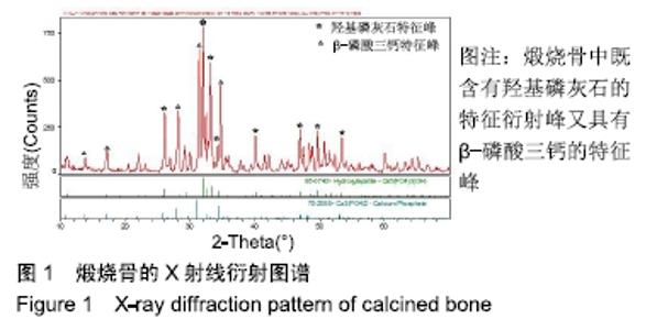
2.1 X射线衍射分析结果 从图1中可见,煅烧骨中既含有羟基磷灰石的特征衍射峰(2θ=32.1°的主峰和33°的次强峰等)又具有β-磷酸三钙的特征峰(2θ=31°的主峰和33.2°的次强峰),煅烧骨与标准的羟基磷灰石/β-磷酸钙峰吻合。X射线衍射定量分析结果表明,两种成分之间的质量比为羟基磷灰石/β-磷酸钙=60/40。 "

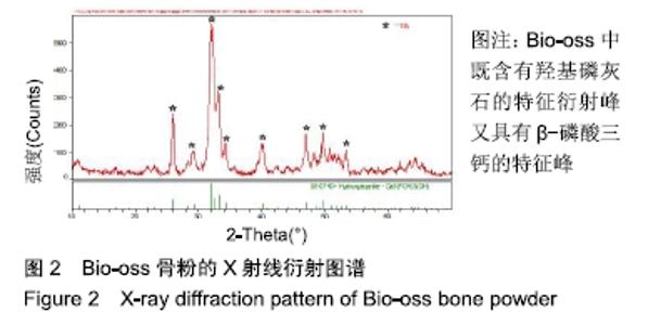
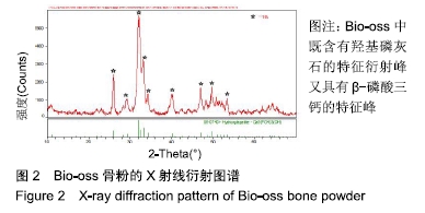
从图2中可见,Bio-oss中既含有羟基磷灰石的特征衍射峰(2θ=32.1°的主峰和33°的次强峰)又具有β-磷酸三钙的特征峰(2θ=31°的主峰和33.2°的次强峰),与标准的羟基磷灰石特征峰基本吻合,说明Bio-oss的成分主要是羟基磷灰石。 "
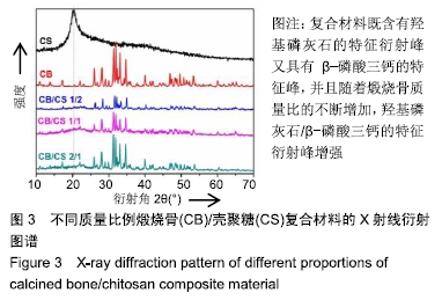
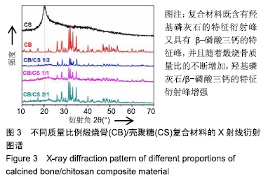
从图3可以看出,在2θ=20.2°处的衍射峰为壳聚糖特征峰,由于壳聚糖是非晶态,故为弥散的宽峰;在2θ=32.1°和33°属于羟基磷灰石的特征衍射峰;在2θ=31°和33.2°属于β-磷酸三钙的特征峰。可以认为采用溶液共混法制备的煅烧骨/壳聚糖复合材料组成成分没有发生变化,保留了煅烧骨和壳聚糖的物质特性;从图3中还可以发现随着煅烧骨质量比的不断增加,羟基磷灰石/β-磷酸三钙的特征衍射峰增强。 "
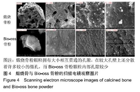
2.2 扫描电镜观察结果 从图4可见,煅烧骨粉颗粒直径在1.2-1.5 mm,颗粒拥有大小相互贯通的孔隙,孔隙由直径为1-300 μm的孔所组成,在较大孔壁上还分散着许多较小的微孔,而Bio-oss骨粉颗粒内部孔隙较少。 "
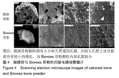
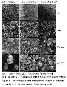
图5显示了不同质量比例煅烧骨/壳聚糖复合材料在低倍和高倍下的扫描电镜图,从图中可见煅烧骨颗粒比较均匀地分散在壳聚糖介质中。 "

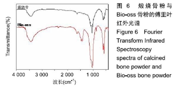
2.3 傅里叶红外光谱分析结果 通过红外光谱图上谱带显示的吸收峰位置、宽度及强度等参数对比纯羟基磷灰石和β-磷酸三钙标准图谱后发现,煅烧骨粉主要由羟基磷灰石和β-磷酸三钙组成,见图6。 "
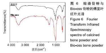
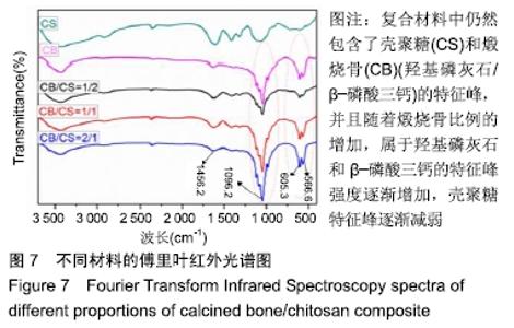
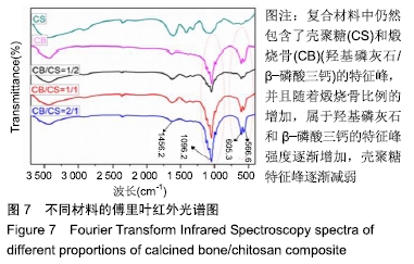
图7分别列出了壳聚糖、煅烧骨及不同质量比例煅烧 骨/壳聚糖复合材料的红外光谱图,由图中可以发现,复合材料中仍然包含了壳聚糖和煅烧骨(羟基磷灰石/β-磷酸三钙)的特征峰,表明材料经过复合后并没有改变原材料的化学成分。随着煅烧骨成分的增加,属于羟基磷灰石和β-磷酸三钙的特征峰(1 048.6,605.3,569.6 cm-1)强度逐渐增加,壳聚糖特征峰(3 431.7,1 605 cm-1)则逐渐减弱,但是基本上没有发生位移,说明煅烧骨和壳聚糖之间属于物理结合,并没有发生化学键的结合。 "
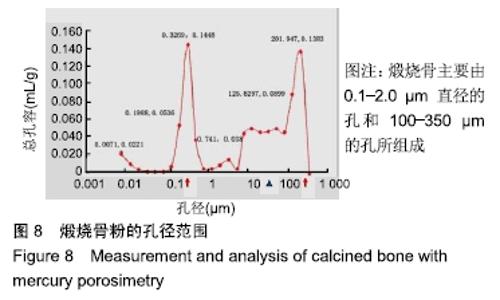
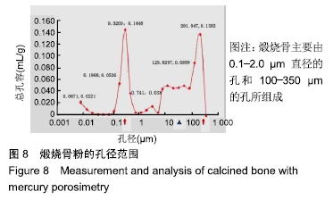
2.4 压汞法检测结果 通过压汞法测得该煅烧骨粉的孔隙率为85%,具有0.841 3 mL/g的总孔容和6.827 m2/g的比表面积,该煅烧骨粉的孔径范围为0.01-350 μm。从图8中可以看出,2个主峰分别位于0.5 μm和250 μm(红色箭头),在所有孔隙中,以0.1-2.0 μm直径的微孔和100-350 μm的大孔为主。另外,还有部分10-90 μm直径的小孔(蓝色三角形指示)。结果表明:该煅烧骨主要由0.1-2.0 μm直径的孔和100-350 μm的孔所组成,这也与扫描电镜结果吻合,进一步证明在较大孔壁上(100-350 μm)分散着大量较小的微孔(0.1-2.0 μm)。 "

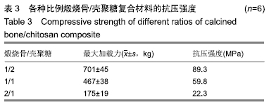
2.5 抗压强度检测结果 从表3中可以看出,复合材料的最大抗压强度达到89.3M Pa,最小强度为22.3 MPa,随着煅烧骨粉成分的增加,抗压强度呈下降趋势。 "
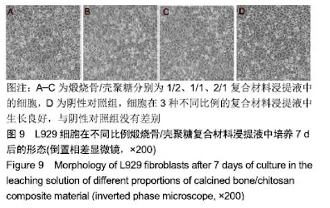
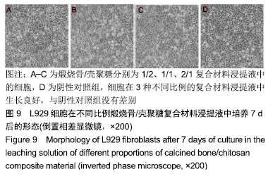
2.6 L929细胞的形态学观察结果 从图9中可以观察到,细胞在3种不同比例的复合材料浸提液中生长良好,与阴性对照组没有差别,说明复合材料对细胞的生长没有影响。 "

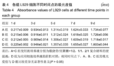
2.7 材料对L929的细胞毒性 细胞在各组浸提液中生长良好,随着培养时间的增加,各组细胞的量也不断增多。从表4中可以看出,细胞在第1-7天增殖较快,其中第1-3天增长最快,从第7天开始增长速度变慢,进入平台期,4组组内5个时间段的细胞增殖比较差异有显著性意义(P < 0.05),相同时间点下各实验组与阴性对照组间的细胞增殖比较差异无显著性意义(P > 0.05)。各复合材料组的细胞相对增殖率为94.2%-98.5%,对应的细胞毒性为1级,见表5,符合生物材料安全级别要求。 "

"

| [1] TRAINI T, PIATTELLI A, CAPUTI S, et al. Regeneration of human bone using different bone substitute biomaterials.Clin Implant Dent Relat Res.2015;17(1):150-162. [2] XI Y, MIAO X, LI Y, et al. BMP2-mimicking peptide modified with E7 coupling to calcined bovine bone enhanced bone regeneration associating with activation of the Runx2/SP7 signaling axis.J Biomed Mater Res B Appl Biomater. 2020;108(1):80-93. [3] IIJIMA K, TSUJI Y, KURIKI I, et al. Control of cell adhesion and proliferation utilizing polysaccharide composite film scaffolds.Colloids Surf B Biointerfaces. 2017;160:228-237. [4] GRAHAM SM, LEONIDOU A, ASLAM-PERVEZ N, et al. Biological therapy of bone defects:the immunology of bone allo-transplantation. Expert opin Biol Ther.2010;10(6):885-901. [5] 赵淑珍,张婷,王彦鹏,等.复合3D支架在修复颅骨缺损中的应用进展[J].中国综合临床,2019,35(2):183-186. [6] RESMIM CM, DALPASQUALE M, VIELMO NIC, et al.Study of physico-chemical properties and in vitro antimicrobial activity of hydroxyapatites obtained from bone calcination.Prog Biomater. 2019; 8(1):1-9. [7] GOTO T, SASAKI K. Synthesis of morphologically controlled hydroxyapatite from fish bone by urea-assisted hydrothermal treatment and its Sr2+ sorption capacity.Powder Technol. 2016;292: 314-322. [8] TEKIYEH RM, NAJAFI M, SHAHRAKI S. Machinability of AA7075-T6/carbon nanotube surface composite fabricated by friction stir processing. Proceedings of the Institution of Mechanical Engineers, Part E: Journal of Process Mechanical Engineering. 2018;233(4): 839-848. [9] ANWAR A, AKBAR S. Novel continuous microwave assisted flow synthesis of nanosized manganese substituted hydroxyapatite.Ceram Int.2018;44(9):10878-10882. [10] DOMÍNGUEZ-TRUJILLO C, PEÓN E, CHICARDI E, et al. Sol-gel deposition of hydroxyapatite coatings on porous titanium for biomedical applications.Surf Coat Technol.2018;333:158-162. [11] 赵宏霞.羟基磷灰石微结构调控的研究进展[J].功能材料, 2017,48(1): 1047-01055. [12] RUIXIN L, CHENG X, YINGJIE L, et al. Degradation behavior and compatibility of micro, nanoHA/chitosan scaffolds with interconnected spherical macropores.Int J Biol Macromol. 2017;103: 385-394. [13] XU AT, QI WT, LIN MN, et al. The optimization of sintering treatment on bovine-derived bone grafts for bone regeneration: in vitro and in vivo evaluation.J Biomed Mater Res B Appl Biomater. 2020;108(1): 272-281. [14] HEIDARI F, RAZAVI M, GHAEDI M, et al. Investigation of mechanical properties of natural hydroxyapatite samples prepared by cold isostatic pressing method.J Alloys Compd. 2017;693: 1150-1156. [15] TONSUAADU K, GROSS KA, PLUDUMA L, et al. A review on the thermal stability of calcium apatites. J Therm Anal Calorim. 2012; 110(2):647-659. [16] AHMAD FARA ANK, ABDULLAH HZ. Influence of Calcination Temperature on the Microstructure and Crystallographic Properties of Hydroxyapatite from Black Tilapia Fish Scale. Mater Sci Forum. 2016; 840:151-155. [17] 徐子昂,刘晨,肖良,等.髓核组织工程支架的构建及其生物相容性[J].中国矫形外科杂志,2019,35(13):1211-1216. [18] 赵献峰.壳聚糖负载TiO2抗菌材料的研究[J].山西化工, 2018,38(3): 17-18,32. [19] XIA YJ, WEI W, XIA H, et al. Effect of recombinant human bone morphogenetic protein delivered by chitosan microspheres on ectopic osteogenesis in rats.Exp Ther Med. 2019;17(5):3891-3898. [20] YUAN H, CHEN N, LU XY, et al. Experimental study of natural hydroxyapatite-chitosan composite on reconstructing bone defect. Stomatology.2005;25(2):65-67. [21] TENG SH, LEE EJ, YOON BH, et al. Chitosan/nanohydroxyapatite composite membranes via dynamic filtration for guided bone regeneration.J Biomed Mater Res A.2009;88(3):569-580. [22] GE Z, BAGUENARD S, LIM LY, et al. Hydroxyapatite–chitin materials as potential tissue engineered bone substitutes. Biomaterials. 2004; 25(6):1049-1058. [23] ITOH S, YAMAGUCHI I, SUZUKI M, et al. Hydroxyapatite-coated tendon chitosan tubes with adsorbed laminin peptides facilitate nerve regeneration in vivo.Brain Res.2003;993(1-2):111-123. [24] LÜ X, ZHENG B, TANG X, et al. In vitro biomechanical and biocompatible evaluation of natural hydroxyapatite/chitosan composite for bone repair.J Appl Biomater Biomech. 2011;9(1):11-18. [25] MARUYAMA M, ITO M. In vitro properties of a chitosan-bonded self-hardening paste with hydroxyapatite granules.J Biomed Mater Res.1996;32(4):527-532. [26] 司徒镇强,吴军正.细胞培养[M].2版.西安:世界图书出版社公司, 2003: 186-187. [27] JIANG SD, JIANG LS, DAI LY. Surgical treatment of calcaneal fractures with use of beta-tricalcium phosphate ceramic grafting.Foot Ankle Int.2008;29(10):1015-1019. [28] 王诗美.类骨质羟基磷灰石植入兔股骨缺损的实验研究[D].成都:四川大学, 2005. [29] JIN HH, KIM DH, KIM TW, et al. In vivo evaluation of porous hydroxyapatite/chitosan-alginate composite scaffolds for bone tissue engineering.Int J Biol Macromol. 2012;51(5):1079-1085. [30] WANG J, QU L, MENG X, et al. Preparation and biological properties of PLLA/beta-TCP composites reinforced by chitosan fibers.Biomed Mater.2008;3(2):025004. [31] 马东洋,薛振恂,刘兰忠,等.天然珊瑚和同种异体脱钙骨基质作为骨组织工程支架的组织相容性研究[J].西北国防医学杂志, 2003,24(4):259-260. [32] HOLLINGER JO, BATTISTONE GC. Biodegradable bone repair materials. Synthetic polymers and ceramics.Clin Orthop Relat Res. 1986;(207):290-305. [33] LU JX, FLAUTRE B, ANSELME K, et al. Role of interconnections in porous bioceramics on bone recolonization in vitro and in vivo.J Mater Sci Mater Med.1999;10(2):111-120. [34] PEÑA J, ROMÁN J, VICTORIA CABAÑAS M, et al. An alternative technique to shape scaffolds with hierarchical porosity at physiological temperature.Acta Biomater. 2010;6(4):1288-1296. [35] VENKATESAN J, QIAN ZJ, RYU B, et al. A comparative study of thermal calcination and an alkaline hydrolysis method in the isolation of hydroxyapatite from Thunnus obesus bone.Biomed Mater. 2011; 6(3):035003. [36] VENKATESAN J, KIM SK. Effect of Temperature on Isolation and Characterization of Hydroxyapatite from Tuna (Thunnus obesus) Bone. Materials (Basel).2010;3(10):4761-4772. [37] ZHANG J, NIE J, ZHANG Q, et al. Preparation and characterization of bionic bone structure chitosan/hydroxyapatite scaffold for bone tissue engineering.J Biomater Sci Polym Ed.2014;25(1):61-74. [38] ROGINA A, IVANKOVIĆ M, IVANKOVIĆ H. Preparation and characterization of nano-hydroxyapatite within chitosan matrix. Mater Sci Eng C Mater Biol Appl.2013;33(8):4539-4544. [39] PU XM, WEI K, ZHANG QQ. In situ forming chitosan/hydroxyapatite rods reinforced via genipin crosslinking.Mater Lett.2013;94:169-171. [40] LIU H, PENG H, WU Y, et al. The promotion of bone regeneration by nanofibrous hydroxyapatite/chitosan scaffolds by effects on integrin-BMP/Smad signaling pathway in BMSCs. Biomaterials. 2013; 34(18):4404-4417. [41] VENKATESAN J, PALLELA R, BHATNAGAR I, et al. Chitosan-amylopectin/hydroxyapatite and chitosan-chondroitin sulphate/hydroxyapatite composite scaffolds for bone tissue engineering.Int J Biol Macromol. 2012;51(5):1033-1042. [42] VENKATESAN J, RYU B, SUDHA PN, et al. Preparation and characterization of chitosan-carbon nanotube scaffolds for bone tissue engineering.Int J Biol Macromol. 2012;50(2):393-402. [43] LIMA PA, RESENDE CX, SOARES GD, et al. Preparation, characterization and biological test of 3D-scaffolds based on chitosan, fibroin and hydroxyapatite for bone tissue engineering.Mater Sci Eng C Mater Biol Appl. 2013;33(6):3389-3395. [44] SUN F, LIM BK, RYU SC, et al. Preparation of multi-layered film of hydroxyapatite and chitosan. Mater Sci Eng C. 2010;30(6):789-794. [45] KIM H, KIM HM, JANG JE, et al. Osteogenic Differentiation of Bone Marrow Stem Cell in Poly(Lactic-co-Glycolic Acid) Scaffold Loaded Various Ratio of Hydroxyapatite.Int J Stem Cells. 2013;6(1):67-74. [46] MOHAMED KR, EL-RASHIDY ZM, SALAMA AA. In vitro properties of nano-hydroxyapatite/chitosan biocomposites. Ceram Int. 2011;37(8): 3265-3271. [47] 张霁文.煅烧骨/羧甲基壳聚糖复合材料修复家兔下颌骨缺损实验研究[D].贵阳:贵州医科大学,2016. [48] COSTA-PINTO AR, CORRELO VM, SOL PC, et al. Chitosan-poly(butylene succinate) scaffolds and human bone marrow stromal cells induce bone repair in a mouse calvaria model.J Tissue Eng Regen Med.2012;6(1):21-28. [49] SHAMELI K, AHMAD MB, YUNUS WM, et al. Green synthesis of silver/montmorillonite/chitosan bionanocomposites using the UV irradiation method and evaluation of antibacterial activity.Int J Nanomedicine. 2010;5:875-887. [50] SHAMELI K, AHMAD MB, YUNUS WM, et al. Silver/poly (lactic acid) nanocomposites: preparation, characterization, and antibacterial activity. Int J Nanomedicine.2010;5:573-579. |
| [1] | Luo Xiaoling, Zhang Li, Yang Maohua, Xu Jie, Xu Xiaomei. Effect of naringenin on osteogenic differentiation of human periodontal ligament stem cells [J]. Chinese Journal of Tissue Engineering Research, 2022, 26(7): 1051-1056. |
| [2] | Zhang Jianguo, Chen Chen, Hu Fengling, Huang Daoyu, Song Liang. Design and biomechanical properties of dental implant pore structure based on three-dimensional finite element analysis [J]. Chinese Journal of Tissue Engineering Research, 2022, 26(4): 585-590. |
| [3] | Kang Kunlong, Wang Xintao. Research hotspot of biological scaffold materials promoting osteogenic differentiation of bone marrow mesenchymal stem cells [J]. Chinese Journal of Tissue Engineering Research, 2022, 26(4): 597-603. |
| [4] | Tan Guozhong, Tu Xinran, Guo Liyang, Zhong Jialin, Zhang Yang, Jiang Qianzhou. Biosafety evaluation of three-dimensional printed gelatin/sodium alginate/58S bioactive glass scaffolds for bone defect repair [J]. Chinese Journal of Tissue Engineering Research, 2022, 26(4): 521-527. |
| [5] | Le Guoping, Zhang Ming, Xi Licheng, Luo Hanwen. Preparation and in vitro evaluation of vancomycin hydrochloride@polylactic acid-glycolic acid copolymer-chitosan-hyaluronic acid composite sustained-release microspheres [J]. Chinese Journal of Tissue Engineering Research, 2022, 26(4): 528-534. |
| [6] | Yun Xiao, Ding Tong, Yang Weiqiang, Guo Xinjun. Nano hydroxyapatite/chitosan scaffold loaded with Akebia saponin D in bone defect repair [J]. Chinese Journal of Tissue Engineering Research, 2022, 26(27): 4293-4299. |
| [7] | Liao Jun, Xu Pu. Effect of nanonized freshwater pearl powder on the expression of osteogenic related genes [J]. Chinese Journal of Tissue Engineering Research, 2022, 26(27): 4325-4329. |
| [8] | Shen Zhen, Guo Ying, Jiang Ziwei, Zhang Yan, Li Zige, Chen Zehua, Ye Xiangling, Chen Guoqian. Comparison of the effects between two routes of total flavones of Rhizoma Drynariae administration on large segmental bone defects in rats based on bone tissue engineering technique [J]. Chinese Journal of Tissue Engineering Research, 2022, 26(27): 4346-4352. |
| [9] | Liu Yin, Liu Qin, Chen Jiao, Gu Xianyang, Chen Jiawen, Ma Minxian. Properties of injectable gluconolactone-sodium alginate/beta-tricalcium phosphate/polyethylene glycol composite hydrogel scaffold [J]. Chinese Journal of Tissue Engineering Research, 2022, 26(27): 4308-4313. |
| [10] | Zheng Haisheng, Lan Yuqing, Zhong Xingwu, Ding Hui. Effect of curcumin nanoparticles on proliferation of human retinal pigment epithelial cells [J]. Chinese Journal of Tissue Engineering Research, 2022, 26(27): 4335-4339. |
| [11] | Wang Wenli, Zhang Zihan, Li Yourui. Application of the socket-shield technique in the implant restoration [J]. Chinese Journal of Tissue Engineering Research, 2022, 26(27): 4406-4410. |
| [12] | Xu Ran, Chen Xingyu, Li Zhiqiang. Antibacterial agents loaded on hydroxyapatite scaffolds: action mechanism between the drug and the scaffold [J]. Chinese Journal of Tissue Engineering Research, 2022, 26(27): 4411-4416. |
| [13] | Ji Hangyu, Gu Jun, Xie Linghan, Bao Junping, Peng Xin, Wu Xiaotao. Chitosan/poly(lactic-co-glycolic acid)/polylactic acid scaffold with sustained release of nerve growth factor promotes the differentiation of bone marrow mesenchymal stem cells into neurons [J]. Chinese Journal of Tissue Engineering Research, 2022, 26(25): 3974-3979. |
| [14] | Zhang Haonan, Wang Xingran, Li Meimei, Ma Jinjin, Ma Yanxia, Saijilafu. Effect of N-arachidonylethanolamine on axon regeneration of the dorsal root ganglion [J]. Chinese Journal of Tissue Engineering Research, 2022, 26(25): 3999-4003. |
| [15] | Meng Lulu, Liu Hao, Liu Han, Zhang Jun, Li Ruixin, Gao Lilan. Mechanical properties of silk fibroin/type I collagen/hydroxyapatite scaffolds based on low-temperature 3D printing [J]. Chinese Journal of Tissue Engineering Research, 2022, 26(22): 3550-3555. |
| Viewed | ||||||
|
Full text |
|
|||||
|
Abstract |
|
|||||