Chinese Journal of Tissue Engineering Research ›› 2020, Vol. 24 ›› Issue (13): 2028-2033.doi: 10.3969/j.issn.2095-4344.2044
Previous Articles Next Articles
Biological characteristics of dental pulp stem cells induced by lipopolysaccharide
Liu Ying1, Gao Jie2, Wu Buling3, 4
- 1Stomatological Hospital of Southern Medical University & Guangdong Provincial Stomatological Hospital, Guangzhou 510280, Guangdong Province, China; 2Deren Stomatological Hospital, Guangzhou 510620, Guangdong Province, China; 3School of Stomatology, Southern Medical University, Guangzhou 510515, Guangdong Province, China; 4Department of Stomatology, Nanfang Hospital, Southern Medical University, Guangzhou 510515, Guangdong Province, China
-
Received:2019-04-08Revised:2019-04-18Accepted:2019-07-15Online:2020-05-08Published:2020-03-09 -
Contact:Wu Buling, MD, Chief physician, Professor, School of Stomatology, Southern Medical University, Guangzhou 510515, Guangdong Province, China; Department of Stomatology, Nanfang Hospital, Southern Medical University, Guangzhou 510515, Guangdong Province, China -
About author:Liu Ying, MD, Attending physician, Stomatological Hospital of Southern Medical University & Guangdong Provincial Stomatological Hospital, Guangzhou 510280, Guangdong Province, China -
Supported by:the Natural Science Foundation of Guangdong Province, No. 2015A030310071; the Medical Science and Technology Research Project of Guangdong Province, No. A2019485
CLC Number:
Cite this article
Liu Ying, Gao Jie, Wu Buling. Biological characteristics of dental pulp stem cells induced by lipopolysaccharide[J]. Chinese Journal of Tissue Engineering Research, 2020, 24(13): 2028-2033.
share this article
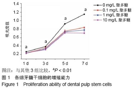
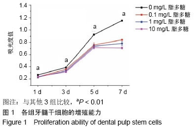
2.1 牙髓干细胞增殖能力 MTT法检测脂多糖对牙髓干细胞增殖能力的影响。结果显示,空白对照组从3 d开始高速增长呈对数生长曲线,5 d生长放缓呈平缓曲线,整个生长曲线呈“S”型。各质量浓度脂多糖刺激组细胞的吸光度值在第1,3,5,7天4个时间点均低于空白对照组(P < 0.01),差异有显著性意义,见图1。 "
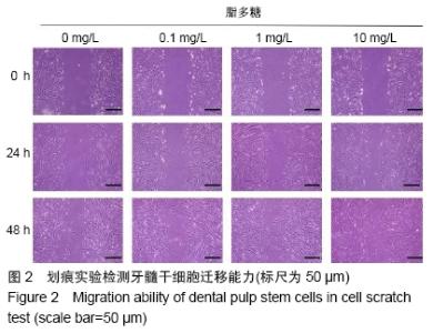
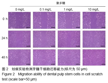
2.2 牙髓干细胞的迁移能力 2.2.1 划痕实验 各质量浓度脂多糖培养牙髓干细胞 24 h后,各组细胞均向划痕中央迁移,但各脂多糖刺激组细胞的迁移速度都高于空白对照组;培养48 h后,迁移速度的差异进一步增大,高质量浓度脂多糖组的划痕可以被牙髓干细胞覆盖,而空白对照组划痕依然清晰。细胞的迁移速度与脂多糖质量浓度呈正相关,见图2。 "
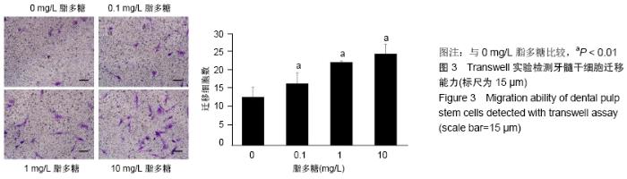
2.2.2 Transwell实验结果 各质量浓度脂多糖刺激牙髓干细胞24 h,细胞迁移的数量均高于空白对照组,并且随脂多糖质量浓度增高,迁移细胞数量增多,见图3。 "

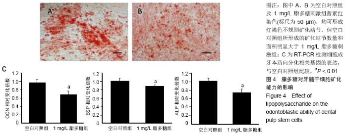
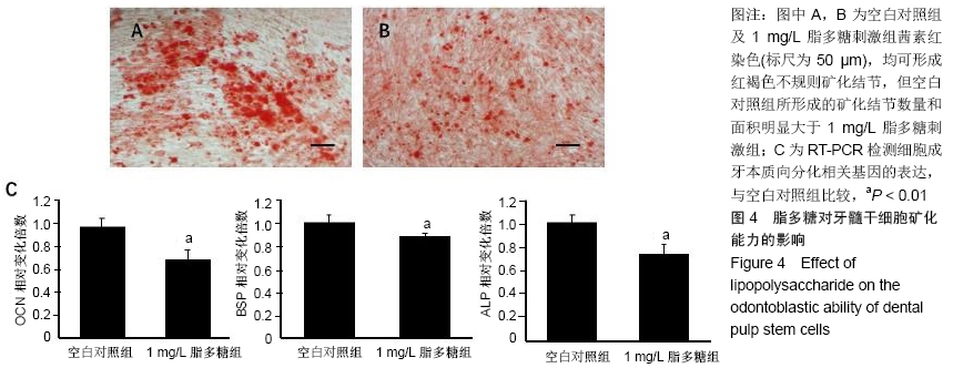
2.3 牙髓干细胞的成牙本质向分化能力 2.3.1 茜素红染色结果 牙髓干细胞矿化诱导21 d后,茜素红染色检测其矿化结节形成能力。空白对照组及脂多糖刺激组均可形成红褐色不规则矿化结节,但空白对照组所形成的矿化结节数量和面积明显大于脂多糖刺激组,见图4A,B。 2.3.2 RT-PCR检测成牙本质相关基因的表达变化 牙髓干细胞矿化诱导21 d后,RT-PCR技术检测细胞成牙本质向分化相关基因的表达。与空白对照组比较,脂多糖组细胞OCN、BSP、ALP表达量显著下调,提示脂多糖刺激牙髓干细胞后成牙本质向分化能力受到抑制,见图4C。 "
| [1] MA D, GAO J, YUE J, et al. Changes in proliferation and osteogenic differentiation of stem cells from deep caries in vitro. J Endod. 2012;38(6):796-802. [2] YANG G, JU Y, LIU S, et al. Lipopolysaccharide upregulates the proliferation, migration, and odontoblastic differentiation of NG2+ cells from human dental pulp in vitro. Cell Biol Int. 2019 Mar 7. doi: 10.1002/cbin.11127. [Epub ahead of print] [3] HORST OV, TOMPKINS KA, COATS SR, et al. TGF-beta1 Inhibits TLR-mediated odontoblast responses to oral bacteria. J Dent Res. 2009;88(4):333-338. [4] LIU Y, GAO Y, ZHAN X, et al. TLR4 activation by lipopolysaccharide and Streptococcus mutans induces differential regulation of proliferation and migration in human dental pulp stem cells. J Endod. 2014;40(9):1375-1381. [5] 刘影,高杰,吴补领.改良组织块酶消化法原代培养人牙髓干细胞的研究[J].口腔疾病防治,2018,26(3):166-170. [6] GRONTHOS S, MANKANI M, BRAHIM J, et al. Postnatal human dental pulp stem cells (DPSCs) in vitro and in vivo. Proc Natl Acad Sci U S A. 2000;97(25):13625-13630. [7] ARTHUR A, RYCHKOV G, SHI S, et al. Adult human dental pulp stem cells differentiate toward functionally active neurons under appropriate environmental cues. Stem Cells. 2008;26(7):1787-1795. [8] KIM JK, BAKER J, NOR JE, et al. mTor plays an important role in odontoblast differentiation. J Endod. 2011;37(8): 1081-1085. [9] SUH JS, KIM KS, LEE JY, et al. A cell-permeable fusion protein for the mineralization of human dental pulp stem cells. J Dent Res. 2012;91(1):90-96. [10] NAM S, WON JE, KIM CH, et al. Odontogenic differentiation of human dental pulp stem cells stimulated by the calcium phosphate porous granules. J Tissue Eng. 2011;2011:812547. [11] GRONTHOS S, BRAHIM J, LI W, et al. Stem cell properties of human dental pulp stem cells. J Dent Res. 2002;81(8):531-535. [12] WANG CY, TANI-ISHII N, STASHENKO P. Bone-resorptive cytokine gene expression in periapical lesions in the rat. Oral Microbiol Immunol. 1997;12(2):65-71. [13] ZHANG X, NING T, WANG H, et al. Stathmin regulates the proliferation and odontoblastic/osteogenic differentiation of human dental pulp stem cells through Wnt/β-catenin signaling pathway. J Proteomics. 2019;202:103364. [14] CHEN Y, GAO Y, TAO Y, et al. Identification of a Calcium-sensing Receptor in Human Dental Pulp Cells That Regulates Mineral Trioxide Aggregate-induced Mineralization. J Endod.2019;45(7):907-916. [15] JIANG HW, ZHANG W, REN BP, et al. Expression of toll like receptor 4 in normal human odontoblasts and dental pulp tissue. J Endod. 2006;32(8):747-751. [16] MUTOH N, TANI-ISHII N, TSUKINOKI K, et al. Expression of toll-like receptor 2 and 4 in dental pulp. J Endod. 2007;33(10): 1183-1186. [17] ZHANG J, WU L, QU JM. Inhibited proliferation of human lung fibroblasts by LPS is through IL-6 and IL-8 release. Cytokine. 2011;54(3):289-295. [18] SARRAZY V, VEDRENNE N, BILLET F, et al. TLR4 signal transduction pathways neutralize the effect of Fas signals on glioblastoma cell proliferation and migration. Cancer Lett. 2011;311(2):195-202. [19] WANG YY, SUN SP, ZHU HS, et al. GABA regulates the proliferation and apoptosis of MAC-T cells through the LPS-induced TLR4 signaling pathway. Res Vet Sci. 2018; 118:395-402. [20] YU L, ZHAO Y, GU X, et al. Dual effect of LPS on murine myeloid leukemia cells: Pro-proliferation and anti-proliferation. Exp Cell Res. 2016;344(2):210-218. [21] PARK YD, KIM YS, JUNG YM, et al. Porphyromonas gingivalis lipopolysaccharide regulates interleukin (IL)-17 and IL-23 expression via SIRT1 modulation in human periodontal ligament cells. Cytokine. 2012;60(1):284-293. [22] LI K, LV G, PAN L. Sirt1 alleviates LPS induced inflammation of periodontal ligament fibroblasts via downregulation of TLR4. Int J Biol Macromol. 2018;119:249-254. [23] BOTERO TM, SHELBURNE CE, HOLLAND GR, et al. TLR4 mediates LPS-induced VEGF expression in odontoblasts. J Endod. 2006;32(10):951-955. [24] NAKAO J, FUJII Y, KUSUYAMA J, et al. Low-intensity pulsed ultrasound (LIPUS) inhibits LPS-induced inflammatory responses of osteoblasts through TLR4-MyD88 dissociation. Bone. 2014;58:17-25. [25] 刘影,高杰,吴补领.脂多糖与变形链球菌刺激DPSCs中Toll样受体4的表达[J].中国组织工程研究,2018,22(33):5333-5337. [26] COIL J, TAM E, WATERFIELD JD. Proinflammatory cytokine profiles in pulp fibroblasts stimulated with lipopolysaccharide and methyl mercaptan. J Endod. 2004;30(2):88-91. [27] NAGAOKA S, TOKUDA M, SAKUTA T, et al. Interleukin-8 gene expression by human dental pulp fibroblast in cultures stimulated with Prevotella intermedia lipopolysaccharide. J Endod.1996;22(1):9-12. [28] TOKUDA M, SAKUTA T, FUSHUKU A, et al. Regulation of interleukin-6 expression in human dental pulp cell cultures stimulated with Prevotella intermedia lipopolysaccharide. J Endod. 2001;27(4):273-277. [29] ADACHI T, NAKANISHI T, YUMOTO H, et al. Caries-related bacteria and cytokines induce CXCL10 in dental pulp. J Dent Res. 2007;86(12):1217-1222. [30] PARK SH, HSIAO GY, HUANG GT. Role of substance P and calcitonin gene-related peptide in the regulation of interleukin-8 and monocyte chemotactic protein-1 expression in human dental pulp. Int Endod J. 2004;37(3):185-192. [31] SILVA AC, FARIA MR, FONTES A, et al. Interleukin-1 beta and interleukin-8 in healthy and inflamed dental pulps. J Appl Oral Sci. 2009;17(5):527-532. [32] LIU J, XU D, WANG Q, et al. LPS induced miR-181a promotes pancreatic cancer cell migration via targeting PTEN and MAP2K4. Dig Dis Sci. 2014;59(7):1452-1460. [33] CHEN HJ, LIANG TM, LEE IJ, et al. Scutellariae radix suppresses LPS-induced liver endothelial cell activation and inhibits hepatic stellate cell migration. J Ethnopharmacol. 2013;150(3):835-842. [34] HORTELANO S, LÓPEZ-FONTAL R, TRAVÉS PG, et al. ILK mediates LPS-induced vascular adhesion receptor expression and subsequent leucocyte trans-endothelial migration. Cardiovasc Res. 2010;86(2):283-292. [35] LIU J, XU D, WANG Q, et al. LPS induced miR-181a promotes pancreatic cancer cell migration via targeting PTEN and MAP2K4. Dig Dis Sci. 2014;59(7):1452-1460. [36] PETYAEV IM, ZIGANGIROVA NA, KOBETS NV, et al. Roquefort cheese proteins inhibit Chlamydia pneumoniae propagation and LPS-induced leukocyte migration. Scientific World Journal. 2013;2013:140591. [37] WANG N, MENG X, LIU Y, et al. LPS promote Osteosarcoma invasion and migration through TLR4/HOTAIR.Gene. 2019; 680:1-8. [38] LI D, FU L, ZHANG Y, et al. The effects of LPS on adhesion and migration of human dental pulp stem cells in vitro. J Dent. 2014;42(10):1327-1334. [39] PARK JH, KWON SM, YOON HE, et al. Lipopolysaccharide promotes adhesion and migration of murine dental papilla-derived MDPC-23 cells via TLR4. Int J Mol Med. 2011; 27(2):277-281. [40] 刘影,高杰,吴补领. TLR4 在人健康和深龋牙髓组织中的定位表达研究[J].口腔疾病防治,2017,25(3):153-158. [41] MORSCZECK CO, DREES J, GOSAU M. Lipopolysaccharide from Escherichia coli but not from Porphyromonas gingivalis induce pro-inflammatory cytokines and alkaline phosphatase in dental follicle cells. Arch Oral Biol. 2012;57(12):1595-1601. [42] GUO C, YUAN L, WANG JG, et al. Lipopolysaccharide (LPS) induces the apoptosis and inhibits osteoblast differentiation through JNK pathway in MC3T3-E1 cells. Inflammation. 2014; 37(2):621-631. [43] PEI J, FAN L, NAN K, et al. Excessive Activation of TLR4/NF-κB Interactively Suppresses the Canonical Wnt/β-catenin Pathway and Induces SANFH in SD Rats. Sci Rep. 2017;7(1):11928. [44] YU H, ZHANG X, SONG W, et al. Effects of 3-dimensional Bioprinting Alginate/Gelatin Hydrogel Scaffold Extract on Proliferation and Differentiation of Human Dental Pulp Stem Cells. J Endod. 2019;45(6):706-715. |
| [1] | Li Cai, Zhao Ting, Tan Ge, Zheng Yulin, Zhang Ruonan, Wu Yan, Tang Junming. Platelet-derived growth factor-BB promotes proliferation, differentiation and migration of skeletal muscle myoblast [J]. Chinese Journal of Tissue Engineering Research, 2021, 25(7): 1050-1055. |
| [2] | Liu Cong, Liu Su. Molecular mechanism of miR-17-5p regulation of hypoxia inducible factor-1α mediated adipocyte differentiation and angiogenesis [J]. Chinese Journal of Tissue Engineering Research, 2021, 25(7): 1069-1074. |
| [3] | Ma Zetao, Zeng Hui, Wang Deli, Weng Jian, Feng Song. MicroRNA-138-5p regulates chondrocyte proliferation and autophagy [J]. Chinese Journal of Tissue Engineering Research, 2021, 25(5): 674-678. |
| [4] | Wang Yujiao, Liu Dan, Sun Song, Sun Yong. Biphasic calcium phosphate loaded with advanced platelet rich fibrin can promote the activity of rabbit bone marrow mesenchymal stem cells [J]. Chinese Journal of Tissue Engineering Research, 2021, 25(4): 504-509. |
| [5] | Zhou Jihui, Yao Meng, Wang Yansong, Li Xinzhi, Zhou You, Huang Wei, Chen Wenyao. Influence of novel nanoscaffolds on biological behaviors of neural stem cells and the related gene expression [J]. Chinese Journal of Tissue Engineering Research, 2021, 25(4): 532-536. |
| [6] | Chen Yang, Huang Denggao, Gao Yuanhui, Wang Shunlan, Cao Hui, Zheng Linlin, He Haowei, Luo Siqin, Xiao Jingchuan, Zhang Yingai, Zhang Shufang. Low-intensity pulsed ultrasound promotes the proliferation and adhesion of human adipose-derived mesenchymal stem cells [J]. Chinese Journal of Tissue Engineering Research, 2021, 25(25): 3949-3955. |
| [7] | Tang Xiaokai, Li Weiming. Role and mechanism of Nel-like molecule-1 in promoting bone fusion after spinal fusion [J]. Chinese Journal of Tissue Engineering Research, 2021, 25(24): 3914-3920. |
| [8] | Zhou Wu, Wang Binping, Wang Yawen, Cheng Yanan, Huang Xieshan. Transforming growth factor beta combined with bone morphogenetic protein-2 induces the proliferation and differentiation of mouse MC3T3-E1 cells [J]. Chinese Journal of Tissue Engineering Research, 2021, 25(23): 3630-3635. |
| [9] | Li Xinping, Cui Qiuju, Zeng Shuguang, Ran Gaoying, Zhang Zhaoqiang, Liu Xianwen, Fang Wei, Xu Shuaimei. Effect of modification of β-tricalcium phosphate/chitosan hydrogel on growth and mineralization of dental pulp stem cells [J]. Chinese Journal of Tissue Engineering Research, 2021, 25(22): 3493-3499. |
| [10] | Yu Chengshuai, Du Gang, Pang Shenning, Lao Shan. Chemerin, a pro-inflammatory adipokine, regulates chondrocyte proliferation and metabolism by increasing production of nitric oxide [J]. Chinese Journal of Tissue Engineering Research, 2021, 25(2): 258-263. |
| [11] | Dai Yaling, Chen Lewen, He Xiaojun, Lin Huawei, Jia Weiwei, Chen Lidian, Tao Jing, Liu Weilin. Construction of miR-146b overexpression lentiviral vector and the effect on the proliferation of hippocampal neural stem cells [J]. Chinese Journal of Tissue Engineering Research, 2021, 25(19): 3024-3030. |
| [12] | Ailimaierdan·Ainiwaer, Wang Ling, Gu Li, Dilidaer•Taxifulati, Wang Shan, Yin Hongbin. Effect of transforming growth factor-beta3 on the proliferation and osteogenic capability of osteoblasts [J]. Chinese Journal of Tissue Engineering Research, 2021, 25(17): 2664-2669. |
| [13] | Wang Renxian, Cao Jingjing, Wang Honggang, Wan Ben, Liu Weifeng. Effects of dispersants on aggregation, intracellular distribution and cell proliferation of nano-hydroxyapatite [J]. Chinese Journal of Tissue Engineering Research, 2021, 25(16): 2500-2505. |
| [14] | Jiang Tao, Wu Shuo, Li Zhiqiang, Shou Xi, Mayire·Nuermaimaiti, Ma Chuang, Wei Qin. Platelet-derived growth factor BB promotes the proliferation of bone marrow mesenchymal stem cells of Sprague-Dawley rats [J]. Chinese Journal of Tissue Engineering Research, 2021, 25(13): 1976-1981. |
| [15] | Yan Nan, Si Xiaofeng, Zeng Liang, Tian Wei, Shan Guangdong, Xiong Lishuo, Yang Weijie, Wang Zhengdong. Nerve growth factor interferes with proliferation and alpha-actin expression of skeletal muscle satellite cells in rats [J]. Chinese Journal of Tissue Engineering Research, 2021, 25(13): 2030-2035. |
| Viewed | ||||||
|
Full text |
|
|||||
|
Abstract |
|
|||||