Chinese Journal of Tissue Engineering Research ›› 2017, Vol. 21 ›› Issue (6): 940-945.doi: 10.3969/j.issn.2095-4344.2017.06.021
Previous Articles Next Articles
Biocompatibility of electrospun carbon nanotubes/poly(L-lactic acid) nanofiber scaffolds with mouse neural stem cells
- Department of Spine Surgery, the Third Affiliated Hospital, Sun Yat-sen University, Guangzhou 510630, Guangdong Province, China
-
Received:2017-02-07Online:2017-02-28Published:2017-03-16 -
Contact:Liu Bin, M.D., Chief physician, Department of Spine Surgery, the Third Affiliated Hospital, Sun Yat-sen University, Guangzhou 510630, Guangdong Province, China -
About author:Lin Cheng-kai, Master, Department of Spine Surgery, the Third Affiliated Hospital, Sun Yat-sen University, Guangzhou 510630, Guangdong Province, China -
Supported by:the National Natural Science Foundation of China, No. 31170947, 31470949, 81472122; Guangdong Natural Science Foundation, No. S2012020011099, S2013010016413; Guangdong Science and Technology Planning Project, No. 2012B060300008; Guangzhou Science and Technology Planning Project, No. 2013J4100062; New Teacher’s Fund for Doctor Stations of Ministry of Education of China, No. 20100171120088
CLC Number:
Cite this article
Lin Cheng-kai, Rong Li-min, Liu Bin. Biocompatibility of electrospun carbon nanotubes/poly(L-lactic acid) nanofiber scaffolds with mouse neural stem cells[J]. Chinese Journal of Tissue Engineering Research, 2017, 21(6): 940-945.
share this article
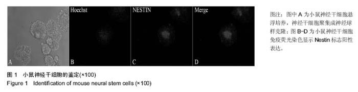
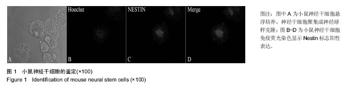
2.1 小鼠神经干细胞的分离培养鉴定结果 从胚鼠中分离得到的小鼠神经干细胞悬浮培养聚集成球。传代培养数次后,光镜下观察可见神经球样克隆形成(图1A)。免疫荧光染色鉴定神经干细胞,可见Nestin染色呈阳性(图1B-D)。"

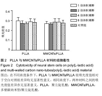
2.2 细胞毒性检测结果 不同浓度条件下,PLLA组和MWCNTs/PLLA组吸光度值相对于空白对照组差异均无显著性意义(P > 0.05)。相同浓度下,两种材料之间的吸光度值差异无显著性意义(P > 0.05,图2)。"
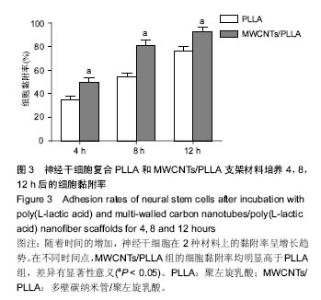
2.3 细胞黏附性 随着培养时间增加,两组材料的细胞黏附率均明显增多。在不同时间点, MWCNTs/CNT的细胞黏附率明显高于PLLA(P < 0.05,图3)。"

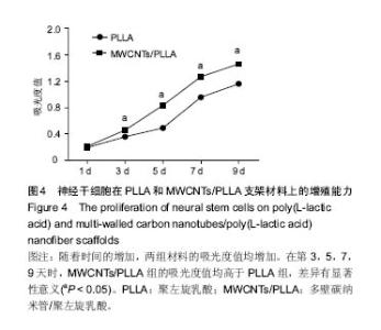
2.4 细胞增殖能力 随着培养时间的延长,两组材料的吸光度值均增高。在第3,5,7,9天时,MWCNTs/PLLA组的吸光度值均高于PLLA组(P < 0.05,图4)。"


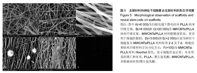
2.5 扫描电镜观察及Hoechst 33342染色结果 扫描电镜结果显示,PLLA材料表面较光滑(图5A),多壁碳纳米管修饰后,MWCNTs/PLLA表面变得粗糙(图5B,C),更有利于细胞的黏附,神经干细胞种植于材料表面,细胞形态正常,且与材料黏附紧密(图5D),神经干细胞复合材料分化培养 7 d后,扫描电镜显示细胞沿着纳米纤维材料方向分化生长(图5E),Hoechst染色显示细胞形态正常,未见明显的凋亡和坏死(图5F)。"

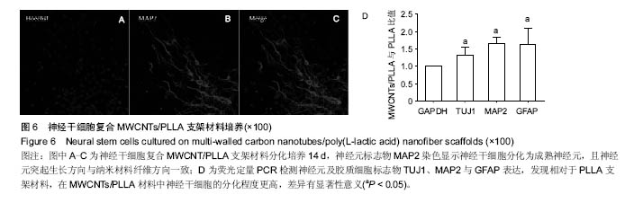
2.6 神经干细胞复合MWCNTs/PLLA分化培养结果 神经干细胞复合MWCNTs/PLLA分化培养14 d后,成熟神经元特异性标志物MAP2染色显示神经干细胞能在材料表面生存及分化为成熟神经元,且纳米材料纤维有引导神经元突起生长方向的作用(图6)。荧光定量PCR检测神经元及胶质细胞标志物TUJ1、MAP2与GFAP,发现在MWCNTs/ PLLA材料中,神经干细胞分化为神经元及星形胶质细胞程度更高(图6)。"
| [1]Nichols CM, Brenner MJ, Fox IK, et al. Effects of motor versus sensory nerve grafts on peripheral nerve regeneration. Exp Neurol. 2004;190(2):347-355.[2]IJkema-Paassen J, Jansen K, Gramsbergen A, et al. Transection of peripheral nerves, bridging strategies and effect evaluation. Biomaterials. 2004;25(9):1583-1592.[3]Houle JD, Tessler A. Repair of chronic spinal cord injury. Exp Neurol. 2003;182(2):247-260.[4]Evans NR, Davies EM, Dare CJ, et al. Tissue engineering strategies in spinal arthrodesis: the clinical imperative and challenges to clinical translation. Regen Med. 2013;8(1):49-64.[5]Quarto R, Giannoni P. Bone Tissue Engineering: Past-Present-Future. Methods Mol Biol. 2016;1416:21-33.[6]Wen X, Wang Y, Guo Z, et al. Cauda equina-derived extracellular matrix for fabrication of nanostructured hybrid scaffolds applied to neural tissue engineering.Tissue Eng Part A. 2015;21(5-6):1095-1105.[7]Bhang SH, Lim JS, Choi CY, et al. The behavior of neural stem cells on biodegradable synthetic polymers. J Biomater Sci Polym Ed. 2007;18(2):223-239.[8]Bini TB, Gao S, Xu X, et al. Peripheral nerve regeneration by microbraided poly(L-lactide-co-glycolide) biodegradable polymer fibers. J Biomed Mater Res A. 2004;68(2):286-295.[9]Xu CY, Inai R, Kotaki M, et al. Aligned biodegradable nanofibrous structure: a potential scaffold for blood vessel engineering. Biomaterials. 2004;25(5):877-886.[10]Hurtado A, Cregg JM, Wang HB, et al. Robust CNS regeneration after complete spinal cord transection using aligned poly-L-lactic acid microfibers. Biomaterials. 2011; 32(26):6068-6079.[11]Wang HB, Mullins ME, Cregg JM, et al. Varying the diameter of aligned electrospun fibers alters neurite outgrowth and Schwann cell migration. Acta Biomater. 2010;6(8):2970-2978.[12]Yu Y, Lü X, Ding F. Influence of Poly(L-Lactic Acid) Aligned Nanofibers on PC12 Differentiation.J Biomed Nanotechnol. 2015;11(5):816-827.[13]Yu Y, Lü X, Ding F. microRNA regulatory mechanism by which PLLA aligned nanofibers influence PC12 cell differentiation. J Neural Eng. 2015;12(4):046010.[14]Jahno VD, Ribeiro GB, dos Santos LA, et al. Chemical synthesis and in vitro biocompatibility tests of poly (L-lactic acid). J Biomed Mater Res A. 2007;83(1):209-215.[15]Gong Y, Zhu Y, Liu Y, et al. Layer-by-layer assembly of chondroitin sulfate and collagen on aminolyzed poly(L-lactic acid) porous scaffolds to enhance their chondrogenesis. Acta Biomater. 2007;3(5):677-685.[16]MacDonald RA, Voge CM, Kariolis M, et al. Carbon nanotubes increase the electrical conductivity of fibroblast-seeded collagen hydrogels. Acta Biomater. 2008; 4(6):1583-1592.[17]Zhang Z, Rouabhia M, Wang Z, et al. Electrically conductive biodegradable polymer composite for nerve regeneration: electricity-stimulated neurite outgrowth and axon regeneration. Artif Organs. 2007;31(1):13-22.[18]He L, Zhao P, Han Q, et al. Surface modification of poly-l-lactic acid fibrous scaffolds by a molecular-designed multi-walled carbon nanotube multilayer for enhancing cell interactions. Carbon. 2013; 56: 224-234.[19]Sun X, Shao H, Xiang K, et al. Poly(dopamine)-modified carbon nanotube multilayered film and its effects on macrophages. Carbon. 2016; 113:176-191.[20]Brewer GJ, Torricelli JR. Isolation and culture of adult neurons and neurospheres. Nat Protoc. 2007;2(6):1490-1498.[21]Ziv Y, Avidan H, Pluchino S, et al. Synergy between immune cells and adult neural stem/progenitor cells promotes functional recovery from spinal cord injury. Proc Natl Acad Sci U S A. 2006;103(35):13174-13179.[22]Darsalia V, Kallur T, Kokaia Z. Survival, migration and neuronal differentiation of human fetal striatal and cortical neural stem cells grafted in stroke-damaged rat striatum. Eur J Neurosci. 2007;26(3):605-614.[23]Paul C, Samdani AF, Betz RR, et al. Grafting of human bone marrow stromal cells into spinal cord injury: a comparison of delivery methods. Spine (Phila Pa 1976). 2009;34(4): 328-334.[24]Li J, Shi R. Fabrication of patterned multi-walled poly-l-lactic acid conduits for nerve regeneration. J Neurosci Methods. 2007;165(2):257-264.[25]Lai BQ, Wang JM, Ling EA, et al. Graft of a tissue-engineered neural scaffold serves as a promising strategy to restore myelination after rat spinal cord transection. Stem Cells Dev. 2014;23(8):910-921.[26]倪石磊,齐宏旭,鲍圣德,等.组织工程支架在犬急性脊髓损伤修复中应用的初步研究[J].中国微侵袭神经外科杂志, 2005,10(9): 404-407.[27]Kabiri M, Oraee-Yazdani S, Shafiee A, et al. Neuroregenerative effects of olfactory ensheathing cells transplanted in a multi-layered conductive nanofibrous conduit in peripheral nerve repair in rats. J Biomed Sci. 2015; 22:35.[28]He L, Liao S, Quan D, et al. Synergistic effects of electrospun PLLA fiber dimension and pattern on neonatal mouse cerebellum C17.2 stem cells. Acta Biomater. 2010;6(8): 2960-2969.[29]Kabiri M, Oraee-Yazdani S, Dodel M, et al. Cytocompatibility of a conductive nanofibrous carbon nanotube/poly (L-Lactic acid) composite scaffold intended for nerve tissue engineering. EXCLI J. 2015;14:851-860.[30]Yoo HS, Kim TG, Park TG. Surface-functionalized electrospun nanofibers for tissue engineering and drug delivery. Adv Drug Deliv Rev. 2009;61(12):1033-1042.[31]Feng ZQ, Wang T, Zhao B, et al. Soft Graphene Nanofibers Designed for the Acceleration of Nerve Growth and Development. Adv Mater. 2015;27(41):6462-6468.[32]Kabiri M, Soleimani M, Shabani I, et al. Neural differentiation of mouse embryonic stem cells on conductive nanofiber scaffolds. Biotechnol Lett. 2012;34(7):1357-1365.[33]Lovat V, Pantarotto D, Lagostena L, et al. Carbon nanotube substrates boost neuronal electrical signaling. Nano Lett. 2005;5(6):1107-1110.[34]Ogawa Y, Sawamoto K, Miyata T, et al. Transplantation of in vitro-expanded fetal neural progenitor cells results in neurogenesis and functional recovery after spinal cord contusion injury in adult rats. J Neurosci Res. 2002;69(6): 925-933.[35]Piltti KM, Salazar DL, Uchida N, et al. Safety of human neural stem cell transplantation in chronic spinal cord injury Stem Cells Transl Med. 2013;2(12):961-974.[36]Tavares I. Human neural stem cell transplantation in spinal cord injury models: how far from clinical application. Stem Cell Res Ther. 2013;4(3):61.Lu Y, Wang MY. Neural stem cell grafts for complete spinal cord injury. Neurosurgery. 20 |
| [1] | Yao Xiaoling, Peng Jiancheng, Xu Yuerong, Yang Zhidong, Zhang Shuncong. Variable-angle zero-notch anterior interbody fusion system in the treatment of cervical spondylotic myelopathy: 30-month follow-up [J]. Chinese Journal of Tissue Engineering Research, 2022, 26(9): 1377-1382. |
| [2] | Zhang Jinglin, Leng Min, Zhu Boheng, Wang Hong. Mechanism and application of stem cell-derived exosomes in promoting diabetic wound healing [J]. Chinese Journal of Tissue Engineering Research, 2022, 26(7): 1113-1118. |
| [3] | An Weizheng, He Xiao, Ren Shuai, Liu Jianyu. Potential of muscle-derived stem cells in peripheral nerve regeneration [J]. Chinese Journal of Tissue Engineering Research, 2022, 26(7): 1130-1136. |
| [4] | Fan Yiming, Liu Fangyu, Zhang Hongyu, Li Shuai, Wang Yansong. Serial questions about endogenous neural stem cell response in the ependymal zone after spinal cord injury [J]. Chinese Journal of Tissue Engineering Research, 2022, 26(7): 1137-1142. |
| [5] | Yang Sidi, Wang Qian, Xu Nuo, Wang Ronghan, Jin Chuanqi, Lu Ying, Dong Ming. Biodentine enhances the proliferation and differentiation of osteoblasts through upregulating bone morphogenetic protein-2 [J]. Chinese Journal of Tissue Engineering Research, 2022, 26(4): 516-520. |
| [6] | Le Guoping, Zhang Ming, Xi Licheng, Luo Hanwen. Preparation and in vitro evaluation of vancomycin hydrochloride@polylactic acid-glycolic acid copolymer-chitosan-hyaluronic acid composite sustained-release microspheres [J]. Chinese Journal of Tissue Engineering Research, 2022, 26(4): 528-534. |
| [7] | Chen Shuo, Xiao Dongqin, Li Xingping, Ran Bin, Shi Feng, Zhang Chengdong, Deng Li, Huang Nanxiang, Liu Kang, Feng Gang, Duan Ke. Preparation and characterization of tantalum functional coating on titanium implant [J]. Chinese Journal of Tissue Engineering Research, 2022, 26(4): 546-552. |
| [8] | He Yunying, Li Lingjie, Zhang Shuqi, Li Yuzhou, Yang Sheng, Ji Ping. Method of constructing cell spheroids based on agarose and polyacrylic molds [J]. Chinese Journal of Tissue Engineering Research, 2022, 26(4): 553-559. |
| [9] | He Guanyu, Xu Baoshan, Du Lilong, Zhang Tongxing, Huo Zhenxin, Shen Li. Biomimetic orientated microchannel annulus fibrosus scaffold constructed by silk fibroin [J]. Chinese Journal of Tissue Engineering Research, 2022, 26(4): 560-566. |
| [10] | Chen Xiaoxu, Luo Yaxin, Bi Haoran, Yang Kun. Preparation and application of acellular scaffold in tissue engineering and regenerative medicine [J]. Chinese Journal of Tissue Engineering Research, 2022, 26(4): 591-596. |
| [11] | Kang Kunlong, Wang Xintao. Research hotspot of biological scaffold materials promoting osteogenic differentiation of bone marrow mesenchymal stem cells [J]. Chinese Journal of Tissue Engineering Research, 2022, 26(4): 597-603. |
| [12] | Shen Jiahua, Fu Yong. Application of graphene-based nanomaterials in stem cells [J]. Chinese Journal of Tissue Engineering Research, 2022, 26(4): 604-609. |
| [13] | Zhang Tong, Cai Jinchi, Yuan Zhifa, Zhao Haiyan, Han Xingwen, Wang Wenji. Hyaluronic acid-based composite hydrogel in cartilage injury caused by osteoarthritis: application and mechanism [J]. Chinese Journal of Tissue Engineering Research, 2022, 26(4): 617-625. |
| [14] | Li Hui, Chen Lianglong. Application and characteristics of bone graft materials in the treatment of spinal tuberculosis [J]. Chinese Journal of Tissue Engineering Research, 2022, 26(4): 626-630. |
| [15] | Gao Cangjian, Yang Zhen, Liu Shuyun, Li Hao, Fu Liwei, Zhao Tianyuan, Chen Wei, Liao Zhiyao, Li Pinxue, Sui Xiang, Guo Quanyi. Electrospinning for rotator cuff repair [J]. Chinese Journal of Tissue Engineering Research, 2022, 26(4): 637-642. |
| Viewed | ||||||
|
Full text |
|
|||||
|
Abstract |
|
|||||