Chinese Journal of Tissue Engineering Research ›› 2016, Vol. 20 ›› Issue (50): 7460-7468.doi: 10.3969/j.issn.2095-4344.2016.50.002
Previous Articles Next Articles
Human umbilical cord mesenchymal stem cells promote the proliferation of HepG-2 cells through interleukin-6/STAT3 signaling pathway
Zheng Sheng1, Yang Juan1, Chen Wen-qin2, Liu Jing2, Zhang Fan1, Wang Yu-bo1
- 1Department of Digestive Diseases, Third People’s Hospital of Yunnan Province, Kunming 650011, Yunnan Province, China
2Department of Obstetrics, Dong Fang Hospital of Kunming, Kunming 650000, Yunnan Province, China
-
Revised:2016-10-09Online:2016-12-02Published:2016-12-02 -
Contact:Yang Juan, Master, Attending physician, Department of Digestive Diseases, Third People’s Hospital of Yunnan Province, Kunming 650011, Yunnan Province, China -
About author:Zheng Sheng, Master, Attending physician, Department of Digestive Diseases, Third People’s Hospital of Yunnan Province, Kunming 650011, Yunnan Province, China -
Supported by:the Natural Science Foundation of Yunnan Province, No. 2012FD095; the Scientific Research Fund of Yunnan Provincial Educational Department, No. 2014Z125, 2015Z146; the Clinical Department Construction of Yunnan Province, No. [2015]18
CLC Number:
Cite this article
Zheng Sheng, Yang Juan, Chen Wen-qin, Liu Jing, Zhang Fan, Wang Yu-bo. Human umbilical cord mesenchymal stem cells promote the proliferation of HepG-2 cells through interleukin-6/STAT3 signaling pathway[J]. Chinese Journal of Tissue Engineering Research, 2016, 20(50): 7460-7468.
share this article
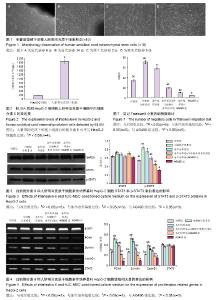
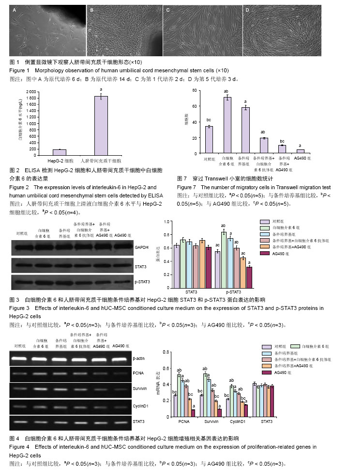
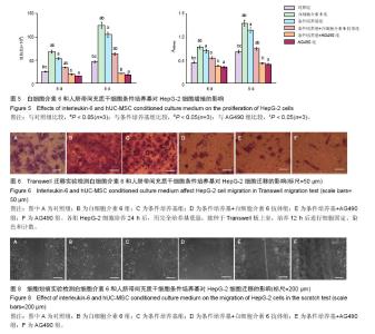
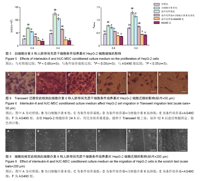
2.1 人脐带间充质干细胞的生物学特性 脐带组织原代培养3 d后,可见少许细胞贴壁生长,细胞多呈短梭形;培养7-10 d后,细胞聚集融合成单层,细胞形态大多为长梭形或多角形;传代至第3、4代,细胞形态均一,胞质透光性良好,细胞形态类似成纤维细胞,呈漩涡状生长(图1)。流式细胞仪检测第5代细胞高表达CD29、CD90、CD105(> 95%),极低表达CD19(0.1%),符合间充质干细胞鉴定标准。 2.2 白细胞介素6和人脐带间充质干细胞条件培养基对HepG-2细胞STAT3和p-STAT3蛋白表达的影响 ELISA结果提示人脐带间充质干细胞条件培养基中白细胞介素6水平为(1 856.2±96.6) ng/L,HepG-2细胞白细胞介素6表达水平仅为(180.8±35.5) ng/L,两组相比较,差异有显著性意义(P < 0.05,图2)。 20 μg/L重人白细胞介素6及40%人脐带间充质干细胞条件培养基分别作用于HepG-2细胞30 min后,p-STAT3表达明显升高。白细胞介素6中和抗体加入条件培养基后STAT3的活性被抑制,同时p-STAT3的表达明显下降。JAK2特异性抑制剂AG490显著抑制了HepG-2细胞STAT3的活化并降低了条件培养基对STAT3的激活作用,组间比较差异有显著性意义(P < 0.05)。各组细胞STAT3蛋白表达差异无显著性意义(P > 0.05,图3)。 2.3 白细胞介素6和人脐带间充质干细胞条件培养基对HepG-2细胞增殖相关基因表达的影响 RT-PCR检测结果显示:条件培养基组增殖相关基因PCNA、CyclinD1、Survivin表达水平均明显增高,白细胞介素6中和抗体可明显减弱条件培养基的促增殖基因表达作用,与对照组相比差异均有显著性意义(P < 0.05)。AG490加入条件培养基后,HepG-2细胞的PCNA、CyclinD1、Survivin mRNA表达明显下调,与对照组相比差异均有显著性意义(P < 0.05)。重组白细胞介素6对各增殖相关基因均具有显著的上调作用,差异均有显著性意义(P < 0.05)。STAT3基因的转录水平在各处理因素作用24 h后,未见明显变化(图4)。 2.4 白细胞介素6和人脐带间充质干细胞条件培养基对HepG-2细胞增殖的影响 细胞计数实验及CCK-8实验结果显示外源性白细胞介素6及人脐带间充质干细胞条件培养基均能促进HepG-2细胞增殖,与对照组相比差异均有显著性意义(P < 0.05)。AG490可明显延缓了HepG-2细胞的增殖,加入条件培养基后HepG-2细胞增殖与其他组比较差异仍有显著性意义(P < 0.05,图5)。 2.5 白细胞介素6和人脐带间充质干细胞条件培养基对HepG-2细胞迁移的影响 20 μg/L重组白细胞介素6和40%人脐带间充质干细胞条件培养基对HepG-2细胞分别预处理24 h,细胞迁移能力明显增强,与对照组相比差异均有显著性意义(P < 0.05);白细胞介素6中和抗体加入条件培养基后,HepG-2细胞迁移能力下降,与对照组相比差异有显著性意义(P < 0.05);条件培养基+ AG490组穿过聚碳酸酯膜的细胞数量明显少于对照组及条件培养基组,组间比较差异显著性意义(P < 0.05,图6,图7)。划痕实验结果显示,白细胞介素6及人脐带间充质干细胞条件培养基均能提高HepG-2细胞的损伤愈合能力,而条件培养基中的白细胞介素6被中和后,细胞的愈合能力明显下降,与Transwell实验结果一致。使用AG490抑制HepG-2细胞JAK2/STAT3信号通路后,无论人脐带间充质干细胞条件培养基是否存在,细胞的愈合能力均严重受损(图8)。"
"
| [1] De Miguel MP, Fuentes-Julián S, Blázquez-Martínez A, et al. Immunosuppressive properties of mesenchymal stem cells: advances and applications. Curr Mol Med. 2012;12(5):574-591.[2] Chamberlain G, Fox J, Ashton B, et al. Concise review: mesenchymal stem cells: their phenotype, differentiation capacity, immunological features, and potential for homing. Stem Cells. 2007;25(11): 2739-2749.[3] Uchibori R, Tsukahara T, Ohmine K, et al. Cancer gene therapy using mesenchymal stem cells. Int J Hematol. 2014;99(4):377-382.[4] Bolontrade MF, Sganga L, Piaggio E, et al. A specific subpopulation of mesenchymal stromal cell carriers overrides melanoma resistance to an oncolytic adenovirus. Stem Cells Dev. 2012;21(14):2689-2702.[5] Ryu CH, Park SH, Park SA, et al. Gene therapy of intracranial glioma using interleukin 12-secreting human umbilical cord blood-derived mesenchymal stem cells. Hum Gene Ther. 2011;22(6):733-743.[6] Petsa A, Gargani S, Felesakis A, et al. Effectiveness of protocol for the isolation of Wharton's Jelly stem cells in large-scale applications. In Vitro Cell Dev Biol Anim. 2009;45(10):573-576.[7] Prasanna SJ, Gopalakrishnan D, Shankar SR, et al. Pro-inflammatory cytokines, IFNgamma and TNFalpha, influence immune properties of human bone marrow and Wharton jelly mesenchymal stem cells differentially. PLoS One. 2010;5(2):e9016.[8] Baksh D, Yao R, Tuan RS. Comparison of proliferative and multilineage differentiation potential of human mesenchymal stem cells derived from umbilical cord and bone marrow. Stem Cells. 2007;25(6):1384-1392.[9] Friedman R, Betancur M, Boissel L, et al. Umbilical cord mesenchymal stem cells: adjuvants for human cell transplantation. Biol Blood Marrow Transplant. 2007;13(12):1477-1486.[10] Candido J, Hagemann T. Cancer-related inflammation. J Clin Immunol. 2013;33 Suppl 1:S79-84.[11] Liu X, Wang J, Wang H, et al. REG3A accelerates pancreatic cancer cell growth under IL-6-associated inflammatory condition: Involvement of a REG3A-JAK2/STAT3 positive feedback loop. Cancer Lett. 2015;362(1):45-60.[12] Garbers C, Aparicio-Siegmund S, Rose-John S. The IL-6/gp130/STAT3 signaling axis: recent advances towards specific inhibition. Curr Opin Immunol. 2015; 34:75-82.[13] Mali SB. Review of STAT3 (Signal Transducers and Activators of Transcription) in head and neck cancer. Oral Oncol. 2015;51(6):565-569.[14] Salas S, Jiguet-Jiglaire C, Campion L, et al. Correlation between ERK1 and STAT3 expression and chemoresistance in patients with conventional osteosarcoma. BMC Cancer. 2014;14:606.[15] Raicevic G, Rouas R, Najar M, et al. Inflammation modifies the pattern and the function of Toll-like receptors expressed by human mesenchymal stromal cells. Hum Immunol. 2010;71(3):235-244.[16] Prockop DJ, Oh JY. Mesenchymal stem/stromal cells (MSCs): role as guardians of inflammation. Mol Ther. 2012;20(1):14-20.[17] Chanthra N, Payungporn S, Chuaypen N, et al. Single Nucleotide Polymorphisms in STAT3 and STAT4 and Risk of Hepatocellular Carcinoma in Thai Patients with Chronic Hepatitis B. Asian Pac J Cancer Prev. 2015; 16(18):8405-8410.[18] Cron L, Allen T, Febbraio MA. The role of gp130 receptor cytokines in the regulation of metabolic homeostasis. J Exp Biol. 2016;219(Pt 2):259-265.[19] Kim JE, Lee JY, Kang MJ, et al. Withaferin A Inhibits Helicobacter pylori-induced Production of IL-1β in Dendritic Cells by Regulating NF-κB and NLRP3 Inflammasome Activation. Immune Netw. 2015;15(6): 269-277.[20] Chang Q, Daly L, Bromberg J. The IL-6 feed-forward loop: a driver of tumorigenesis. Semin Immunol. 2014;26(1):48-53.[21] Bournazou E, Bromberg J. Targeting the tumor microenvironment: JAK-STAT3 signaling. JAKSTAT. 2013;2(2):e23828.[22] Rokavec M, Wu W, Luo JL. IL6-mediated suppression of miR-200c directs constitutive activation of inflammatory signaling circuit driving transformation and tumorigenesis. Mol Cell. 2012;45(6):777-789.[23] Meydan N, Grunberger T, Dadi H, et al. Inhibition of acute lymphoblastic leukaemia by a Jak-2 inhibitor. Nature. 1996;379(6566):645-648.[24] Teng Y, Ghoshal P, Ngoka L, et al. Critical role of the WASF3 gene in JAK2/STAT3 regulation of cancer cell motility. Carcinogenesis. 2013;34(9):1994-1999.[25] Wang H, Su X, Yang M, et al. Reciprocal control of miR-197 and IL-6/STAT3 pathway reveals miR-197 as potential therapeutic target for hepatocellular carcinoma. Oncoimmunology. 2015;4(10):e1031440.[26] Chanthra N, Payungporn S, Chuaypen N, et al. Association of Single Nucleotide Polymorphism rs1053004 in Signal Transducer and Activator of Transcription 3 (STAT3) with Susceptibility to Hepatocellular Carcinoma in Thai Patients with Chronic Hepatitis B. Asian Pac J Cancer Prev. 2015; 16(12):5069-5073.[27] 朱丽华,徐刚,龚爱华,等. STAT3抑制剂Stattic对肝癌细胞Bel-7402生长、迁移、侵袭和放射敏感性的影响[J].中华放射医学与防护杂志,2015,35(6):413-418.[28] 王亚群,韩秋菊,庞敏,等. 靶向阻断STAT3增强肝癌细胞H22对化疗药物阿霉素的敏感性[J].中国免疫学杂志 2015,31(10):1304-1309,1314.[29] 张毅,张君薇,谢淑丽,等.RNAi沉默STAT3基因联合mTOR抑制剂rapamycin诱导BEL-7402肝癌细胞凋亡[J].吉林大学学报:医学版,2013,39(5):903-908.[30] 张巨波,郭坤,陈漪,等.信号转导转录激活因子3抑制剂WP1066对肝细胞肝癌生物学特性影响的研究[J].中国临床医学,2012,19(4):339-342.[31] Li HJ, Sun QM, Liu LZ. High expression of IL-9R promotes the progression of human hepatocellular carcinoma and indicates a poor clinical outcome. Oncol Rep. 2015;34(2):795-802.[32] Yang YM, Lee CG, Koo JH, et al. Gα12 overexpressed in hepatocellular carcinoma reduces microRNA-122 expression via HNF4α inactivation, which causes c-Met induction. Oncotarget. 2015;6(22):19055-19069. |
| [1] | Pu Rui, Chen Ziyang, Yuan Lingyan. Characteristics and effects of exosomes from different cell sources in cardioprotection [J]. Chinese Journal of Tissue Engineering Research, 2021, 25(在线): 1-. |
| [2] | Lin Qingfan, Xie Yixin, Chen Wanqing, Ye Zhenzhong, Chen Youfang. Human placenta-derived mesenchymal stem cell conditioned medium can upregulate BeWo cell viability and zonula occludens expression under hypoxia [J]. Chinese Journal of Tissue Engineering Research, 2021, 25(在线): 4970-4975. |
| [3] | Zhang Tongtong, Wang Zhonghua, Wen Jie, Song Yuxin, Liu Lin. Application of three-dimensional printing model in surgical resection and reconstruction of cervical tumor [J]. Chinese Journal of Tissue Engineering Research, 2021, 25(9): 1335-1339. |
| [4] | Hou Jingying, Yu Menglei, Guo Tianzhu, Long Huibao, Wu Hao. Hypoxia preconditioning promotes bone marrow mesenchymal stem cells survival and vascularization through the activation of HIF-1α/MALAT1/VEGFA pathway [J]. Chinese Journal of Tissue Engineering Research, 2021, 25(7): 985-990. |
| [5] | Shi Yangyang, Qin Yingfei, Wu Fuling, He Xiao, Zhang Xuejing. Pretreatment of placental mesenchymal stem cells to prevent bronchiolitis in mice [J]. Chinese Journal of Tissue Engineering Research, 2021, 25(7): 991-995. |
| [6] | Liang Xueqi, Guo Lijiao, Chen Hejie, Wu Jie, Sun Yaqi, Xing Zhikun, Zou Hailiang, Chen Xueling, Wu Xiangwei. Alveolar echinococcosis protoscolices inhibits the differentiation of bone marrow mesenchymal stem cells into fibroblasts [J]. Chinese Journal of Tissue Engineering Research, 2021, 25(7): 996-1001. |
| [7] | Fan Quanbao, Luo Huina, Wang Bingyun, Chen Shengfeng, Cui Lianxu, Jiang Wenkang, Zhao Mingming, Wang Jingjing, Luo Dongzhang, Chen Zhisheng, Bai Yinshan, Liu Canying, Zhang Hui. Biological characteristics of canine adipose-derived mesenchymal stem cells cultured in hypoxia [J]. Chinese Journal of Tissue Engineering Research, 2021, 25(7): 1002-1007. |
| [8] | Geng Yao, Yin Zhiliang, Li Xingping, Xiao Dongqin, Hou Weiguang. Role of hsa-miRNA-223-3p in regulating osteogenic differentiation of human bone marrow mesenchymal stem cells [J]. Chinese Journal of Tissue Engineering Research, 2021, 25(7): 1008-1013. |
| [9] | Lun Zhigang, Jin Jing, Wang Tianyan, Li Aimin. Effect of peroxiredoxin 6 on proliferation and differentiation of bone marrow mesenchymal stem cells into neural lineage in vitro [J]. Chinese Journal of Tissue Engineering Research, 2021, 25(7): 1014-1018. |
| [10] | Zhu Xuefen, Huang Cheng, Ding Jian, Dai Yongping, Liu Yuanbing, Le Lixiang, Wang Liangliang, Yang Jiandong. Mechanism of bone marrow mesenchymal stem cells differentiation into functional neurons induced by glial cell line derived neurotrophic factor [J]. Chinese Journal of Tissue Engineering Research, 2021, 25(7): 1019-1025. |
| [11] | Duan Liyun, Cao Xiaocang. Human placenta mesenchymal stem cells-derived extracellular vesicles regulate collagen deposition in intestinal mucosa of mice with colitis [J]. Chinese Journal of Tissue Engineering Research, 2021, 25(7): 1026-1031. |
| [12] | Pei Lili, Sun Guicai, Wang Di. Salvianolic acid B inhibits oxidative damage of bone marrow mesenchymal stem cells and promotes differentiation into cardiomyocytes [J]. Chinese Journal of Tissue Engineering Research, 2021, 25(7): 1032-1036. |
| [13] | Wang Xianyao, Guan Yalin, Liu Zhongshan. Strategies for improving the therapeutic efficacy of mesenchymal stem cells in the treatment of nonhealing wounds [J]. Chinese Journal of Tissue Engineering Research, 2021, 25(7): 1081-1087. |
| [14] | Wang Shiqi, Zhang Jinsheng. Effects of Chinese medicine on proliferation, differentiation and aging of bone marrow mesenchymal stem cells regulating ischemia-hypoxia microenvironment [J]. Chinese Journal of Tissue Engineering Research, 2021, 25(7): 1129-1134. |
| [15] | Zeng Yanhua, Hao Yanlei. In vitro culture and purification of Schwann cells: a systematic review [J]. Chinese Journal of Tissue Engineering Research, 2021, 25(7): 1135-1141. |
| Viewed | ||||||
|
Full text |
|
|||||
|
Abstract |
|
|||||