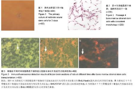
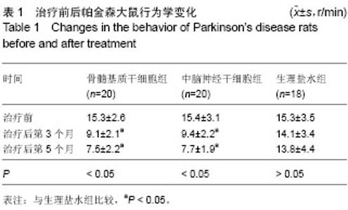
| [1] 顾平,王坤,王彦永,等.骨髓基质干细胞和神经干细胞移植治疗帕金森病大鼠的效果比较[J].中国组织工程研究与临床康复,2008, 12(12):2221-2226.
[2] 叶民,陈生弟,汪锡金,等.体外诱导骨髓基质干细胞治疗帕金森病大鼠模型的研究[J].中华神经科杂志,2005,38(10):624-627.
[3] 王坤.骨髓基质干细胞和神经干细胞移植治疗帕金森病大鼠的疗效比较研究[D].石家庄:河北医科大学,2008.
[4] 王诚.菲立磁标记骨髓基质细胞移植治疗帕金森病的实验研究[D]. 苏州:苏州大学,2007.
[5] 王安民,晁福玲.细胞移植治疗帕金森病的理论研究与应用技术[J].中国临床康复,2006,10(5):114-116.
[6] 陈生弟,王刚,刘军,等.帕金森病发病机制与诊治的基础与临床研究进展[J].上海交通大学学报:医学版,2012,32(9):1221- 1226.
[7] 叶民,陈生弟.骨髓基质干细胞诱导分化及治疗潜能的研究进展[J].临床神经病学杂志,2004,17(4):311-312.
[8] 邹志浩.TH基因转染的骨髓源性神经干细胞在不同移植途径下对PD大鼠模型治疗作用的实验研究[D].广州:第一军医大学, 2006.
[9] Chen X, Liu W, Guoyuan Y, et al. Protective effects of intracerebral adenoviral-mediated GDNF gene transfer in a rat model of Parkinson's disease. Parkinsonism Relat Disord. 2003;10(1):1-7.
[10] Sanchez-Ramos JR. Neural cells derived from adult bone marrow and umbilical cord blood. J Neurosci Res. 2002;69(6): 880-893.
[11] 叶民,陈生弟,陆国强,等.成年大鼠骨髓基质干细胞诱导分化为神经元样细胞不同方法比较的研究[J].中国神经科学杂志,2004, 20(4):275-280.
[12] Guglielmetti C, Praet J, Rangarajan JR, et al. Multimodal imaging of subventricular zone neural stem/progenitor cells in the cuprizone mouse model reveals increased neurogenic potential for the olfactory bulb pathway, but no contribution to remyelination of the corpus callosum. Neuroimage. 2014;86: 99-110.
[13] Xia JG,Miu JL,Ding HB,et al.Changes of brain gray matter structure in Parkinson’s disease patients with dementia. Neural Regen Res. 2013;8(14):1276-128.
[14] 郑晴雯.小鼠神经发育基因Sox1诱导大鼠骨髓基质干细胞神经分化[D].广州:广州医学院,2005.
[15] 蒋明,陈新成,吴旻,等.鼠胚神经干细胞移植对帕金森模型大鼠的治疗作用[J].中国组织工程研究与临床康复,2011,15(49): 9178-9181.
[16] 邹志浩,张世忠,姜晓丹,等.基因修饰骨髓源性神经元样干细胞治疗帕金森大鼠的研究[J].中华神经外科杂志,2010,26(2): 179-182.
[17] 卢爱丽.脐带间充质干细胞治疗帕金森病的疗效[J].中国老年学杂志,2013,33(21):5245-5247.
[18] 王磊,曾水林,李涛,等.帕金森模型大鼠海马区神经干细胞增殖情况的实验研究[J].军医进修学院学报,2005,26(3):211-213.
[19] Hölscher C.New drug treatments show neuroprotective effects in Alzheimer’s and Parkinson’s diseases. Neural Regen Res. 2014;9 (21):1870-1873.
[20] 柯春龙,陈白莉,余振华,等.白藜芦醇对神经干细胞诱导分化为多巴胺能神经元移植治疗帕金森模型鼠的影响[J].中国组织工程研究与临床康复,2009,13(27):5281-5285.
[21] Yoon HH,Min J,Shin N,et al.Are human dental papilla-derived stem cell and human brain-derived neural stem cell transplantations suitable for treatment of Parkinson’s disease? Neural Regen Res. 2013;8 (13): 1190-1200.
[22] 王海军,罗烨,刘自英,等.神经干细胞移植对帕金森大鼠的疗效观察[J].齐齐哈尔医学院学报,2014,35(21):3126-3127.
[23] 冯德朋.脐血间充质干细胞移植治疗帕金森大鼠的实验研究[D]. 泰安:泰山医学院,2010.
[24] 王梓葳.大鼠胚胎干细胞向神经干细胞的体外分化及大鼠帕金森疾病模型的建立[D].北京:中国科学院研究生院,2011.
[25] 葸瑞,白海,王存邦,等.脐带间充质干细胞治疗帕金森症临床疗效观察[J].中华细胞与干细胞杂志:电子版,2012,2(4):274-276.
[26] 王晓梅.重组大鼠质粒pEGFP-GDNF转染大鼠胚胎神经干细胞治疗帕金森模型鼠的实验研究[D]. 上海:上海交通大学,2005.
[27] 陈勇.人羊膜间充质干细胞和人羊膜上皮细胞移植治疗帕金森病模型大鼠的效应比较[D]. 遵义:遵义医学院,2012.
[28] 张平,赵钢勇,宋月平,等.转染pIRESneo-EGFP-BDNF的骨髓间充质干细胞侧脑室注射对帕金森大鼠行为学的影响[J].山东医药,2012,52(26):23-26.
[29] 李美霖,杨慧. 自分化法培养大鼠骨髓间充质干细胞移植治疗大鼠帕金森病的研究[J].中国输血杂志,2014,27(10):1000-1005.
[30] 王旭,张志彬,贾芙蓉,等. 自体骨髓间充质干细胞移植治疗帕金森病的疗效[J].中国老年学杂志,2014,34(1):22-23.
[31] 毕涌,洪娟,林晓滨,等. 人β-NGF基因修饰的骨髓间充质干细胞对帕金森病大鼠行为学的影响[J].中国病理生理杂志,2014, 30(3): 473-478.
[32] 陈丹丹,付文玉,庄宝祥,等. 骨髓间充质干细胞黑质内移植对帕金森病大鼠的治疗作用[J].中国实验动物学报,2013,21(1): 22-26.
[33] 张晓英.神经干细胞移植治疗帕金森病的有效性与安全性[J].中国组织工程研究,2013,17(27):5033-5040.
[34] 徐胜利,周明,陈彪. 神经营养因子基因修饰的神经干细胞在帕金森病大鼠模型中的治疗作用[J].中华老年医学杂志,2010, 29(1):58-62.
[35] 顾平,王坤,王彦永,等. 骨髓基质干细胞和神经干细胞移植治疗帕金森病大鼠的效果比较[J].中国组织工程研究与临床康复, 2008,12(12):2221-2226.
[36] 郑敏,王亚平,裴雪涛. 神经干细胞移植对帕金森病模型大鼠的治疗作用[J].基础医学与临床,2007,27(8):841-845.
[37] Svendsen CN, Clarke DJ, Rosser AE, et al. Survival and differentiation of rat and human epidermal growth factor-responsive precursor cells following grafting into the lesioned adult central nervous system. Exp Neurol. 1996; 137(2):376-388.
[38] Zhang Y, Yang Y, Tang H, et al. Hyperbaric oxygen therapy ameliorates local brain metabolism, brain edema and inflammatory response in a blast-induced traumatic brain injury model in rabbits. Neurochem Res. 2014;39(5):950-960.
[39] 王兰.尿嘧啶脱氧核苷和Resovist双标记骨髓基质细胞移植治疗帕金森病大鼠的实验研究[D]. 苏州:苏州大学,2009.
[40] 贾丛康.骨髓基质干细胞治疗帕金森病模型鼠的研究[D].乌鲁木齐:新疆医科大学,2009. |


