Chinese Journal of Tissue Engineering Research ›› 2015, Vol. 19 ›› Issue (14): 2133-2137.doi: 10.3969/j.issn.2095-4344.2015.14.001
Optimized effect of connexin 43 gene silencing on the proliferation of fetal liver stem cells
Zhang Zeng-guang1, Qin Ming-fang2
- 1Department of Hepatobiliary Surgery, Tianjin 4th Centre Hospital, Tianjin 300143, China; 2Department of Minimally Invasive Surgery, Nankai Hospital, Tianjin 300100, China
-
Revised:2015-03-14Online:2015-04-02Published:2015-04-02 -
Contact:Qin Ming-fang, Professor, Chief physician, Doctoral supervisor, Department of Minimally Invasive Surgery, Nankai Hospital, Tianjin 300100, China -
About author:Zhang Zeng-guang, Master, Attending physician, Department of Hepatobiliary Surgery, Tianjin 4th Centre Hospital, Tianjin 300143, China
CLC Number:
Cite this article
Zhang Zeng-guang, Qin Ming-fang. Optimized effect of connexin 43 gene silencing on the proliferation of fetal liver stem cells[J]. Chinese Journal of Tissue Engineering Research, 2015, 19(14): 2133-2137.
share this article
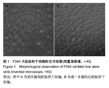
2.1 胎肝干细胞形态 原代培养1 d后细胞呈贴壁性生长,圆形或者卵圆形,胞质清晰,核质比高,核大。在特殊培养条件下胎肝干细胞不断的增殖分裂,速度较快,第3天形成了3-5个胎肝干细胞组成的细胞团,1周时细胞团数量增至6-10个,培养30 d左右时形成了肉眼可见的细胞集落,见图1。采用流式细胞仪对胎肝干细胞进行鉴定,CD133和上皮细胞黏附分子呈现高表达,证实了培养细胞为胎肝干细胞。 "


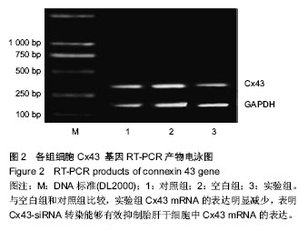
2.2 Cx43-siRNA转染抑制胎肝干细胞中Cx43 mRNA的表达 RT-PCR检测结果显示,与空白组和对照组比较(0.70±0.07,0.71±0.04),实验组Cx43 mRNA的表达明显减少(0.47±0.06),差异均有显著性意义(P < 0.05)。空白组和对照组比较Cx43 mRNA的表达差异无显著性意义(P > 0.05),表明Cx43-siRNA转染能够有效抑制胎肝干细胞中Cx43 mRNA的表达,见图2。"

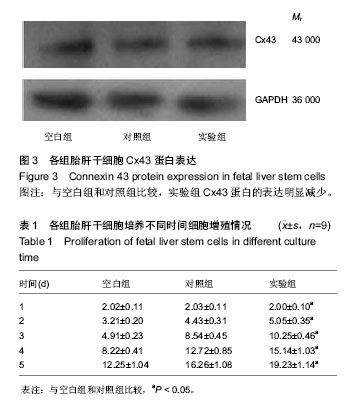
2.3 Cx43-siRNA转染抑制胎肝干细胞中Cx43蛋白的表达 与空白组和对照组比较(0.97±0.14,0.99±0.16),实验组Cx43蛋白的表达明显减少(0.65±0.12),差异均有显著性意义(P < 0.05)。空白组与对照组比较Cx43蛋白的表达差异无显著性意义(P > 0.05),见图3。 2.4 细胞生长曲线 Cx43-siRNA转染后,实验组鼠胎肝干细胞增殖能力较对照组、空白组明显增强,差异有显著性意义(P < 0.05),见表1。 2.5 CCK-8检测结果 Cx43-siRNA转染48 h后,实验组鼠胎肝干细胞吸光度值(0.29±0.06)较对照组、空白组(0.17± 0.03,0.17±0.02)明显升高,差异有显著性意义(P < 0.05)。"
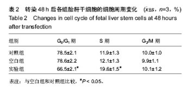
2.6 细胞周期分布 Cx43-siRNA转染48 h后,实验组S期细胞明显多于对照组、空白组,实验组G1期细胞明显少于对照组、空白组,差异有显著性意义(P < 0.05),M期细胞无明显变化,见表2。"

| [1] Park Y, Chen Y, Ordovas L, et al. Hepatic differentiation of human embryonic stem cells on microcarriers. J Biotechnol. 2014;174:39-48. [2] Zamule SM, Coslo DM, Chen F, et al. Differentiation of human embryonic stem cells along a hepatic lineage. Chem Biol Interact. 2011;190(1):62-72. [3] Laird DW. Syndromic and non-syndromic disease-linked Cx43 mutations. FEBS Lett. 2014;588(8):1339-1348. [4] 王凌云,田衍平,牛建钦,等.Cx43基因干扰对大鼠星形胶质细胞谷氨酸基础释放的影响[J].第三军医大学学报,2012,34(8): 701-704. [5] Ishkitiev N, Yaegaki K, Imai T, et al. High-purity hepatic lineage differentiated from dental pulp stem cells in serum-free medium. J Endod. 2012;38(4):475-480. [6] Mohsin S, Shams S, Ali Nasir G, et al. Enhanced hepatic differentiation of mesenchymal stem cells after pretreatment with injured liver tissue. Differentiation. 2011;81(1):42-48. [7] Nishiofuku M, Yoshikawa M, Ouji Y, et al. Modulated differentiation of embryonic stem cells into hepatocyte-like cells by coculture with hepatic stellate cells. J Biosci Bioeng. 2011;111(1):71-77. [8] Augustine TN, Kramer B. Signals from pancreatic mesoderm influence the expression of a pancreatic phenotype in hepatic stem-like cell line PHeSC-A2 in vitro: a preliminary study. Acta Histochem. 2011;113(3):349-352. [9] Ordovás L, Park Y, Verfaillie CM. Stem cells and liver engineering. Biotechnol Adv. 2013;31(7):1094-1107. [10] Baxter MA, Rowe C, Alder J, et al. Generating hepatic cell lineages from pluripotent stem cells for drug toxicity screening. Stem Cell Res. 2010;5(1):4-22. [11] Behrens J, Kameritsch P, Wallner S, et al. The carboxyl tail of Cx43 augments p38 mediated cell migration in a gap junction-independent manner. Eur J Cell Biol. 2010;89(11): 828-838. [12] Ton QV, Kathryn Iovine M. Semaphorin3d mediates Cx43-dependent phenotypes during fin regeneration. Dev Biol. 2012;366(2):195-203. [13] Yuan MJ, Huang H, Tang YH, et al. Effects of ghrelin on Cx43 regulation and electrical remodeling after myocardial infarction in rats. Peptides. 2011;32(11):2357-2361. [14] Feng C, Cao L, Zuo Z. RNA interference-produced autoregulation of inducible nitric oxide synthase expression. FEBS Lett. 2011;585(15):2488-2492. [15] Pisarev AV, Skabkin MA, Pisareva VP, et al. The role of ABCE1 in eukaryotic posttermination ribosomal recycling. Mol Cell. 2010;37(2):196-210. [16] 龚建平,韩本立.肝脏细胞的分离、培养和鉴定技术[J].世界华人消化杂志, 1999,7(5):417-419. [17] Cienfuegos JA, Baixauli J. Stem cells and liver regeneration: looking toward the future. Gastroenterol Hepatol. 2012;35(9): 675. [18] Rao MS, Khan AA, Parveen N, et al. Characterization of hepatic progenitors from human fetal liver during second trimester. World J Gastroenterol. 2008;14(37):5730-5737. [19] 黄利华,郭姣,黄珮,等. 新生大鼠肝干细胞的分离、培养与鉴定[J].广东药学院学报,2010,26(4):416-419. [20] Dollé L, Best J, Empsen C, et al. Successful isolation of liver progenitor cells by aldehyde dehydrogenase activity in naïve mice. Hepatology. 2012;55(2):540-552. [21] Ling L, Ni Y, Wang Q, et al. Transdifferentiation of mesenchymal stem cells derived from human fetal lung to hepatocyte-like cells. Cell Biol Int. 2008;32(9):1091-1098. [22] Evarts RP, Hu Z, Fujio K, et al. Activation of hepatic stem cell compartment in the rat: role of transforming growth factor alpha, hepatocyte growth factor, and acidic fibroblast growth factor in early proliferation. Cell Growth Differ. 1993;4(7): 555-561. [23] Parveen N, Aleem AK, Habeeb MA, et al. An update on hepatic stem cells: bench to bedside. Curr Pharm Biotechnol. 2011;12(2):226-230. [24] Dan YY, Yeoh GC. Liver stem cells: a scientific and clinical perspective. J Gastroenterol Hepatol. 2008;23(5):687-698. [25] He ZP, Tan WQ, Tang YF, et al. Activation, isolation, identification and in vitro proliferation of oval cells from adult rat livers. Cell Prolif. 2004;37(2):177-187. [26] Zhang Y, Bai XF, Huang CX. Hepatic stem cells: existence and origin. World J Gastroenterol. 2003;9(2):201-204. [27] Shupe TD, Piscaglia AC, Oh SH, et al. Isolation and characterization of hepatic stem cells, or "oval cells," from rat livers. Methods Mol Biol. 2009;482:387-405. [28] Rountree CB, Ding W, Dang H, et al. Isolation of CD133+ liver stem cells for clonal expansion. J Vis Exp. 2011;(56). pii: 3183. [29] Côrte-Real J, Duarte N, Tavares L, et al. Innate stimulation of B1a cells enhances the autoreactive IgM repertoire in the NOD mouse: implications for type 1 diabetes. Diabetologia. 2012;55(6):1761-1772. [30] Hardy RR, Hayakawa K. Positive and negative selection of natural autoreactive B cells. Adv Exp Med Biol. 2012;750: 227-238. [31] Kendall PL, Woodward EJ, Hulbert C, et al. Peritoneal B cells govern the outcome of diabetes in non-obese diabetic mice. Eur J Immunol. 2004;34(9):2387-2395. [32] Casali P, Schettino EW. Structure and function of natural antibodies. Curr Top Microbiol Immunol. 1996;210:167-179. [33] Côrte-Real J, Duarte N, Tavares L, et al. Autoimmunity triggers in the NOD mouse: a role for natural auto-antibody reactivities in type 1 diabetes. Ann N Y Acad Sci. 2009;1173: 442-448. [34] 赵戈,刘卫辉,王涛,等. 小分子干扰RNA介导的RhoA 沉默优化胎肝干细胞培养[J].中国组织工程研究,2012,16(10): 1803-1807. [35] Nakamura T, Mizuno S. The discovery of hepatocyte growth factor (HGF) and its significance for cell biology, life sciences and clinical medicine. Proc Jpn Acad Ser B Phys Biol Sci. 2010; 86(6):588-610. [36] Rongo C. Epidermal growth factor and aging: a signaling molecule reveals a new eye opening function. Aging (Albany NY). 2011;3(9):896-905. [37] Bhave VS, Paranjpe S, Bowen WC, et al. Genes inducing iPS phenotype play a role in hepatocyte survival and proliferation in vitro and liver regeneration in vivo. Hepatology. 2011;54(4): 1360-1370. [38] Shi XL, Gu JY, Zhang Y, et al. Protective effects of ACLF sera on metabolic functions and proliferation of hepatocytes co-cultured with bone marrow MSCs in vitro. World J Gastroenterol. 2011;17(19):2397-2406. [39] Thomas JW, Kendall PL, Mitchell HG. The natural autoantibody repertoire of nonobese diabetic mice is highly active. J Immunol. 2002;169(11):6617-6624. |
| [1] | Xu Xiang, Yin He-ping. Platelet-rich plasma accelerates the proliferation of bone marrow mesenchymal stem cells [J]. Chinese Journal of Tissue Engineering Research, 2015, 19(14): 2144-2148. |
| [2] | Lin Shu-zhong, Liu Jun. Effects of different doses of puerarin on osteoblasts in vitro [J]. Chinese Journal of Tissue Engineering Research, 2015, 19(11): 1658-1662. |
| [3] | Yao Ming-zhi. Effects of Jiegu Qili Tablet on proliferation and mineralization of MC-3T3 cells [J]. Chinese Journal of Tissue Engineering Research, 2015, 19(11): 1694-1698. |
| [4] | Feng Chao, Li Zhe, Lv Xiang-guo, Xu Yue-min, Fu Qiang. In vitro preparation and biochemical evaluation of oxygen generative keratin/silk fibroin compound biomaterial [J]. Chinese Journal of Tissue Engineering Research, 2014, 18(52): 8480-8486. |
| [5] | Yan Zhi-dong, Yan Jia, Zhuansun Yong-xun, Chen Rui, Zhang Wei, Feng Su-ling, Li Jian-guo . Small interfering RNA inhibits the expression of surface antigens CD80/CD86 from mature dendritic cells [J]. Chinese Journal of Tissue Engineering Research, 2014, 18(5): 754-760. |
| [6] | Li Dan, Hui Rui, Hu Yong-wu, Han Yan, Guo Shu-zhong. Effects of Dragon’s blood extracts on fibroblast proliferation and procollagen type III [J]. Chinese Journal of Tissue Engineering Research, 2014, 18(46): 7437-7441. |
| [7] | Zhang Hu-lin, Zhang Zi-qiang, Xu Hong-bin, Zhang Xiao-gang. Drynaria freeze-dried powder at different dosages influences proliferation and differentiation of rabbit bone marrow mesenchymal stem cells [J]. Chinese Journal of Tissue Engineering Research, 2014, 18(41): 6649-6654. |
| [8] | Shi Jian-ming, Wu Ya-hua, Geng Shu-guo, Yin Ming. Proliferation, senescence and differentiation of mesenchymal stem cells: canonical and non-canonical regulations of Wnt signaling pathway [J]. Chinese Journal of Tissue Engineering Research, 2014, 18(41): 6719-6724. |
| [9] | Yin Shou-xin, Zhou Zhen, Ma Mei-xue, Yang Jing-bo, Yang Dong-ye. Long-term culture of hepatic oval cells in rats in vitro [J]. Chinese Journal of Tissue Engineering Research, 2014, 18(29): 4663-4668. |
| [10] | Wang Ding, Song Bing, Zhong Xuan, Sun Xiao-fang, Fan Yong . Phytohaemagglutinin stimulates the proliferation of peripheral blood mononuclear cells and expression of secretory cytokines [J]. Chinese Journal of Tissue Engineering Research, 2014, 18(23): 3707-3714. |
| [11] | Chen Peng, Zhang Jie, Rong Dong-ming, Han Zhong-yu, Yuan Si-jie, Tian Jing. Effects of non-dextran coated superparamagnetic iron oxide nanoparticles on proliferation of bone marrow mesenchymal stem cells [J]. Chinese Journal of Tissue Engineering Research, 2014, 18(16): 2526-2531. |
| [12] | Zhang Yuan-yu, Liu Xia, Li Kun, Guo Yong-rong, Bai Jing-ping. Effect of recombinant Mycobacterium tuberculosis heat shock protein 10 on proliferation of human osteoblasts and regulation of bone metabolism [J]. Chinese Journal of Tissue Engineering Research, 2014, 18(11): 1665-1671. |
| [13] | Li Ying, Gong Ya-fei, Chen Xin-jie. Biological properties of C57BL/6 mouse embryonic fibroblasts and preparation of feeder layers [J]. Chinese Journal of Tissue Engineering Research, 2014, 18(11): 1737-. |
| Viewed | ||||||
|
Full text |
|
|||||
|
Abstract |
|
|||||