Chinese Journal of Tissue Engineering Research ›› 2015, Vol. 19 ›› Issue (9): 1457-1462.doi: 10.3969/j.issn.2095-4344.2015.09.025
Previous Articles Next Articles
Applications and prospects of digital orthopedics: more precise, individual and intuitional outlook
Huo Li-feng, Ni Heng-jian
- Department of Human Anatomy, College of Medicine, Nantong University, Nantong 226000, Jiangsu Province, China
-
Revised:2015-01-26Online:2015-02-26Published:2015-02-26 -
Contact:Ni Heng-jian, Professor, Department of Human Anatomy, College of Medicine, Nantong University, Nantong 226000, Jiangsu Province, China -
About author:Huo Li-feng, Studying for master’s degree, Department of Human Anatomy, College of Medicine, Nantong University, Nantong 226000, Jiangsu Province, China
CLC Number:
Cite this article
Huo Li-feng, Ni Heng-jian. Applications and prospects of digital orthopedics: more precise, individual and intuitional outlook[J]. Chinese Journal of Tissue Engineering Research, 2015, 19(9): 1457-1462.
share this article
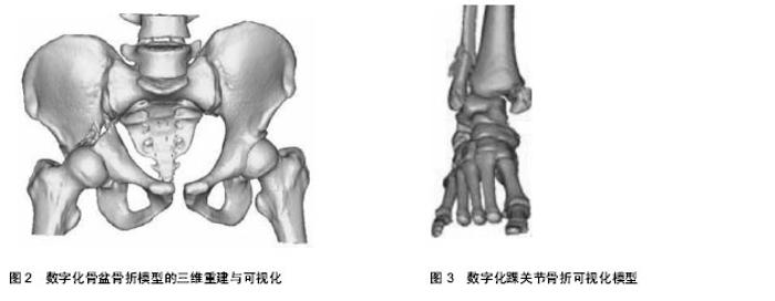
2.1 计算机生物力学 采用三维图像重建后有限元分析的虚拟仿真实验可以解决传统生物力学实验难以解决的需要大量人体标本,以及全面的解释标本各个部分内在的相互作用机制等问题。 有限元法通过利用计算机把不规则的、复杂力学分析对像离散化成有限个几何单元体进行分解计算,能够反映机体内部的应力变化情况,对形状、载荷、材料力学性能和结构较复杂的物体进行应力、应变分析。并可在持续性研究中改变部分参数或者重复模拟实验,并反映变化后的情况,这是其他实验技术难以实现的。目前有限元法已广泛运用于运动损伤的机制分析、手术治疗规划、术后效果评价、矫形及器械优化设计等众多方面[8-12]。通过人体虚拟技术,撞击性损伤的应力应变分布的有限元研究,在骨科基础理论研究上已经初步取得成效,对预防损伤和损伤机制的研究具有重要的指导意义[13-17]。 目前,模型软组织(韧带、肌肉等)大多是参考解剖学结论并根据个人的理解添加的,这与实际存在一定的偏差,而国内材料的性质也有相当大一部分是参考国外资料,但是外国人与国人在体格方面有很大的差异,且生物材料性能等测试工作欠缺,这些缺陷都要求进一步深入了解与分析。实验生物力学技术与计算生物力学技术都在不断地发展,二者互为补充、相辅相成[18]。计算生物力学为实验力学提供了新的发展空间,应用起来快捷、经济;而实验生物力学则是基础,为计算生物力学提供辅助条件。由于理论始终有假设和简化的成分存在,因此计算生物力学必须与临床资料进行比较、与大体标本力学实验进行对照分析,从而互相促进,使二者结合更为紧密。 2.2 术前骨折的影像处理和三维建模 计算机CT三维建模技术即在骨折手术前利用患者的影像学数据,通过 Mimics 等三维操控软件系统在计算机中建立相应的骨关节三维模型。 在常规的骨折诊疗过程中,手术复位及固定的参考标准主要包括CT 扫描图像,术前X射线片以及术中透视结果等。但在复杂粉碎性骨折中,建立CT三维重建骨关节模型可以使术者在术前即对复杂骨折形成一个直观的立体的概念,掌握骨折的细节和全面情况,在此基础上作出准确判断;从而术者有更充分的证据来进行手术设计,能够更大限度地提高复位和固定效果,保护骨折部位的血运。 具体到某个部位的骨折,骨盆髋臼骨折的情况常常比较复杂。对于不稳定性骨盆骨折尤其情况较严重者,确定治疗方案的关键是正确的诊断分型[19]。以往常通过拍摄闭孔斜位及髂骨斜位X射线片来判断臼壁的骨折情况,目前通过采用计算机三维建模技术可以更直观地展示骨盆髋臼骨折的实际情况包括骨折的位置、类型、骨折部位的解剖结构等,为复杂骨折的诊断和治疗提供直观精准的的依据(图2)。 在 Mimics 软件中,Object为可操控的独立的三维实体,术前对于重要的骨单元或骨折块均应建立 Object,以便在模拟手术中进行操作,即建立单元(简称建元)。在各部位骨折中如涉及关节面,往往都是最重要的骨折,因此凡是涉及关节面且面积> 0.5 cm2者都应单独建立单元。因此应由熟悉软件性能的技术人员来执行骨折三维CT重建,并仔细观察,不可遗漏重要的碎小骨块。具体到胫骨平台骨折和股骨髁上骨折,这类骨折损伤较重,涉及关节面的骨折往往有多块,同时常伴有关节面的塌陷,恢复膝关节功能是其主要治疗目的[20-25]。在进行CT三维重建时,应注意对以上相关的骨折块尤其是塌陷的关节面部分单独建元分析,以便在计算机辅助设计时可以撬拔复位模拟。对于后踝骨折,一般认为当负重面< 25%时骨折对胫距关节的生物力学特性影响十分微小,可不予手术复位固定[26-32]。 有学者认为当骨折块关节面≥胫骨远端关节面10%时,需切开复位固定,否则将改变关节内原有的接触应力,从而增加创伤性关节炎的发生率[33-38]。因此对于明确的后踝骨折,在进行三维重建时均应予以建元(图3)。"

| [1] 王瑞雄,陈夏平,刘志强,等.改良Stoppa入路在髋臼及骨盆骨折手术治疗中的应用[J]. 中国骨与关节损伤杂志,2014,29(2): 108-110. [2] Zielinski SM, Bouwmans CA, Heetveld MJ, et al. FAITH trialinvestigators.The societal costs of femoral neck fracture patients treated with internal fixation. Osteoporos Int. 2014; 25(3):875-885. [3] 罗永祥, Akkineni AR, Anja Lode W, et al. 3-D打印:一种个性化制备复杂支架和组织工程植入物的多功能快速成型技术(英文)[J]. 中国修复重建外科杂志,2014,28(3):279-285. [4] 倪明,沈燕国,胡晓亮,等.经改良Stoppa入路治疗骨盆髋臼骨折的临床体会[J].中国骨与关节损伤杂志,2013,28(2):101-103. [5] Xie A,Fang C,Huang Y,et al. Application ofthree- dimensionalreconstruction and visible simulationtechnique in reoperation of hepatolithiasis. Gastroenterol Hepatol. 2013;28(2):248-254. [6] Bose S, Vahabzadeh S, Bandyopadhyay A. Bone tissueengineering using 3D printing. Mater Today. 2013; 16(12): 496-504. [7] Georges L,Carlvan L,Luiza MO,et al.Trabecular bonestrains around a dental implant and associated micromotions-A micro-CT-based three-dimensional finite element study. J Biomech. 2010;43 (7):1251-1261. [8] 章莹,尹庆水,万磊,等.数字技术在创伤骨科的应用[J].中国骨科临床与基础研究杂志,2011, 3(2):113- 119. [9] Wong MS. Computer- aided design and computer- aided manufact ure (CAD/CAM)system for construction of spinal orthosis for patients with adolescent idiopat hicscoliosis. Physiother Theory Pract. 2011;27(1):74-79. [10] 赵德伟.共同推进人工关节植入物与数字化骨科领域科研与临床的发展[J].中国组织工程研究与临床康复杂志,2011, 15(17): 3041-3042. [11] 张元智,赵建民,李志军,等.数字化技术在腓肠神经筋膜皮瓣移植中的应用[J].内蒙古医学院学报,2010,32(5):445- 448. [12] Beaupre GS. Effect of fracture gap on stability of compression plate fixation: a finite element study. J Orthop Res. 2011;29(1): 152-153. [13] Kim KK, Heo YM, Won YY, et al. Navigation-assisted total knee arthroplasty for the knee retaining femoral intramedullary nail, and distal femoral plate and screws. Clin Orthop Surg. 2011;3(1): 77-80. [14] Wang B, Xia Q, Miao J, et al.Application of digital orthopedic technology for observing degenerative lumbar segmental instability of three-dimensional kinematic characteristics in vivo.Zhonghua Yi Xue Za Zhi. 2014; 94(29):2264-2268. [15] Klingler JH, Sircar R, Scheiwe C,et al.Comparative Study of C-Arms for Intraoperative 3-Dimensional Imaging and Navigation in Minimally Invasive Spine Surgery Part I - Applicability and Image Quality.J Spinal Disord Tech. 2014. [16] Veli I, Yuksel B, Uysal T.Longitudinal evaluation of dental arch asymmetry in Class II subdivision malocclusion with 3-dimensional digital models.Am J Orthod Dentofacial Orthop. 2014;145(6):763-770. [17] Feinglass NG, Clendenen SR, Shine TS,et al. Real-time two-dimensional and three-dimensional echocardiographic imaging of the thoracic spinal cord: a possible new window into the central neuraxis.J Clin Monit Comput. 2015;29(1): 121-125. [18] Yang B, Fang SB, Li CS,et al. Digital three-dimensional model of lumbar region 4-5 and its adjacent structures based on a virtual Chinese human.Orthop Surg. 2013;5(2): 130-134. [19] Takai K, Kin T, Oyama H,et al. Three-dimensional angioarchitecture of spinal dural arteriovenous fistulas, with special reference to the intradural retrograde venous drainage system.J Neurosurg Spine. 2013;18(4):398-408. [20] Foss K, da Costa RC, Moore S,et al.Three-dimensional kinematic gait analysis of Doberman Pinschers with and without cervical spondylomyelopathy.J Vet Intern Med. 2013; 7(1):112-119. [21] Ding J, Sun G, Lu Y, et al.Evaluation of anterior ethmoidal artery by 320-slice CT angiography with comparison to three-dimensional spin digital subtraction angiography: initial experiences.Korean J Radiol. 2012;13(6): 667-673. [22] Zhu QG, Fang M, Pan L.Effects of tuina manipulation on the three-dimensional space of cervical vertebral segments of cervical spondylosis patients.Zhongguo Zhong Xi Yi Jie He Za Zhi. 2012;32(7):922-925. [23] Wade R, Yang H, McKenna C,et al. A systematic review of the clinical effectiveness of EOS 2D/3D X-ray imaging system.Eur Spine J. 2013;22(2):296-304. [24] Abdullah KG, Bishop FS, Lubelski D,et al.Radiation exposure to the spine surgeon in lumbar and thoracolumbar fusions with the use of an intraoperative computed tomographic 3-dimensional imaging system.Spine (Phila Pa 1976). 2012;37(17):E1074-1078. [25] Yoshihara M, Terajima M, Yanagita N, et al.Three-dimensional analysis of the pharyngeal airway morphology in growing Japanese girls with and without cleft lip and palate.Am J Orthod Dentofacial Orthop. 2012;141(4 Suppl):S92-101 [26] Glaser DA, Doan J, Newton PO.Comparison of 3-dimensional spinal reconstruction accuracy: biplanar radiographs with EOS versus computed tomography.Spine (Phila Pa 1976). 2012;37(16):1391-1397. [27] Liu GJ, Zhang SX, Qiu MG,et al. A novel technique for three-dimensional reconstruction for surgical simulation around the craniocervical junction region.Int Surg. 2011; 96(3):274-280. [28] Fujimori T, Iwasaki M, Nagamoto Y,et al.Three-dimensional measurement of growth of ossification of the posterior longitudinal ligament.J Neurosurg Spine. 2012;16(3):289-295. [29] Wang F, Song H, Zhao F,et al.Supra-acetabular external fixation for pelvic fractures: a digital anatomical study.Clin Anat. 2012;25(4):503-508. [30] Behrendt D, Mmtze M, Steinke H,et al.Evaluation of 2D and 3D navigation for iliosacral screw fixation.Int J Comput Assist Radiol Surg. 2012;7(2):249-255. [31] Takai K, Kin T, Oyama H,et al.The use of 3D computer graphics in the diagnosis and treatment of spinal vascular malformations.J Neurosurg Spine. 2011;15(6):654-659. [32] Hartwig T, Streitparth F, Gross C,et al.Digital 3-dimensional analysis of the paravertebral lumbar muscles after circumferential single-level fusion.J Spinal Disord Tech. 2011;24(7):451-454. [33] Labelle H, Aubin CE, Jackson R,et al. Seeing the spine in 3D: how will it change what we do? J Pediatr Orthop. 2011;31(1 Suppl):S37-45. [34] Lagravmre MO, Low C,Flores-Mir C, et al.Intraexaminer and interexaminer reliabilities of landmark identification on digitized lateral cephalograms and formatted 3-dimensional cone-beam computerized tomography images.Am J Orthod Dentofacial Orthop. 2010;137(5):598-604. [35] Fu D, Jin AM, Tian J,et al.Three-dimensional visualization of the structures related to the anterior cervical segment approach.Nan Fang Yi Ke Da Xue Xue Bao. 2010;30 (4): 888-890. [36] Anderson K, Yamamoto E, Kaplan J,et al.Neurolucida Lucivid versus Neurolucida camera: A quantitative and qualitative comparison of three-dimensional neuronal reconstructions.J Neurosci Methods. 2010;186(2):209-214. [37] Janssen MM, Drevelle X, Humbert L,et al.Differences in male and female spino-pelvic alignment in asymptomatic young adults: a three-dimensional analysis using upright low-dose digital biplanar X-rays.Spine (Phila Pa 1976). 2009;34(23): E826-832. [38] Aadland TD, Thielen KR, Kaufmann TJ,et al.3D C-arm conebeam CT angiography as an adjunct in the precise anatomic characterization of spinal dural arteriovenous fistulas.AJNR Am J Neuroradiol. 2010;31(3):476-480. [39] Lu S, Xu YQ, Zhang YZ,et al.Primary clinical result of digital template as navigation to supper cervical pedicle instrumentation.Zhonghua Wai Ke Za Zhi. 2009;47(5): 359-362. [40] Chien PC, Parks ET, Eraso F, et al.Comparison of reliability in anatomical landmark identification using two-dimensional digital cephalometrics and three-dimensional cone beam computed tomography in vivo.Dentomaxillofac Radiol. 2009; 38(5):262-273. [41] Oz U, Orhan K, Abe N. Comparison of linear and angular measurements using two-dimensional conventional methods and three-dimensional cone beam CT images reconstructed from a volumetric rendering program in vivo. Dentomaxillofac Radiol. 2011;40(8):492-500. [42] Moshiri M, Scarfe WC, Hilgers ML, et al. Accuracy of linear measurements from imaging plate and lateral cephalometric images derived from cone-beam computed tomography. Am J Orthod Dentofacial Orthop. 2007;132(4):550-560. [43] Oberlaender M, Bruno RM, Sakmann B, et al. Transmitted light brightfield mosaic microscopy for three-dimensional tracing of single neuron morphology. J Biomed Opt. 2007; 12(6):064029. [44] Cattaneo PM, Bloch CB, Calmar D, et al. Comparison between conventional and cone-beam computed tomography-generated cephalograms. Am J Orthod Dentofacial Orthop. 2008;134(6):798-802. |
| [1] | Xu Feng, Kang Hui, Wei Tanjun, Xi Jintao. Biomechanical analysis of different fixation methods of pedicle screws for thoracolumbar fracture [J]. Chinese Journal of Tissue Engineering Research, 2021, 25(9): 1313-1317. |
| [2] | Chen Xinmin, Li Wenbiao, Xiong Kaikai, Xiong Xiaoyan, Zheng Liqin, Li Musheng, Zheng Yongze, Lin Ziling. Type A3.3 femoral intertrochanteric fracture with augmented proximal femoral nail anti-rotation in the elderly: finite element analysis of the optimal amount of bone cement [J]. Chinese Journal of Tissue Engineering Research, 2021, 25(9): 1404-1409. |
| [3] | Zhou Jihui, Li Xinzhi, Zhou You, Huang Wei, Chen Wenyao. Multiple problems in the selection of implants for patellar fracture [J]. Chinese Journal of Tissue Engineering Research, 2021, 25(9): 1440-1445. |
| [4] | Xu Yulin, Shen Shi, Zhuo Naiqiang, Yang Huilin, Yang Chao, Li Yang, Zhao Heng, Zhao Lu. Biomechanical comparison of three different plate fixation methods for acetabular posterior column fractures in standing and sitting positions [J]. Chinese Journal of Tissue Engineering Research, 2021, 25(6): 826-830. |
| [5] | Cai Qunbin, Zou Xia, Hu Jiantao, Chen Xinmin, Zheng Liqin, Huang Peizhen, Lin Ziling, Jiang Ziwei. Relationship between tip-apex distance and stability of intertrochanteric femoral fractures with proximal femoral anti-rotation nail: a finite element analysis [J]. Chinese Journal of Tissue Engineering Research, 2021, 25(6): 831-836. |
| [6] | Song Chengjie, Chang Hengrui, Shi Mingxin, Meng Xianzhong. Research progress in biomechanical stability of lateral lumbar interbody fusion [J]. Chinese Journal of Tissue Engineering Research, 2021, 25(6): 923-928. |
| [7] | Liu Zhao, Xu Xilin, Shen Yiwei, Zhang Xiaofeng, Lü Hang, Zhao Jun, Wang Zhengchun, Liu Xuzhuo, Wang Haitao. Guiding role and prospect of staging and classification combined collapse prediction method for osteonecrosis of femoral head [J]. Chinese Journal of Tissue Engineering Research, 2021, 25(6): 929-934. |
| [8] | Xie Chongxin, Zhang Lei. Comparison of knee degeneration after anterior cruciate ligament reconstruction with or without remnant preservation [J]. Chinese Journal of Tissue Engineering Research, 2021, 25(5): 735-740. |
| [9] | Nie Shaobo, Li Jiantao, Sun Jien, Zhao Zhe, Zhao Yanpeng, Zhang Licheng, Tang Peifu. Mechanical stability of medial support nail in treatment of severe osteoporotic intertrochanteric fracture [J]. Chinese Journal of Tissue Engineering Research, 2021, 25(3): 329-333. |
| [10] | Tan Jiachang, Yuan Zhenchao, Wu Zhenjie, Liu Bin, Zhao Jinmin. Biomechanical analysis of elastic nail combined with end caps and wire fixation for long oblique femoral shaft fractures [J]. Chinese Journal of Tissue Engineering Research, 2021, 25(3): 334-338. |
| [11] | Chen Lu, Zhang Jianguang, Deng Changgong, Yan Caiping, Zhang Wei, Zhang Yuan. Finite element analysis of locking screw assisted acetabular cup fixation [J]. Chinese Journal of Tissue Engineering Research, 2021, 25(3): 356-361. |
| [12] | Zhou Jihui, Li Xinzhi, Zhou You, Huang Wei, Chen Wenyao. Comparison of the advantages and disadvantages of multiple implants in treatment of traumatic dislocation of sternoclavicular joint [J]. Chinese Journal of Tissue Engineering Research, 2021, 25(3): 443-448. |
| [13] | Li Kun, Li Zhijun, Zhang Shaojie, Gao Shang, Sun Hao, Yang Xi, Wang Xing, Dai Lina . A 4-year-old child model of occipito-atlanto-axial joints established by finite element dynamic simulation [J]. Chinese Journal of Tissue Engineering Research, 2021, 25(24): 3773-3778. |
| [14] | Shu Qihang, Liao Yijia, Xue Jingbo, Yan Yiguo, Wang Cheng. Three-dimensional finite element analysis of a new three-dimensional printed porous fusion cage for cervical vertebra [J]. Chinese Journal of Tissue Engineering Research, 2021, 25(24): 3810-3815. |
| [15] | Sun Maji, Wang Qiuan, Zhang Xingchen, Guo Chong, Yuan Feng, Guo Kaijin. Development and biomechanical analysis of a new anterior cervical pedicle screw fixation system [J]. Chinese Journal of Tissue Engineering Research, 2021, 25(24): 3821-3825. |
| Viewed | ||||||
|
Full text |
|
|||||
|
Abstract |
|
|||||