Chinese Journal of Tissue Engineering Research ›› 2014, Vol. 18 ›› Issue (43): 6990-6995.doi: 10.3969/j.issn.2095-4344.2014.43.017
Previous Articles Next Articles
Preparation and characterization of poly D,L-lactide-co-glycolide CXCR4-miRNA nanoparticles
Gao Feng, Dong Qin, Cui Jie, Chen Pei, Wang Shao-liang
- Department of Nephrology, Armed Police Hospital of Shanghai, Shanghai 201103, China
-
Received:2014-09-03Online:2014-10-15Published:2014-10-15 -
Contact:Dong Qin, Attending physician, Department of Nephrology, Armed Police Hospital of Shanghai, Shanghai 201103, China -
About author:Gao Feng, Attending physician, Department of Nephrology, Armed Police Hospital of Shanghai, Shanghai 201103, China -
Supported by:the Youth Fund of Shanghai Health Bureau, No. 2011Y126
CLC Number:
Cite this article
Gao Feng, Dong Qin, Cui Jie, Chen Pei, Wang Shao-liang. Preparation and characterization of poly D,L-lactide-co-glycolide CXCR4-miRNA nanoparticles[J]. Chinese Journal of Tissue Engineering Research, 2014, 18(43): 6990-6995.
share this article
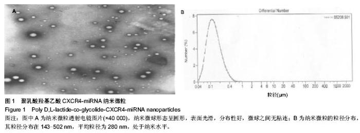
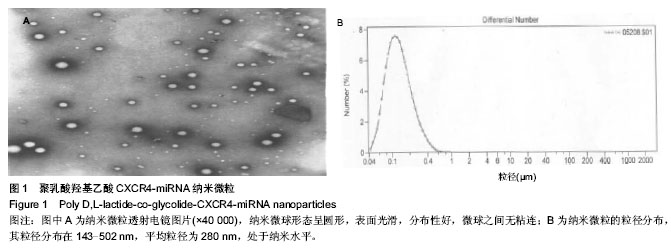
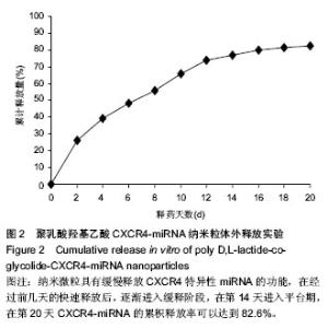
2.1 纳米复合微粒包封率和载药量的测定 分别对不同时间制备的3批次纳米微球进行CXCR4-miRNA含量测定,其平均载药量为(0.515±0.023)%,平均包封率为50.2%,批间差异小,制备工艺简单,充分说明本方法工艺稳定,重现性好。与国内外其他研究相比[26-27],纳米微粒的载药量、包封率类似。 2.2 纳米微粒的形态、粒径和分布观察 结果表明,本实验所制备的纳米微球形态呈圆形,表面光滑,分布性好,微球之间无粘连(图1A),其粒径分布在143-502 nm,平均粒径为280 nm(图1B),处于纳米水平。 2.3 纳米微粒体外释放实验 由聚乳酸羟基乙酸CXCR4-miRNA纳米微粒体外释放实验结果(图2)可以看出,纳米微粒具有缓慢释放CXCR4特异性miRNA的功能,在经过前几天的快速释放后,逐渐进入缓释阶段,在第14天进入平台期,在第20天CXCR4-miRNA的累积释放率可以达到82.6%。"
"

| [1] Wei F,Moore DC,Li Y,et al.Attenuation of osteoarthritis via blockade of the SDF-1/CXCR4 signaling pathway.Arthritis Res Ther.2012;14(4):R177. [2] Bikash D,Shili X,Fedora G,et al.Small Molecule Inhibitors of CXCR4.Theranostics.2013;3(1):47-75. [3] Jin F,Brockmeier U,Otterbach F,et al.New insight into the SDF-1/CXCR4 axis in a breast carcinoma model: hypoxia-induced endothelial SDF-1 and tumor cell CXCR4 are required for tumor cell intravasation.Mol Cancer Res. 2012;10(8):1021-1031. [4] Heckmann D,Maier P,Laufs S,et al.The disparate twins: a comparative study of CXCR4 and CXCR7 in SDF-1α-induced gene expression, invasion and chemosensitivity of colon cancer.Clin Cancer Res.2014;20(3):604-616. [5] Seku?a M,Miekus K,Majka M.Downregulation of the CXCR4 receptor inhibits cervical carcinoma metastatic behavior in vitro and in vivo.Int J Oncol.2014;44(6):1853-1860. [6] Zhao Y,Guo J,Gu S,et al.SDF-1/CXCR4 signal is involved in decreased expression of p57kip2 in de novo MDS patients. Hematology.2012;17(4):220-228. [7] Wang L, Huang T, Chen W, et al. Silencing of CXCR4 by RNA interference inhibits cell growth and metastasis in human renal cancer cells.Oncol Rep. 2012;28(6): 2043-2048. [8] D'Alterio C,Consales C,Polimeno M,et al.Concomitant CXCR4 and CXCR7 expression predicts poor prognosis in renal cancer.Curr Cancer Drug Targets. 2010;10(7):772-781. [9] Bhardwaj A,Singh S,Singh AP.MicroRNA-based Cancer Therapeutics: Big Hope from Small RNAs.Mol Cell Pharmacol. 2010;2(5):213-219. [10] Zhang G,Wang Q,Xu R.Therapeutics Based on microRNA: A New Approach for Liver Cancer.Curr Genomics.2010;11(5): 311-325. [11] Ranganath SH,Fu Y,Arifin DY,et al.The use of submicron/nanoscale PLGA implants to deliver paclitaxel with enhanced pharmacokinetics and therapeutic efficacy in intracranial glioblastoma in mice.Biomaterials. 2010;31(19): 5199-5207. [12] de Martimprey H,Bertrand JR,Malvy C,et al.New core-shell nanoparticules for the intravenous delivery of siRNA to experimental thyroid papillary carcinoma. Pharm Res. 2010; 27(3):498-509. [13] 董勤,崔杰,高烽,等.STAT3反义寡核苷酸纳米复合微粒的制备及特性研究[J].武警医学院学报,2011,20(9):723-726. [14] Liu JW,Zhao Q,Wan CX.Research progresses on degradation mechanism in vivo and medical applications of polylactic acid.Space Med Eng.2001;14(4):308-312. [15] Sahoo SK,Ma Labhasetwar V.Efficacy of transferrin- conjugated paclitaxel-loaded nanoparticles in a murine model of prostate cancer.Int J Cancer.2004;112(2): 335-340. [16] Rivera PA,Martinez-Oharriz MC,Rubio M,et al.Fluconazole encapsulation in PLGA microspheres by spray-drying.J Microencaspsul.2004;21(2):203-211. [17] Cheng FY,Wang SP,Su CH,et al. Stabilizer-free poly(lactide-co-glycolide)nanoparticles for multimodal biomedical probes.Biomaterials.2008;29(13):2014-2112. [18] Amjadi I,Rabice M,Hosseini MS,et al.Synthesis and characterization of doxorubicin-loaded poly(lactide-co- glycolide)nanoparticles as a sustained-release anticancer drug delivery system.Appl Biochem Biotechnol.2012;168(6): 1434-1447. [19] Nair KL,Thulasidasan AKT,Deepa G,et al.Purely aqueous PLGA nanoparticulate formulations of curcumin exhibit enhanced anticancer activity with dependence on the combination of the carrier.Int J Pharm.2012;425(1/2):44-52. [20] Anand P,Nair HB,Sung B,et al.Design of Curcumin Loaded PLGA Nanoparticles Formulation with Enhanced Cellular Uptake, and Increased Bioactivity in vitro and Superior Bioavailabilityin vivo.Biochem Pharmacol.2010;79(3): 330-338. [21] Wang YF,Liu PF,Qiu LH,et al.Toxicity and therapy of cisplatin- loaded EGF modified mPEG-PLGA-PLL nanoparticles for SKOV3 cancer in mice.Biomaterials. 2013;34(16):4068-4077. [22] Chen J,Li S,Shen Q.Folic acid and cell-penetrating peptide conjugated PLGA-PEG bifunctional nanoparticles for vincristine sulfate delivery.Eur J Pharm Sci. 2012;47(2): 430-443. [23] Graf N,Bielenberg DR,Kolishetti N,et al.alpha(V)beta(3) Integrin- Targeted PLGA-PEG Nanoparticles for Enhanced Anti-tumor Efficacy of a Pt(IV) Prodrug.ACS Nano. 2012;6(5): 4530-4539. [24] Wang G,Yu B,Wu Y,et al.Controlled preparation and antitumor efficacy of vitamin E TPGS-functionalized PLGA nanoparticles for delivery of paclitaxel.Int J Pharm. 2013;446(1/2):24-33. [25] Chakravarthi SS, Robinson DH.Enhanced cellular association of paclitaxel delivered in chitosan-PLGA particles. Int J Pharm. 2011;409(1-2):111-120. [26] 马立坤,叶鹏,黄文良,等.骨形态发生蛋白2/聚乳酸缓释微球的制备及表征[J].中国组织工程研究,2014,18(3):395-400. [27] Dangi R,Hurkat P,Jain A,et al.Targeting liver cancer via ASGP receptor using 5-FU-loaded surface-modified PLGA nanoparticles.J Microencapsul.2014;31 (5):479-487. [28] Zanin H,Hollanda LM,Ceragioli HJ,et al.Carbon nanoparticles for gene transfection in eukaryotic cell lines. Mater Sci Eng C Mater Biol Appl.2014;39:359-370. [29] Apaolaza PS,Delgado D,del Pozo-Rodríguez A,et al.A novel gene therapy vector based on hyaluronic acid and solid lipid nanoparticles for ocular diseases. Int J Pharm.2014;465(1-2): 413-426. [30] Ji XT,Huang L,Huang HQ.Construction of nanometer cisplatin core-ferritin (NCC-F) and proteomic analysis of gastric cancer cell apoptosis induced with cisplatin released from the NCC-F. J Proteomics.2012;75(11):3145-3157. [31] Liu J,Jiang Z,Zhou J,et al.Enzyme-synthesized poly (amine-co-esters) as nonviral vectors for gene delivery.J Biomed Mater Res A.2011;96(2):456-465. [32] Chen M,Ouyang H, Zhou S,et al.PLGA-nanoparticle mediated delivery of anti-OX40 monoclonal antibody enhances anti-tumor cytotoxic T cell responses. Cell Immunol. 2014;287(2):91-99. [33] Feng L,Ward JA,Li SK,et al.Assessment of PLGA-PEG-PLGA copolymer hydrogel for sustained drug delivery in the ear.Curr Drug Deliv,.2014;11(2):279-286. [34] Jagani HV,Josyula VR,Palanimuthu VR,et al.Improvement of therapeutic efficacy of PLGA nanoformulation of siRNA targeting anti-apoptotic Bcl-2 through chitosan coating. Eur J Pharm Sci.2013;48(4-5):611-618. [35] Chen YS,Alany RG,Young SA,et al.In vitro release characteristics and cellular uptake of poly (D,L-lactic-co-glycolic acid) nanoparticles for topical delivery of antisense oligodeoxynucleotides.Drug Deliv.2011;18(7):493-501. [36] Lane ME,Brennan FS,Corrigan OI.Comparison of post-emulsification freeze drying or spray drying processes for the microencapsulation of plasmid DNA. J Pharm Pharmacol. 2005;57(7):831-838. [37] Qi F,Wu J,Fan Q,et al.Preparation of uniform-sized exenatide-loaded PLGA microspheres as long-effective release system with high encapsulation efficiency and bio-stability.Colloids Surf B Biointerfaces.2013;112:492-498. [38] 郑延波,刘胜,李丽君,等.包载缺氧诱导因子-1ɑ反义寡核苷酸纳米粒子的制备和特性评价[J].生物医学工程与临床,2011, 15(6): 579-563. [39] Matsumoto A,Matsukawa Y,Suzuki T,et al.Drug release characteristics of multi-reservoir type microspheres with poly(dl-lactide-co-glycolide) and poly(dl-lactide). J Control Release.2005;106(1-2):172-180. [40] 叶向阳,孙湘,贾会文,等.利福平/聚乳酸-聚羟基乙酸缓释微球的制备及特性[J].中国组织工程研究与临床康复,2011,15(51): 9608-9612. [41] Lee YS, Lowe JP,Gilby E,et al.The initial release of cisplatin from poly(lactide-co-glycolide) microspheres.Int J Pharm. 2010;383(1-2):244-254 |
| [1] | Zhang Shuang, Tan Rui, Wang Chunxiao, Wu Fengyu, Guo Hongyu. MicroRNAs for assessing the motion control of human skeletal muscles [J]. Chinese Journal of Tissue Engineering Research, 2021, 25(17): 2755-2760. |
| [2] | Shi Zhengliang, Zhang Hua, Fan Zhiyong, Ma Wei, Yuan He, Yang Bing. Nerve conduits of chitosan/polyvinyl alcohol with brain-derived neurotrophic factor microspheres for peripheral nerve defects in rats [J]. Chinese Journal of Tissue Engineering Research, 2021, 25(10): 1555-1559. |
| [3] | Ren Chunmei, Liu Yufang, Xu Nuo, Shao Miaomiao, He Jianya, Li Xiaojie. Non-coding RNAs in human dental pulp stem cells: regulations and mechanisms [J]. Chinese Journal of Tissue Engineering Research, 2020, 24(7): 1130-1137. |
| [4] | Feng Xiaoxia, Hou Weiwei, Jin Xiaoting, Wang Xinhua. Construction of periodontal biomimetic membrane with electrospun poly(lactic-co-glycolic acid) nanofibers and electrosprayed chitosan microspheres [J]. Chinese Journal of Tissue Engineering Research, 2020, 24(4): 511-516. |
| [5] | Lei Senlin, Liu Hongyuan, Yang Hongsheng, Xiong Yan, Duan Hong. In vivo biosafety and histocompatibility of absorbable poly-D,L-lactic acid screws implanted with ultrasound-assisted technology [J]. Chinese Journal of Tissue Engineering Research, 2020, 24(34): 5454-5460. |
| [6] | Liu Xin, Du Bin, Sun Guangquan, Cao Jinxing, Jiang Xiaohong. Porous beta-tricalcium phosphate-polypyrrole-biotin-icariin composite scaffold promotes recruitment of bone marrow mesenchymal stem cells [J]. Chinese Journal of Tissue Engineering Research, 2020, 24(34): 5532-5537. |
| [7] | Wang Le, Hui Min, Dong Xiling, Zhou Han, Dong Hongliang, Zhang Xiaoming, Liu Tongbin. Effects of sustained-release atorvastatin calcium nanofiber scaffold on cell adhesion and proliferation [J]. Chinese Journal of Tissue Engineering Research, 2020, 24(28): 4492-4497. |
| [8] | Yang Zhen, Li Hao, Gao Cangjian, Fu Liwei, Tian Guangzhao, Zha Kangkang, Sun Zhiqiang, Li Xu, Guo Weimin, Sui Xiang, Huang Jingxiang, Liu Shuyun, Lu Shibi, Guo Quanyi . Regulation of stem cells by transforming growth factor β3/polylactic acid-glycolic acid microspheres [J]. Chinese Journal of Tissue Engineering Research, 2020, 24(28): 4540-4546. |
| [9] | Zhang Helong, Wang Huiyan, Li Zhuo, Gao Jianguo, Zhai Qian. In vitro anti-tuberculosis effect of chitosan-gelatin/poly(lactic acid co-glycolic acid) combined with drug-loaded hydrogel [J]. Chinese Journal of Tissue Engineering Research, 2020, 24(22): 3480-3485. |
| [10] | Yuan Tao, Jiang Hui, Lai Zhendeng, Qian Hong, Yang Shaoqiang, Feng Zhangqi, Zhao Jianning, Bao Nirong. A novel hydroxyapatite fiber scaffold for three-dimensional cell culture [J]. Chinese Journal of Tissue Engineering Research, 2020, 24(22): 3491-3497. |
| [11] | Ma Rui, Wang Jialin, Li Yongwei, Wang Wei. In vitro antibacterial activity and biocompatibility of a porous scaffold containing magnesium [J]. Chinese Journal of Tissue Engineering Research, 2020, 24(22): 3534-3539. |
| [12] | He Jianwei, Zheng Hanshan, Xu Jian, Zhang Ying. Changes in three physical indicators of the muscle after far-infrared ceramic microsphere intervention [J]. Chinese Journal of Tissue Engineering Research, 2020, 24(22): 3527-3533. |
| [13] | Zhang Zhongyan, Li Yubo, Qi Tongning, Chang Tao. Three-dimensional printed polylactic acid resin humerus combined with bioactive coating promotes osteoblast adhesion and increases antibacterial ability [J]. Chinese Journal of Tissue Engineering Research, 2020, 24(16): 2485-2492. |
| [14] | Cheng Lei, Jin Jian, Hu Jingguo, Lu Yusong. Inflammatory reaction and lactic acid concentration after implantation of polylactic acid rib nail versus pure titanium embracing fixator in animals#br# [J]. Chinese Journal of Tissue Engineering Research, 2020, 24(16): 2567-2571. |
| [15] | Qu Renfei, Mai Yuying, Chen Xiaowei, Hu Huanying, Chen Guozhi, Li Dongdong, Liao Hongbing. Relationship between lactic acid concentration and osteoclast differentiation of mouse monocytes [J]. Chinese Journal of Tissue Engineering Research, 2020, 24(14): 2177-2183. |
| Viewed | ||||||
|
Full text |
|
|||||
|
Abstract |
|
|||||