Chinese Journal of Tissue Engineering Research ›› 2014, Vol. 18 ›› Issue (38): 6099-6104.doi: 10.3969/j.issn.2095-4344.2014.38.006
Previous Articles Next Articles
Bone morphogenetic protein-4 compounded with platelet-rich plasma promotes bone healing
Shi Jian-jie, Luo Zhi-bin, Chen Wen-xiong
- Department of Stomatology, the Third Affiliated Hospital of Guangzhou Medical University, Guangzhou 510150, Guangdong Province, China
-
Received:2014-08-21Online:2014-09-10Published:2014-09-10 -
Contact:Luo Zhi-bin, Master, Attending physician, Department of Stomatology, the Third Affiliated Hospital of Guangzhou Medical University, Guangzhou 510150, Guangdong Province, China -
About author:Shi Jian-jie, Chief physician, Department of Stomatology, the Third Affiliated Hospital of Guangzhou Medical University, Guangzhou 510150, Guangdong Province, China -
Supported by:the Science and Technology Development Program of Guangdong Province, No. 2010B060900087
CLC Number:
Cite this article
Shi Jian-jie, Luo Zhi-bin, Chen Wen-xiong . Bone morphogenetic protein-4 compounded with platelet-rich plasma promotes bone healing[J]. Chinese Journal of Tissue Engineering Research, 2014, 18(38): 6099-6104.
share this article
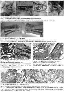
2.1 实验动物数量分析 实验选用大白兔24只,分为4组,实验过程无脱失,全部进入结果分析。 2.2 兔骨缺损区大体观察 大体标本可见12周时空白对照组成骨较差,缺损区中间凹陷,表面纤维组织增生;余各组成骨作用好,成骨饱满,表面光滑(图2)。 2.3 兔骨缺损区组织学观察 4周:富血小板血浆组、富血小板血浆+骨形态发生蛋白4组两组骨缺损处见新生血管丰富,其中富血小板血浆+骨形态发生蛋白4组可见较多的纤维性骨痂形成,局部见较多成骨细胞及软骨细胞增生活跃,骨移植颗粒开始降解,可见与骨干平行的骨样组织,富血小板血浆组见纤维组织生长,材料降解为网状,周围有炎性细胞和少量骨母细胞(见图3)。空白对照组、β-磷酸三钙+Bio-gide生物膜组两组骨缺损区见少量新生血管,β-磷酸三钙+Bio-gide生物膜组周可见大量散在骨粉颗粒及纤维组织结构,其间散在数量不等的成骨细胞和软骨细胞,成骨作用较对照组强。 8周:富血小板血浆+骨形态发生蛋白4组缺损区内骨粉颗粒明显较β-磷酸三钙+Bio-gide生物膜组、富血小板血浆组减少,伴随着大量骨样组织形成,钙化、沉积明显;β-磷酸三钙+Bio-gide生物膜组、富血小板血浆组局部见新生骨组织,少量破骨细胞,少量钙盐沉积,两组间差异无显著性意义;空白对照组出现以纤维性骨痂为主,自体骨边缘见少量新生骨组织,明显较β-磷酸三钙+Bio-gide生物膜组、富血小板血浆组少(图4)。 12周:富血小板血浆+骨形态发生蛋白4组主要为成熟骨,为板层骨,可见哈佛氏小管形成,散在少量编织骨,骨粉颗粒明显减少,少量破骨细胞(图5)。 2.4 组织学图像分析缺损区新生骨结果 计算机图像分析显示,伴随愈合时间推移,新骨形成逐渐增多,β-磷酸三钙颗粒逐渐降解,其中富血小板血浆+骨形态发生蛋白4组12周时新生骨略高于8周但两者差异无显著性意义(P > 0.05)。 术后4 周,富血小板血浆+骨形态发生蛋白4组新生骨略多于富血小板血浆组,但两组间差异无显著性意义 (P > 0.05) ,富血小板血浆组、富血小板血浆+骨形态发生蛋白4组两组骨缺损区新生骨以及新生血管均多于β-磷酸三钙+Bio-gide组实验侧(P < 0.01),β-磷酸三钙+Bio-gide组新成骨多于空白对照组(P < 0.01);术后4,8,12周,富血小板血浆+骨形态发生蛋白4组骨缺损区新生骨均多于β-磷酸三钙+Bio-gide组、富血小板血浆组两组骨缺损区(P < 0.01),富血小板血浆组略多于β-磷酸三钙+Bio-gide组,但两者差异无显著性意义(P > 0.05),β-磷酸三钙+ Bio-gide组、富血小板血浆组两组均多于空白对照组(P < 0.01)。"

| [1] Carlsson L,Röstlund T,Albrektsson B,et al.Osseointegration of titanium implants. Acta Orthop Scand.1986;57(4):285-289. [2] Joos U, Kleinheinz J. Reconstruction of the severely resorbed (Class VI) jaws: routine or exception?J Craniomaxillofac Surg.2000;28(1): 1-4. [3] Balk SD.Calcium as a regulator of the proliferation of normal, but not of transformed, chicken fibroblasts in a plasma-containing medium. Proc Natl Acad Sci USA. 1971;68(2): 271-276. [4] Hildner F, Albrecht C, Gabriel C, et al. State of the art and future perspectives of articular cartilage regeneration: a focus on adipose-derived stem cells and platelet-derived products. J Tissue Eng Regen Med. 2011;5(4):e36-51. [5] Lee KS, Wilson JJ, Rabago DP, et al.Musculoskeletal applications of platelet-rich plasma: fad or future? AJR Am J Roentgenol.2011; 196:628–636 [6] Raghoebar GM, Schortinghuis J, Liem RS, et al.Does platelet-rich plasma promote remodeling of autologous bone grafts used for augmentation of the maxillary sinus floor?. Clin Oral Implants Res.2005;16(3): 349-356. [7] Por YC, Barceló CR, Salyer KE, et al. Bone generation in the reconstruction of a critical size calvarial defect in an experimental model.J Craniofac Surg.2008;19(2): 383-392. [8] Urist MR,Strates BS.Bone morphogenetic protein.J Dent Res.1971;50(6): 1392-1406. [9] Reichert JC, Woodruff MA, Friis T, et al.Ovine bone- and marrowderived progenitor cells and their potential for scaffold-based bone tissue engineering applications in vitro and in vivo. J Tissue Eng Regen Med.2010;4:565-576. [10] De Biase P, Capanna R. Clinical applications of MBP. Injury. 2005;36(Suppl 3):S43-S46. [11] Deschaseaux F, Sensebe L, Heymann D . Mechanisms of bone repair and regeneration. Trends Mol Med.2009;15: 417-429. [12] Wallace AL, Makki R, Weiss JB, et al. Measurement of serum angiogenic factor in devascularized experimental tibial fractures. J Orthop Trauma.1995;9(4): 324-332. [13] Carreira AC1, Lojudice FH, Halcsik E, et al. Bone morphogenetic proteins: facts, challenges, and future perspectives. J Dent Res.2014;93(4):335-380. [14] Ali IH, Brazil DP. Bone morphogenetic proteins and their antagonists: current and emerging clinical uses. Br J Pharmacol.2014;171(15):3620-3652. [15] Boerckel JD, Kolambkar YM, Dupont KM, et al. Effects of protein dose and delivery system on BMP-mediated bone regeneration. Biomaterials.2011;32: 5241-5251. [16] Karsenty G. The complexities of skeletal biology. Nature. 2003; 423: 316-318. [17] Nakamura J, Yanagita M. Bmp modulators in kidney disease. Discov Med.2012; 13: 57-63. [18] Wang EA, Rosen V, D'Alessandro JS,et al.Recombinant human bone morphogenetic protein induces bone formation. Proc Natl Acad Sci U S A.1990;87(6): 2220-2224. [19] Nakahara H, Takaoka K, Koezuka M, et al.Periosteal bone formation elicited by partially purified bone morphogenetic protein. Clin Orthop Relat Res.1989;(239): 299-305. [20] Kim CS, Kim JI, Kim J, et al. Ectopic bone formation associated with recombinant human bone morphogenetic proteins-2 using absorbable collagen sponge and beta tricalcium phosphate as carriers. Biomaterials.2005;26(15): 2501-2507. [21] Degat MC, Ferreira E, Logeart-Avramoglou D. Use of growth factors in the repair of bone. Pathol Biol (Paris). 2005;53: 131-141. [22] Ben-David D, Srouji S, Shapira-Schweitzer K, et al. Low dose BMP-2 treatment for bone repair using a PEGylated fibrinogen hydrogel matrix. Biomaterials. 2013;34:2902-2910. [23] Winn SR, Uludag H, Hollinger JO. Carrier systems for bone morphogenetic proteins. Clin Orthop Relat Res.1999; 367 (Suppl):95-106. [24] Yang X, Han G, Pang X, et al. Chitosan/collagen scaffold containing bone morphogenetic protein-7 DNA supports dental pulp stem cell differentiation in vitro and in vivo. J Biomed Mater Res A. 2012; Feb 18. doi: 10.1002/jbm. a.34064. [Epub ahead of print] [25] Polimeni G, Koo KT, Pringle GA, et al. Histopathological observations of a polylactic acid-based device intended for guided bone/tissue regeneration. Clin Implant Dent Relat Res.2008;10:99-105. [26] Cestari TM, Granjeiro JM, de Assis GF,et al. Bone repair and augmentation using block of sintered bovine-derived anorganic bone graft in cranial bone defect model. Clin Oral Implants Res.2009;20:340-350. [27] Salgado PC, Sathler PC, Castro HC, et al. Bone remodeling, biomaterials and technological applications: revisiting basic concepts.J Biomater Nanobiotechnol.2011;2:318-328. [28] Hagi TT, Wu G, Liu Y, et al. Cell-mediated BMP-2 liberation promotes bone formation in a mechanically unstable implant environment. Bone.2010;46:1322-1327. [29] Berner A, Boerckel JD, Saifzadeh S, et al. Biomimetic tubular nanofiber mesh and platelet rich plasma-mediated delivery of bmp-7 for large bone defect regeneration. Cell Tissue Res. 2012;347(3):603-615. [30] 罗应伟,李松,沈丽宁. BMP-4 mRNA在Meckel’s软骨中的表达及意义.[J].昆明医学院学报,2004;25(3):9-12. [31] Dimitriou R, Tsiridis E, Giannoudis PV. Current concepts of molecular aspects of bone healing. Injury. 2005;36(12): 1392-1404. [32] Gale NW, Yancopoulos GD. Growth factors acting via endothelial cell-specific receptor tyrosine kinases: VEGFs, angiopoietins, and ephrins in vascular development. Genes Dev. 1999;13(9):1055-1066. [33] Alvarez RH, Kantarjian HM, Cortes JE. Biology of platelet- derived growth factor and its involvement in disease. Mayo Clin Proc.2006;81(9):1241-1257. [34] Conover CA. In vitro studies of insulin-like growth factor I and bone. Growth Horm IGF Res. 2000; 10 Suppl B:S107-10. Review. [35] Canalis E. Effect of insulinlike growth factor I on DNA and protein synthesis in cultured rat calvaria. J Clin Invest. 1980; 66(4):709-719. [36] Tanaka H, Quarto R,Williams S,et al.In vivo and in vitro effects of insulin-like growth factor-I (IGF-I) on femoral mRNA expression in old rats. Bone.1994; 15(6):647-53. [37] Meraw SJ, Reeve CM, Lohse CM,et al.Treatment of peri-implant defects with combination growth factor cement. J Periodontol.2000;71(1):8-13. [38] Sipe JB, Zhang J, Waits C, et al.Localization of bone morphogenetic proteins (MBP)-2,-4, and -6 within megakaryocytes and platelets. Bone.2004;35(6):1316-1322. [39] Glowacki J. Angiogenesis in fracture repair. Clin Orthop Relat Res.1998;(355 Suppl): S82-89. 23. [40] Salim A, Nacamuli RP, Morgan EF,et al.Transient changes in oxygen tension inhibit osteogenic differentiation and Runx2 expression in osteoblasts. J Biol Chem.2004;279(38): 40007-40016. |
| [1] | Jiang Hongying, Zhu Liang, Yu Xi, Huang Jing, Xiang Xiaona, Lan Zhengyan, He Hongchen. Effect of platelet-rich plasma on pressure ulcers after spinal cord injury [J]. Chinese Journal of Tissue Engineering Research, 2021, 25(8): 1149-1153. |
| [2] | Liu Yafei, Wang Yalin, Zuo Yanping, Sun Qi, Wei Jing, Zhao Lixia. Structural changes of the temporomandibular joint in adolescents with skeletal Class III malocclusions after maxillary protraction: an X-ray measurement analysis [J]. Chinese Journal of Tissue Engineering Research, 2021, 25(8): 1154-1159. |
| [3] | He Xiangzhong, Chen Haiyun, Liu Jun, Lü Yang, Pan Jianke, Yang Wenbin, He Jingwen, Huang Junhan. Platelet-rich plasma combined with microfracture versus microfracture in the treatment of knee cartilage lesions: a meta-analysis [J]. Chinese Journal of Tissue Engineering Research, 2021, 25(6): 964-969. |
| [4] | Zhang Bin, Sun Lihua, Zhang Junhua, Liu Yusan, Cui Caiyun. A modified flap immediate implant is beneficial to soft tissue reconstruction in maxillary aesthetic area [J]. Chinese Journal of Tissue Engineering Research, 2021, 25(5): 707-712. |
| [5] | Li Yuanyuan, Lu Yingjuan, Ye Yushan, Mustafa M.M Weldali, Chang Shaohai. Constructing finite element models of three maxillary arch forms [J]. Chinese Journal of Tissue Engineering Research, 2021, 25(20): 3125-3129. |
| [6] | Zuo Xiuqin, Yin Sasa, Xie Huimin, Jia Zishan, Zhang Lining. Applicability and specifications of platelet-rich plasma in musculoskeletal repair [J]. Chinese Journal of Tissue Engineering Research, 2021, 25(20): 3239-3245. |
| [7] | Nie Jing, Shi Xiaoyu. Cone-beam CT measurement of alveolar bone thickness of the maxillary anterior area at implant anchorage site in different sexes [J]. Chinese Journal of Tissue Engineering Research, 2021, 25(14): 2133-2136. |
| [8] | Chen Jufang, Tian Yulou, Hao Xin. Role and potential of adipose-derived stem cells in cranio-maxillofacial bone regeneration [J]. Chinese Journal of Tissue Engineering Research, 2021, 25(13): 2087-2096. |
| [9] | Tian Yanping, Li Juan, Liu Xiaobo, Zhang Huiling, Shi Lihong, Jin Rongjiang. Knowledge network mapping of literature regarding platelet-rich plasma in recent 5 years: a visual analysis [J]. Chinese Journal of Tissue Engineering Research, 2021, 25(11): 1745-1752. |
| [10] | Wang Guangping, Li Mingxia, Han Yu, Xu Xiaomei, Xu Jie, Huang Suhua. Comparison of the impacts of two kinds of brackets on external apical root resorption in orthodontic treatment of bimaxillary protrusion patients [J]. Chinese Journal of Tissue Engineering Research, 2021, 25(10): 1539-1544. |
| [11] | Yu Chenghao, Zhang Yi, Qi Chao, Chen Jinli, Gao Jiake, Yu Tengbo. Effect of cytokines and platelet-rich plasma on tendon derived stem cells [J]. Chinese Journal of Tissue Engineering Research, 2021, 25(1): 133-140. |
| [12] | Wang Ning, Zhong Weijian. Application and function of autologous blood concentrate in tissue regeneration [J]. Chinese Journal of Tissue Engineering Research, 2021, 25(1): 146-151. |
| [13] | Xiu Yiping, Zhang Liyan, Qian Xueyi, Li Yan, Li Wantong. The clinical application of platelet-rich plasma to repair chronic refractory wounds: a retrospective study and literature retrieval evidence analysis [J]. Chinese Journal of Tissue Engineering Research, 2020, 24(8): 1231-1237. |
| [14] | Qiu Guorong, He Benxiang, Wang Chun. Biological mechanism of platelet-rich plasma for tendinopathy repair: a visual study based on scientific knowledge map [J]. Chinese Journal of Tissue Engineering Research, 2020, 24(5): 787-795. |
| [15] | Ren Jun, Zhao Yan, Xiao Bin, Ma Chao, Wang Xinke, Hao Yabin, Cheng Jie. Platelet-rich plasma promotes the healing of tibial fracture in rabbits [J]. Chinese Journal of Tissue Engineering Research, 2020, 24(35): 5595-5599. |
| Viewed | ||||||
|
Full text |
|
|||||
|
Abstract |
|
|||||