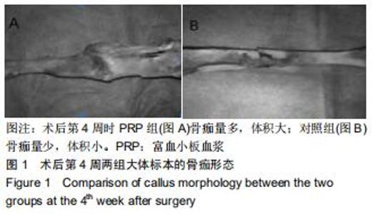
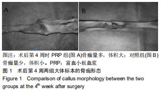
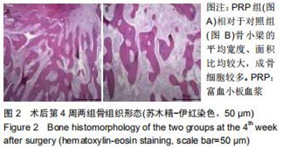
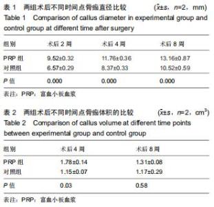
|
[1] AURÉGAN JC, BÉGUÉ T, RIGOULOT G, et al. Success rate and risk factors of failure of the induced membrane technique in children: a systematic review. Injury. 2016;47:S62-S67.
[2] MORELLI I, DRAGO L, GEORGE DA, et al. Masquelet technique: myth or reality? A systematic review and meta-analysis. Injury. 2016; 47:S68-S76.
[3] LE ADK, ENWEZE L, DEBAUN MR, et al. Platelet-Rich Plasma. Clin Sports Med. 2019;38:17-44.
[4] BAEYENS W, GLINEUR R, EVRARD L. The use of platelet concentrates: platelet-rich plasma (PRP) and platelet-rich fibrin (PRF) in bone reconstruction prior to dental implant surgery. Rev Med Brux. 2010;31:521-527.
[5] SCALA M, GIPPONI M, MEREU P, et al. Regeneration of mandibular osteoradionecrosis defect with platelet rich plasma gel. In vivo (Athens, Greece). 2010;24(6):889-893.
[6] JUNGBLUTH P, WILD M, GRASSMANN JP, et al. Platelet-rich plasma on calcium phosphate granules promotes metaphyseal bone healing in mini-pigs. J Orthop Res. 2010; 28(11):1448-1455.
[7] 袁霆,张长青,陆男吉,等.富血小板血浆修复皮肤缺损的实验研究[J].中华创伤骨科杂志,2008,10(7):611-614.
[8] CHANDRA RK, HANDORF C, WEST M, et al. Histologic effects of autologous platelet gel in skin flap healing. Arch Facial Plast Surg. 2007;9:260-263.
[9] 宋扬,宫苹.传统二次离心法和改良法制备富血小板血浆的比较研究[J].实用口腔医学杂志,2005,21(5):629-632.
[10] 付小兵.进一步重视体表慢性难愈合创面发生机制与防治研究[J].中华创伤杂志,2004,20(8):449-451.
[11] 刘建忠,周元国,程天民,等.不同剂量软X射线照射对大鼠伤口愈合影响规律的研究[J].中华放射医学与防护杂志,2002,22(3):183-186.
[12] WAGNER W, WEHRMANN M. Differential cytokine activity and morphology during wound healing in the neonatal and adult rat skin. J Cell Mol Med. 2007;11(6):1342-1351.
[13] 马乐园,赵岩,乔万庆,等.创伤性脑损伤合并骨折中加速骨折愈合过程中相关因子的表达[J].中国组织工程研究,2017,21(32):5115-5121.
[14] 石长贵,蔡筑韵,鲍哲明,等.骨生长因子促进骨折愈合研究进展[J].国际骨科学杂志,2010,31(3):58-60.
[15] 中国医疗保健国际交流促进会骨科分会.富血小板血浆在骨关节外科临床应用专家共识(2018年版)[J].中华关节外科杂志(电子版), 2018,12(5):596-600.
[16] 黄若昆,林月秋,滕寿发,等.复合富血小板血浆的酶处理异种骨修复兔桡骨缺损[J].中国组织工程研究与临床康复,2007,11(49): 9898-9901.
[17] BOSWELL SG, COLE BJ, SUNDMAN EA, et al. Platelet-rich plasma: a milieu of bioactive factors. Arthroscopy. 2012; 28: 429-439.
[18] FERNANDES G, YANG S. Application of platelet-rich plasma with stem cells in bone and periodontal tissue engineering. Bone Res. 2016;4:16036.
[19] RUEHLE MA, KRISHNAN L, VANTUCCI CE, et al. Effects of BMP-2 dose and delivery of microvascular fragments on healing of bone defects with concomitant volumetric muscle loss. J Orthop Res. 2019;37:553-561.
[20] LIAO JCY, HE M, GAN AWT, et al. The effects of autologous platelet-rich fibrin on flexor tendon healing in a rabbit model. J Hand Surg. 2017;42(11):9281-9287.
|