Chinese Journal of Tissue Engineering Research ›› 2014, Vol. 18 ›› Issue (37): 5935-5941.doi: 10.3969/j.issn.2095-4344.2014.37.006
Previous Articles Next Articles
Different pro-angiogenesis ability of bone marrow mesenchymal stem cells in multiple myeloma in active and remissive state
Li Xiang-li, Zhang Xiao-ying, Zang Li, Wang Xiao-fang
- Department of Hematology, Tianjin Medical University Cancer Institute and Hospital, National Clinical Research Center for Cancer, Key Laboratory of Cancer Prevention and Therapy, Tianjin 300060, China
-
Revised:2014-07-30Online:2014-09-03Published:2014-09-03 -
Contact:Wang Xiao-fang, M.D., Master’s supervisor, Department of Hematology, Tianjin Medical University Cancer Institute and Hospital, National Clinical Research Center for Cancer, Key Laboratory of Cancer Prevention and Therapy, Tianjin 300060, China -
About author:Li Xiang-li, Studying for master’s degree, Department of Hematology, Tianjin Medical University Cancer Institute and Hospital, National Clinical Research Center for Cancer, Key Laboratory of Cancer Prevention and Therapy, Tianjin 300060, China -
Supported by:the National Natural Science Foundation of China, No. 81272562
CLC Number:
Cite this article
Li Xiang-li, Zhang Xiao-ying, Zang Li, Wang Xiao-fang. Different pro-angiogenesis ability of bone marrow mesenchymal stem cells in multiple myeloma in active and remissive state[J]. Chinese Journal of Tissue Engineering Research, 2014, 18(37): 5935-5941.
share this article
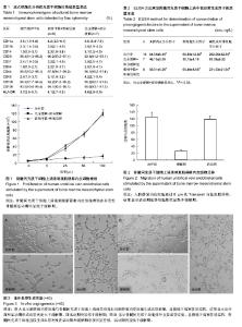
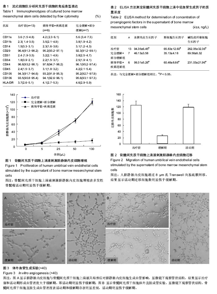
2.1 患者特征 收集天津市肿瘤医院化疗前后的13例多发性骨髓瘤患者,其中男9例,女4例;年龄51-62岁,中位年龄为56岁;根据国际分期系统,Ⅰ、Ⅱ、Ⅲ期患者分别为1例(7.7%)、2例(15.4%)、10例(76.9%);根据免疫球蛋白分型,IgG,IgA,λ轻链型分别为9例,3例,1例。根据国际骨髓瘤工作组统一标准[13],疗效评价分为完全缓解(Complete response,CR)、部分缓解(partial response,PR)、病情平稳(stable disease,SD)、疾病进展(Progressive disease,PD),完全缓解+部分缓解和病情平稳+疾病进展分别为6例,7例。 2.2 流式细胞仪分析骨髓间充质干细胞的免疫表型 多发性骨髓瘤在活动期(治疗前+前病情平稳+疾病进展)及缓解期(完全缓解+部分缓解)骨髓间充质干细胞的免疫表型基本相似。都表达 CD106,CD105,CD29,CD44,不表达CD11a,CD11b,CD14,CD31,CD34,CD45,HLA-DR (表1)。 2.3 ELISA检测血管生成因子水平 多发性骨髓瘤活动期患者骨髓间充质干细胞培养上清中血管内皮生长因子、肝细胞生长因子、碱性成纤维细胞生长因子的质量(P < 0.05,表2);血管生成因子质量浓度在治疗前和活动期之间差异无显著性意义(P > 0.05)。浓度明显高于缓解期 2.4 骨髓间充质干细胞上清液对人脐静脉内皮细胞活性及增殖影响 用MTT实验测定骨髓间充质干细胞培养上清液在不同的体积下对人脐静脉内皮细胞生长的影响。结果显示,骨髓瘤患者骨髓间充质干细胞上清对脐静脉内皮细胞增殖作用呈剂量依赖性,活动期明显强于缓解期(P < 0.05)。且治疗前组与病情平稳+疾病进展组间差异无显著性意义(P > 0.05,图1)。 2.5 骨髓间充质干细胞上清液对内皮细胞迁移影响 通过Transwell小室实验发现骨髓间充质干细胞上清液影响人脐静脉内皮细胞向基底膜迁移,迁移数量在骨髓瘤活动期明显大于缓解期;统计学结果为骨髓瘤活动期患者骨髓间充质干细胞上清对脐静脉内皮细胞迁移作用明显强于缓解期(P < 0.05,图2)。且治疗前组与病情平稳+疾病进展组间差异无显著性意义(P > 0.05)。 2.6 体外血管形成能力 体外成血管实验显示,骨髓瘤患者骨髓间充质干细胞上清在体外具有明显成血管作用,活动期明显强于缓解期(P < 0.05)。骨髓间充质干细胞上清的体外成血管能力在治疗前组与病情平稳+疾病进展组间差异无显著性意义(P > 0.05,图3A)。此外,多发性骨髓瘤活动期骨髓间充质干细胞体外直接成管作用明显强于缓解期(P < 0.05,图3B),且治疗前组与病情平稳+疾病进展组间差异无显著性意义(P > 0.05)。"
| [1] Manier S, Sacco A, Leleu X,et al.Bone marrow microenvironment in multiple myeloma progression.J Biomed Biotechnol. 2012;2012:157496. [2] Ribatti D, Nico B, Vacca A.Importance of the bone marrow microenvironment in inducing the angiogenic response in multiple myeloma.Oncogene. 2006;25(31):4257-4266. [3] Zhang T, Lee YW, Rui YF,et al.Bone marrow-derived mesenchymal stem cells promote growth and angiogenesis of breast and prostate tumors.Stem Cell Res Ther. 2013; 4(3):70. [4] Kéramidas M, de Fraipont F, Karageorgis A,et al.The dual effect of mesenchymal stem cells on tumour growth and tumour angiogenesis.Stem Cell Res Ther. 2013;4(2):41. [5] Pittenger MF, Mackay AM, Beck SC,et al. Multilineage potential of adult human mesenchymal stem cells.Science. 1999;284(5411):143-147. [6] Kinnaird T, Stabile E, Burnett MS,et al. Marrow-derived stromal cells express genes encoding a broad spectrum of arteriogenic cytokines and promote in vitro and in vivo arteriogenesis through paracrine mechanisms.Circ Res. 2004;94(5):678-685. [7] Otsu K, Das S, Houser SD,et al. Concentration-dependent inhibition of angiogenesis by mesenchymal stem cells.Blood. 2009;113(18):4197-4205. [8] Beckermann BM, Kallifatidis G, Groth A,et al.VEGF expression by mesenchymal stem cells contributes to angiogenesis in pancreatic carcinoma.Br J Cancer. 2008; 99(4):622-631. [9] Zhang K, Shi B, Chen J,et al. Bone marrow mesenchymal stem cells induce angiogenesis and promote bladder cancer growth in a rabbit model.Urol Int. 2010;84(1):94-99. [10] Wang X, Zhang Z, Yao C.Angiogenic activity of mesenchymal stem cells in multiple myeloma.Cancer Invest. 2011;29(1): 37-41. [11] Podar K, Tai YT, Davies FE,et al.Vascular endothelial growth factor triggers signaling cascades mediating multiple myeloma cell growth and migration.Blood. 2001;98(2): 428-435. [12] Arnulf B, Lecourt S, Soulier J,et al. Phenotypic and functional characterization of bone marrow mesenchymal stem cells derived from patients with multiple myeloma.Leukemia. 2007; 21(1):158-163. [13] Kyle RA, Rajkumar SV.Criteria for diagnosis, staging, risk stratification and response assessment of multiple myeloma. Leukemia. 2009;23(1):3-9. [14] Carmeliet P.Angiogenesis in health and disease.Nat Med. 2003; 9(6):653-660. [15] Carmeliet P, Jain RK.Angiogenesis in cancer and other diseases.Nature. 2000;407(6801):249-257. [16] Bray F, Sankila R, Ferlay J,et al. Estimates of cancer incidence and mortality in Europe in 1995.Eur J Cancer. 2002;38(1):99-166. [17] Vacca A, Ribatti D, Roncali L,et al. Bone marrow angiogenesis and progression in multiple myeloma.Br J Haematol. 1994;87(3):503-508. [18] Alexandrakis MG, Passam FH, Sfiridaki A,et al. Elevated serum concentration of hepatocyte growth factor in patients with multiple myeloma: correlation with markers of disease activity.Am J Hematol. 2003;72(4):229-233. [19] Bolkun L, Lemancewicz D, Sobolewski K,et al.The evaluation of angiogenesis and matrix metalloproteinase-2 secretion in bone marrow of multiple myeloma patients before and after the treatment.Adv Med Sci. 2013;58(1):118-125. [20] Salvolini E, Orciani M, Vignini A,et al.Skin-derived mesenchymal stem cells (S-MSCs) induce endothelial cell activation by paracrine mechanisms.Exp Dermatol. 2010; 19(9):848-850. [21] Vacca A, Ria R, Ribatti D,et al.A paracrine loop in the vascular endothelial growth factor pathway triggers tumor angiogenesis and growth in multiple myeloma.Haematologica. 2003;88(2):176-185. [22] Menu E, Kooijman R, Van Valckenborgh E,et al.Specific roles for the PI3K and the MEK-ERK pathway in IGF-1-stimulated chemotaxis, VEGF secretion and proliferation of multiple myeloma cells: study in the 5T33MM model.Br J Cancer. 2004; 90(5):1076-1083. [23] Dankbar B, Padró T, Leo R,et al.Vascular endothelial growth factor and interleukin-6 in paracrine tumor-stromal cell interactions in multiple myeloma.Blood. 2000;95(8): 2630-2636. [24] Borset M, Lien E, Espevik T,et al.Concomitant expression of hepatocyte growth factor/scatter factor and the receptor c-MET in human myeloma cell lines.J Biol Chem. 1996; 271(40):24655-24661. [25] Com?a S, Ciuculescu F, Henschler R,et al.MCF7-MSC co-culture assay: approach to assess the co-operation between MCF-7s and MSCs in tumor-induced angiogenesis. Rom J Morphol Embryol. 2011;52(3 Suppl): 1071-1076. [26] Honczarenko M, Le Y, Swierkowski M,et al. Human bone marrow stromal cells express a distinct set of biologically functional chemokine receptors.Stem Cells. 2006;24(4): 1030-1041. [27] Com?a S, Ciuculescu F, Raica M.Mesenchymal stem cell-tumor cell cooperation in breast cancer vasculogenesis. Mol Med Rep. 2012;5(5):1175-1180. [28] Corre J, Mahtouk K, Attal M,et al. Bone marrow mesenchymal stem cells are abnormal in multiple myeloma.Leukemia. 2007; 21(5):1079-1088. [29] Li B, Shi M, Li J, et al. Elevated tumor necrosis factor-alpha suppresses TAZ expression and impairs osteogenic potential of Flk-1+ mesenchymal stem cells in patients with multiple myeloma.Stem Cells Dev. 2007;16(6):921-930. [30] Arnulf B, Lecourt S, Soulier J,et al. Phenotypic and functional characterization of bone marrow mesenchymal stem cells derived from patients with multiple myeloma.Leukemia. 2007; 21(1):158-163. [31] Garayoa M, Garcia JL, Santamaria C,et al. Mesenchymal stem cells from multiple myeloma patients display distinct genomic profile as compared with those from normal donors. Leukemia. 2009;23(8):1515-1527. [32] Sun B, Zhang S, Ni C,et al. Correlation between melanoma angiogenesis and the mesenchymal stem cells and endothelial progenitor cells derived from bone marrow.Stem Cells Dev. 2005;14(3):292-298. [33] Imaizumi T, Itaya H, Nasu S,et al. Expression of vascular endothelial growth factor in human umbilical vein endothelial cells stimulated with interleukin-1alpha--an autocrine regulation of angiogenesis and inflammatory reactions. Thromb Haemost. 2000;83(6):949-955. [34] Kojima-Yuasa A, Hua JJ, Kennedy DO,et al. Green tea extract inhibits angiogenesis of human umbilical vein endothelial cells through reduction of expression of VEGF receptors. Life Sci. 2003;73(10):1299-1313. [35] Aparicio S, Sawant S, Lara N,et al. Expression of angiogenesis factors in human umbilical vein endothelial cells and their regulation by PEDF.Biochem Biophys Res Commun. 2005;326(2):387-394. [36] Zhang B, Yang S, Zhang Y,et al.Co-culture of mesenchymal stem cells with umbilical vein endothelial cells under hypoxic condition.J Huazhong Univ Sci Technolog Med Sci. 2012; 32(2):173-180. [37] Oswald J, Boxberger S, Jørgensen B,et al. Mesenchymal stem cells can be differentiated into endothelial cells in vitro. Stem Cells. 2004;22(3):377-384. [38] Guo X, Li YL.Cytological basis and significance of mesenchymal stem cells differentiated into endothelial cells.Sheng Li Ke Xue Jin Zhan. 2005;36(3):204-208. [39] Pankajakshan D, Kansal V, Agrawal DK.In vitro differentiation of bone marrow derived porcine mesenchymal stem cells to endothelial cells.J Tissue Eng Regen Med. 2013;7(11): 911-920. [40] Rahbarghazi R, Nassiri SM, Khazraiinia P,et al.Juxtacrine and paracrine interactions of rat marrow-derived mesenchymal stem cells, muscle-derived satellite cells, and neonatal cardiomyocytes with endothelial cells in angiogenesis dynamics. Stem Cells Dev. 2013;22(6):855-865. [41] Feng B, Chen L.Review of mesenchymal stem cells and tumors: executioner or coconspirator. Cancer Biother Radiopharm. 2009;24(6):717-721. |
| [1] | Pu Rui, Chen Ziyang, Yuan Lingyan. Characteristics and effects of exosomes from different cell sources in cardioprotection [J]. Chinese Journal of Tissue Engineering Research, 2021, 25(在线): 1-. |
| [2] | Lin Qingfan, Xie Yixin, Chen Wanqing, Ye Zhenzhong, Chen Youfang. Human placenta-derived mesenchymal stem cell conditioned medium can upregulate BeWo cell viability and zonula occludens expression under hypoxia [J]. Chinese Journal of Tissue Engineering Research, 2021, 25(在线): 4970-4975. |
| [3] | Hou Jingying, Yu Menglei, Guo Tianzhu, Long Huibao, Wu Hao. Hypoxia preconditioning promotes bone marrow mesenchymal stem cells survival and vascularization through the activation of HIF-1α/MALAT1/VEGFA pathway [J]. Chinese Journal of Tissue Engineering Research, 2021, 25(7): 985-990. |
| [4] | Shi Yangyang, Qin Yingfei, Wu Fuling, He Xiao, Zhang Xuejing. Pretreatment of placental mesenchymal stem cells to prevent bronchiolitis in mice [J]. Chinese Journal of Tissue Engineering Research, 2021, 25(7): 991-995. |
| [5] | Liang Xueqi, Guo Lijiao, Chen Hejie, Wu Jie, Sun Yaqi, Xing Zhikun, Zou Hailiang, Chen Xueling, Wu Xiangwei. Alveolar echinococcosis protoscolices inhibits the differentiation of bone marrow mesenchymal stem cells into fibroblasts [J]. Chinese Journal of Tissue Engineering Research, 2021, 25(7): 996-1001. |
| [6] | Fan Quanbao, Luo Huina, Wang Bingyun, Chen Shengfeng, Cui Lianxu, Jiang Wenkang, Zhao Mingming, Wang Jingjing, Luo Dongzhang, Chen Zhisheng, Bai Yinshan, Liu Canying, Zhang Hui. Biological characteristics of canine adipose-derived mesenchymal stem cells cultured in hypoxia [J]. Chinese Journal of Tissue Engineering Research, 2021, 25(7): 1002-1007. |
| [7] | Geng Yao, Yin Zhiliang, Li Xingping, Xiao Dongqin, Hou Weiguang. Role of hsa-miRNA-223-3p in regulating osteogenic differentiation of human bone marrow mesenchymal stem cells [J]. Chinese Journal of Tissue Engineering Research, 2021, 25(7): 1008-1013. |
| [8] | Lun Zhigang, Jin Jing, Wang Tianyan, Li Aimin. Effect of peroxiredoxin 6 on proliferation and differentiation of bone marrow mesenchymal stem cells into neural lineage in vitro [J]. Chinese Journal of Tissue Engineering Research, 2021, 25(7): 1014-1018. |
| [9] | Zhu Xuefen, Huang Cheng, Ding Jian, Dai Yongping, Liu Yuanbing, Le Lixiang, Wang Liangliang, Yang Jiandong. Mechanism of bone marrow mesenchymal stem cells differentiation into functional neurons induced by glial cell line derived neurotrophic factor [J]. Chinese Journal of Tissue Engineering Research, 2021, 25(7): 1019-1025. |
| [10] | Duan Liyun, Cao Xiaocang. Human placenta mesenchymal stem cells-derived extracellular vesicles regulate collagen deposition in intestinal mucosa of mice with colitis [J]. Chinese Journal of Tissue Engineering Research, 2021, 25(7): 1026-1031. |
| [11] | Pei Lili, Sun Guicai, Wang Di. Salvianolic acid B inhibits oxidative damage of bone marrow mesenchymal stem cells and promotes differentiation into cardiomyocytes [J]. Chinese Journal of Tissue Engineering Research, 2021, 25(7): 1032-1036. |
| [12] | Wang Xianyao, Guan Yalin, Liu Zhongshan. Strategies for improving the therapeutic efficacy of mesenchymal stem cells in the treatment of nonhealing wounds [J]. Chinese Journal of Tissue Engineering Research, 2021, 25(7): 1081-1087. |
| [13] | Wang Shiqi, Zhang Jinsheng. Effects of Chinese medicine on proliferation, differentiation and aging of bone marrow mesenchymal stem cells regulating ischemia-hypoxia microenvironment [J]. Chinese Journal of Tissue Engineering Research, 2021, 25(7): 1129-1134. |
| [14] | Kong Desheng, He Jingjing, Feng Baofeng, Guo Ruiyun, Asiamah Ernest Amponsah, Lü Fei, Zhang Shuhan, Zhang Xiaolin, Ma Jun, Cui Huixian. Efficacy of mesenchymal stem cells in the spinal cord injury of large animal models: a meta-analysis [J]. Chinese Journal of Tissue Engineering Research, 2021, 25(7): 1142-1148. |
| [15] | Chen Junyi, Wang Ning, Peng Chengfei, Zhu Lunjing, Duan Jiangtao, Wang Ye, Bei Chaoyong. Decalcified bone matrix and lentivirus-mediated silencing of P75 neurotrophin receptor transfected bone marrow mesenchymal stem cells to construct tissue-engineered bone [J]. Chinese Journal of Tissue Engineering Research, 2021, 25(4): 510-515. |
| Viewed | ||||||
|
Full text |
|
|||||
|
Abstract |
|
|||||