| [1]Jayakumar P, Di Silvio L.Osteoblasts in bone tissue engineering.Proc Inst Mech Eng H.2010;224(12):1415-1440.
[2]Yin X, Chen Z, Liu Z, et al.Tissue transglutaminase (TG2) activity regulates osteoblast differentiation and mineralization in the SAOS-2 cell line.Braz J Med Biol Res. 2012;45(8): 693-700.
[3]Makowski AJ, Uppuganti S, Wadeer SA, et al.The loss of activating transcription factor 4 (ATF4) reduces bone toughness and fracturetoughness. Bone. 2014;46(62):1-9.
[4]Guanabens N, Gifre L, Peris P.The role of wnt signaling and sclerostin in the pathogenesis of glucocorticoid-induced osteoporosis. Curr Osteoporos Rep. 2014;12(1):90-97.
[5]Zambuzzi WF, Fernandes GV, Iano FG, et al.Exploring anorganic bovine bone granules as osteoblast carriers for bone bioengineering: a study in rat critical-size calvarial defects. Braz Dent J.2012;23(4):315-321.
[6]Premnath P, Tan B, Venkatakrishnan K. Direct patterning of free standing three dimensional silicon nanofibrous network to facilitate multi-dimensional growth of fibroblasts and osteoblasts.J Biomed Nanotechnol. 2013;9(11):1875-81.
[7]Gao A, Hang R, Huang X, et al. The effects of titania nanotubes with embedded silver oxide nanoparticles on bacteria andosteoblasts. Biomaterials;35(13):4223-4235.
[8]Sidney LE, Kirkham GR, Buttery LD. Comparison of Osteogenic Differentiation of Embryonic Stem Cells and Primary OsteoblastsRevealed by Responses to IL-1β, TNF-α, and IFN-γ. Stem Cells Dev.2014;23(6):605-617.
[9]Lieder R, Sigurjonsson OE. The Effect of Recombinant Human Interleukin-6 on Osteogenic Differentiation and YKL-40 Expression in Human, Bone Marrow-Derived Mesenchymal Stem Cells. Biores Open Access. 2014; 3(1):29-34.
[10]Han DS, Chang HK, Kim KR, et al.Consideration of bone regeneration effect of stem cells: comparison of bone regeneration between bone marrow stem cells and adipose-derived stem cells. J Craniofac Surg. 2014;25(1): 196-201.
[11]Abnosi MH, Soleimani Mehranjani M, Shariatzadeh MA, et al, Para-nonylphenol impairs osteogenic differentiation of rat bone marrow mesenchymal stem cells by influencing the osteoblasts mineralization. Iran J Basic Med Sci. 2012;15(6): 1131-1139.
[12]Hu Y, Tang XX, He HY. Gene expression during induced differentiation of sheep bone marrow mesenchymal stem cells intoosteoblasts. Genet Mol Res. 2013;12(4):6527-6534.
[13]Wang PP, Zhu XF, Yang L, et al. Puerarin stimulates osteoblasts differentiation and bone formation through estrogen receptor, p38 MAPK, and Wnt/ beta-catenin pathways. J Asian Nat Prod Res. 2012;14(9):897-905.
[14]孔祥鹤,牛银波,武祥龙,等.黄芪总黄酮对大鼠原代成骨细胞的影响及其机制研究[J].化学与生物工程,2012,(6):26-30,45.
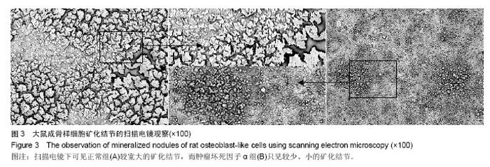
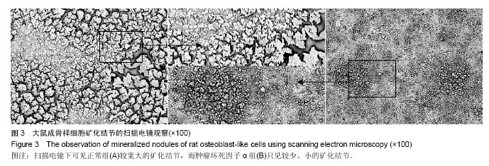
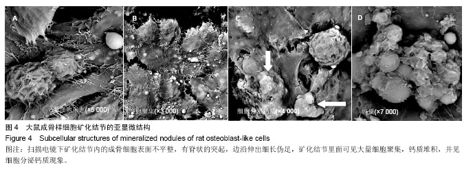
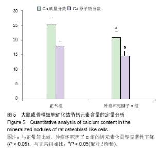
[15]石建伟.成骨不全患者骨胶原结构的扫描电镜和透射电镜研究[D].郑州大学,2011.
[16]章晓霜,唐键,屈菊兰,等.不同强度运动对去卵巢大鼠股骨下端松质骨影响的扫描电镜观察[J].中国运动医学杂志, 2002,21(1): 48-50.
[17]戴如春,姚学锋,刘沐荣,等.是否存在超微小梁?--骨小梁微破裂及微骨痂的扫描电镜观察[J].中国骨质疏松杂志,2005,11(4): 434-436.
[18]Tsukasaki M, Yamada A, Suzuki D, et al. Expression of POEM,a positive regulator of osteoblast differentiation is suppressed by TNF-alpha. Biochem Biophys Res Commun. 2011;410(4):766-770.
[19]Li W, Yu B, Li M, et al. NEMO-binding domain peptide promotes osteoblast differentiation impaired by tumor necrosis factor alpha. Biochem Biophys Res Commun. 2010; 391(2):1228-1233.
[20]Dutta J, Fan Y, Gupta N, et al.Current insights into the regulation of programmed cell death by NF-kappaB. Oncogene. 2006;25(51):6800-6816.
[21]Tran SE, Holmstrom TH, Ahonen M, et al. MAPK/ERK overrides the apoptotic signaling from Fas,TNF,and TRAIL receptors. J Biol Chem. 2001;276(19):16484-16490.
[22]朱建华,张沁,刘继光.肿瘤坏死因子α干预小鼠成骨细胞cbfa1/runx2基因的表达[J].中国组织工程研究与临床康复, 2011,15(37):6851-6854.
[23]Amizuka N, Hasegawa T, Yamamoto T, et al. Microscopic aspects on biomineralization in bone. Clin Calcium. 2014; 24(2):203-214.
[24]Querido W, Farina M. Strontium ranelate increases the formation of bone-like mineralized nodules in osteoblast cell cultures and leads to Sr incorporation into the intact nodules. Cell Tissue Res. 2013;354(2):573-580.
[25]Raouf A, Seth A. Ets transcription factors and targets in osteogenesis. Oncogene. 2000;19(55):6455-6463. |