Chinese Journal of Tissue Engineering Research ›› 2014, Vol. 18 ›› Issue (33): 5257-5265.doi: 10.3969/j.issn.2095-4344.2014.33.002
Previous Articles Next Articles
Endoplasmic reticulum stress-induced apoptosis of osteoblasts within the osteolytic craniums
Liu Guo-yin1, Wang Rui1, Dong Lei2, Zhang Jun-feng2, Zhao Jian-ning1
- 1 Nanjing General Hospital of Nanjing Military Area Command of PLA, School of Medicine of Nanjing University, Nanjing 210002, Jiangsu Province, China; 2 State Key Laboratory of Pharmaceutical Biotechnology, School of Life Sciences, Nanjing University, Nanjing 210093, Jiangsu Province, China
-
Online:2014-08-13Published:2014-08-13 -
Contact:Zhao Jian-ning, Master, Chief physician, Professor, Doctoral supervisor, Nanjing General Hospital of Nanjing Military Area Command of PLA, School of Medicine of Nanjing University, Nanjing 210002, Jiangsu Province, China -
About author:Liu Guo-yin, Studying for master’s degree, Nanjing General Hospital of Nanjing Military Area Command of PLA, School of Medicine of Nanjing University, Nanjing 210002, Jiangsu Province, China -
Supported by:the National Natural Science Foundation of China for Youths, No. 81000792
CLC Number:
Cite this article
Liu Guo-yin, Wang Rui, Dong Lei, Zhang Jun-feng, Zhao Jian-ning. Endoplasmic reticulum stress-induced apoptosis of osteoblasts within the osteolytic craniums[J]. Chinese Journal of Tissue Engineering Research, 2014, 18(33): 5257-5265.
share this article
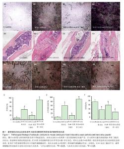
2.1 实验动物数量分析 实验选用小鼠28只,分为4组,实验过程无脱失,全部进入结果分析。 2.2 动物一般情况 各组动物造模后送返原笼,麻醉苏醒后常规饲养。各组动物均存活至实验结束,其间动物活动、进食情况基本正常,切口愈合良好,无皮下积液及切口感染等并发症发生。 2.3 内质网应激反应介导的骨溶解结果 2.3.1 小鼠颅骨骨片经甲苯胺蓝染色后置于显微镜下观察 见图1A和B。在小鼠颅骨植入磨损微粒纳米合金粉末后,可见大量圆形,椭圆形或梭形的骨吸收陷窝,陷窝深浅不等,着色浓度不一。对骨吸收陷窝面积进行积分显示:相对于空白对照组,纳米合金粉末组骨陷窝吸收率为3.3。还发现,在纳米合金粉末植入的基础上,加入内质网应激的诱导剂毒胡萝卜素后,骨吸收的陷窝最密集,骨溶解症状最严重;经内质网应激的抑制剂(4-苯基丁酸)的治疗后,陷窝的数量和大小均明显减少,骨溶解症状得到明显改善。统计学分析显示,磨损微粒纳米合金粉末组以及纳米合金粉末+毒胡萝卜素组,相对于空白对照组,差异均有显著性意义(P < 0.05),纳米合金粉末+4-苯基丁酸组相对于纳米合金粉末组差异也有显著性意义(P < 0.05)。结果表明内质网应激参与骨溶解的形成。 2.3.2 小鼠颅骨的苏木精-伊红染色结果 根据苏木精-伊红染色结果对颅骨骨溶解和炎症浸润进行评判,见图1C和1D。正常小鼠的颅骨结构清晰,黏膜和骨皮质完整,细胞构成大小、形态相同,排列整齐,极向正常,无炎症细胞浸润;而当小鼠颅骨受到纳米合金粉末的刺激后,表现出严重的浸润破坏,颅骨结构紊乱,黏膜层严重受损,骨皮质受到破坏,细胞构成大小、形态不一,排列多层,极向消失;髓腔大小不一、排列紊乱,炎症细胞浸润至颅深层甚至穿透颅骨,造成髓腔内成骨细胞丢失。经内质网应激的抑制剂(4-苯基丁酸)的治疗后,骨形态完整性得到保持,骨侵蚀和炎症浸润症状明显改善。在纳米合金粉末组中加入内质网应激的诱导剂毒胡萝卜素后,得到相反的结果。毒胡萝卜素加入后,骨形态的完好性受到进一步的破坏,骨侵蚀和炎症浸润程度也急剧加重。结果表明内质网应激在骨溶解的发生发展中起着一定的作用。 2.3.3 小鼠颅骨成骨细胞碱性磷酸酶染色结果 见图1E和1F。颅骨中成骨细胞的阳性反应呈现红黑色颗粒或块状沉淀,骨形成隧道呈现深色条带。植入纳米合金粉末后,小鼠颅骨的成骨细胞数量显著减少,骨形成隧道随着磨损微粒纳米合金粉末的加入而逐渐降低。应用4-苯基丁酸后,成骨细胞的数量和骨形成较纳米合金粉末组明显增加;而加入内质网应激的诱导剂毒胡萝卜素后,成骨细胞和骨形成受到进一步损伤。"
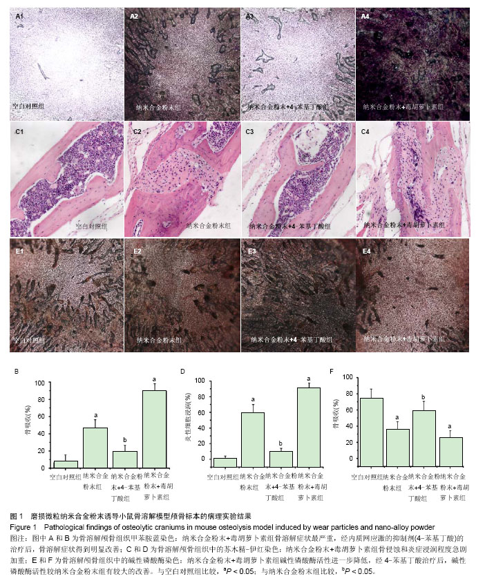
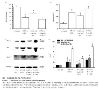
骨溶解骨组织中的碱性磷酸酶活性的趋势与碱性磷酸酶染色的实验结果一致。见图2A,在纳米合金粉末的刺激条件下,碱性磷酸酶的活性较空白对照组明显降低;加入毒胡萝卜素后,碱性磷酸酶活性进一步降低;而经4-苯基丁酸治疗后,碱性磷酸酶活性较纳米合金粉末组有较大的改善。统计学分析显示,纳米合金粉末组以及纳米合金粉末+毒胡萝卜素组,相对于空白对照组,差异均有显著性意义(P < 0.05),纳米合金粉末+4-苯基丁酸组相对于纳米合金粉末组差异也有显著性意义(P < 0.05)。 2.4 骨溶解颅骨组织中的内质网应激反应 见图2C和2D,当受到纳米合金粉末的刺激后,小鼠颅骨的内质网应激反应标志蛋白(GRP78/Bip,IRE1-α以及CHOP)的表达显著升高;在纳米合金粉末的基础上应用毒胡萝卜素后,内质网应激反应标志蛋白的表达得到进一步的提高;然而,腹腔注射4-苯基丁酸后,内质网应激反应标志蛋白的表达较纳米合金粉末组明显降低。此外还发现,这些内质网应激反应标志蛋白的变化趋势与上面提到的骨溶解结果一致。结果表明纳米合金粉末诱导的内质网应激反应参与了骨溶解的发生。 "


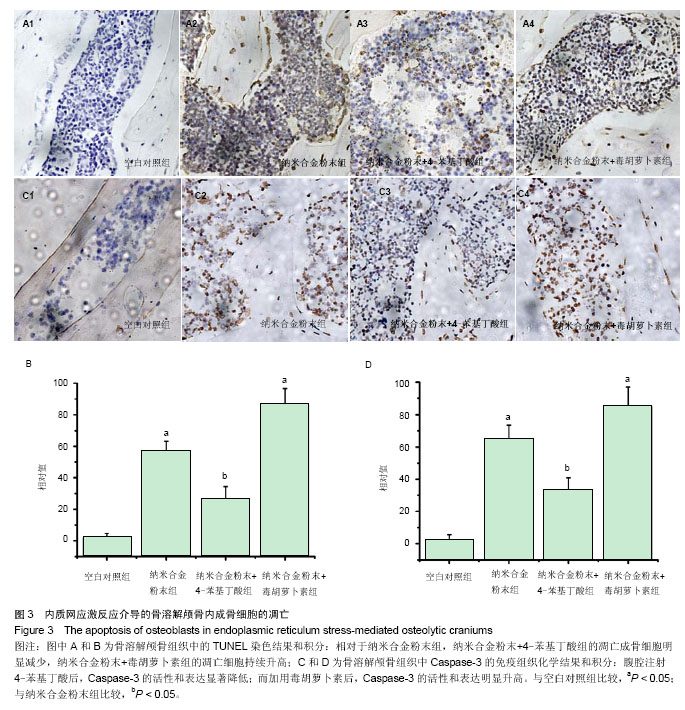
2.5 内质网应激反应介导骨溶解颅骨内成骨细胞的凋亡 2.5.1 骨溶解颅骨组织中成骨细胞的凋亡 TUNEL实验结果见图3A和3B。纳米合金粉末组的颅骨组织中有明显的成骨细胞凋亡,可以看到大量的棕黄色颗粒着色,并有细胞核碎裂等形态学表现。而正常颅骨组织仅发现少量的凋亡成骨细胞。然而,相对于纳米合金粉末组,纳米合金粉末+4-苯基丁酸组的凋亡成骨细胞明显减少,同时,纳米合金粉末+毒胡萝卜素组的凋亡细胞持续升高。 2.5.2 凋亡蛋白Caspase-3在骨溶解颅骨组织中的表达 研究表明,凋亡蛋白Caspase-3的激活不仅可以促进死亡受体的活化和线粒体途径关键蛋白的释放,还与内质网应激反应紧密相连。因此,Caspase-3是凋亡级联反应中的关键蛋白酶,是多种凋亡途径的共同下游效应部分。为明确凋亡蛋白Caspase-3在骨溶解发生发展中的作用,对骨溶解颅骨组织中凋亡蛋白Caspase-3的活性和表达情况进行了检测,通过图2B,3C和3D的实验结果发现,Caspase-3在纳米合金粉末组中高表达并表现出高活性,而在正常颅骨组织中,Caspase-3的活性和表达均很低。同样,与TUNEL实验结果一致,腹腔注射4-苯基丁酸后,Caspase-3的活性和表达显著降低;而加用毒胡萝卜素后,Caspase-3的活性和表达明显升高。"
| [1]Del Buono A, Denaro V, Maffulli N.Genetic susceptibility to aseptic loosening following total hip arthroplasty: a systematic review. Br Med Bull. 2012;101:39-55.
[2]Kadoya Y, Kobayashi A, Ohashi H. Wear and osteolysis in total joint replacements. Acta Orthop Scand Suppl. 1998;278:1-16.
[3]Kaddick C, Catelas I, Pennekamp PH, et al.[Implant wear and aseptic loosening. An overview]. Orthopade.2009.38(8): 690-697.
[4]Goodman SB, Gibon E, Yao Z. The basic science of periprosthetic osteolysis.Instr Course Lect. 2013;62: 201-206.
[5]Gallo J, Goodman SB, Konttinen YT, et al.Particle disease: biologic mechanisms of periprosthetic osteolysis in total hip arthroplasty. Innate Immun.2013; 19(2): 213-224.
[6]P. Edward Purdue, Panagiotis Koulouvaris, Bryan J. Nestor, et al.The central role of wear debris in periprosthetic osteolysis. HSS J.2006;2(2): 102-113.
[7]Goodman SB, Lind M, Song Y, Smith RL.et al.In vitro, in vivo, and tissue retrieval studies on particulate debris. Clin Orthop Relat Res.1998;(352): 25-34.
[8]O'Neill SC, Queally JM, Devitt BM, et al.The role of osteoblasts in peri-prosthetic osteolysis. Bone Joint J.2013. 95-B(8): 1022-1026.
[9]Vermes C, Glant TT, Hallab NJ, et al.The potential role of the osteoblast in the development of periprosthetic osteolysis: review of in vitro osteoblast responses to wear debris, corrosion products, and cytokines and growth factors. J Arthroplasty.2001;16(8 Suppl 1): 95-100.
[10]Jiang Y, Jia T, Wooley PH, et al.Current research in the pathogenesis of aseptic implant loosening associated with particulate wear debris. Acta Orthop Belg.2013;79(1):1-9.
[11]Noordin S, Masri B. Periprosthetic osteolysis: genetics, mechanisms and potential therapeutic interventions. Can J Surg.2012;55(6):408-417.
[12]Goodman SB, Trindade M, Ma T, et al.Pharmacologic modulation of periprosthetic osteolysis. Clin Orthop Relat Res.2005;(430):39-45.
[13]Lochner K, Fritsche A, Jonitz A, et al.The potential role of human osteoblasts for periprosthetic osteolysis following exposure to wear particles. Int J Mol Med.2011;28(6): 1055-1063.
[14]Gallo J, Raska M, Mrázek F,et al.Bone remodeling, particle disease and individual susceptibility to periprosthetic osteolysis. Physiol Res.2008;57(3):339-349.
[15]Jiang Y, Jia T, Gong W, et al.Titanium particle-challenged osteoblasts promote osteoclastogenesis and osteolysis in a murine model of periprosthestic osteolysis. Acta Biomater. 2013;9(7):7564-7572.
[16]Xu C, Bailly-Maitre B, Reed JC. Endoplasmic reticulum stress: cell life and death decisions. J Clin Invest.2005;115(10): p. 2656-2664.
[17]Kim I, Xu W, Reed JC. Cell death and endoplasmic reticulum stress: disease relevance and therapeutic opportunities. Nat Rev Drug Discov.2008;7(12): 1013-1030.
[18]Lai E, Teodoro T, Volchuk A.Endoplasmic reticulum stress: signaling the unfolded protein response. Physiology (Bethesda). 2007;22:193-201.
[19]Dong L, Wang R, Zhu YA, et al.Antisense oligonucleotide targeting TNF-alpha can suppress Co-Cr-Mo particle-induced osteolysis. J Orthop Res.2008;26(8):1114-1120.
[20]Purdue PE, Koulouvaris P, Potter HG, et al.The cellular and molecular biology of periprosthetic osteolysis. Clin Orthop Relat Res.2007;454:251-261.
[21]Beck RT, Illingworth KD, Saleh KJ. Review of periprosthetic osteolysis in total joint arthroplasty: an emphasis on host factors and future directions. J Orthop Res.2012;30(4): 541-546.
[22]Howie DW, Neale SD, Haynes DR,et al.Periprosthetic osteolysis after total hip replacement: molecular pathology and clinical management. Inflammopharmacology.2013;21(6): 389-396.
[23]Wu J, Wang Y, Ren H, et al.[Influence of titanium particles loading on releases of cytokines of osteoblasts]. Sheng Wu Yi Xue Gong Cheng Xue Za Zhi.2011;28(4):748-752.
[24]Wooley PH, Schwarz EM.Aseptic loosening.Gene Ther.2004;11(4): 402-407.
[25]Yang F, Wu W, Cao L, et al.Pathways of macrophage apoptosis within the interface membrane in aseptic loosening of prostheses. Biomaterials. 2011;32(35):9159-9167.
[26]Landgraeber S, von Knoch M, Löer F, et al.Extrinsic and intrinsic pathways of apoptosis in aseptic loosening after total hip replacement. Biomaterials. 2008;29(24-25):3444-3450.
[27]Yousef Abu-Amer, Isra Darwech, John C Clohisy.Aseptic loosening of total joint replacements: mechanisms underlying osteolysis and potential therapies. Arthritis Res Ther.2007;9 Suppl 1: S6.
[28]Jacobs JJ, Roebuck KA, Archibeck M, et al.Osteolysis: basic science. Clin Orthop Relat Res. 2001;(393): 71-77.
[29]Landgraeber S, Jaeckel S, Löer F,et al.Pan-caspase inhibition suppresses polyethylene particle-induced osteolysis. Apoptosis. 2009;14(2):173-181.
[30]Urano F.[Stress signaling from the endoplasmic reticulum]. Tanpakushitsu Kakusan Koso.2004;49(7 Suppl):1002-1005.
[31]Shen X, Zhang K, Kaufman RJ. The unfolded protein response--a stress signaling pathway of the endoplasmic reticulum. J Chem Neuroanat.2004;28(1-2):79-92.
[32]Wang R, Wang Z, Ma Y, et al.Particle-induced osteolysis mediated by endoplasmic reticulum stress in prosthesis loosening. Biomaterials.2013;34(11): 2611-2623.
[33]Hotamisligil GS.Endoplasmic reticulum stress and the inflammatory basis of metabolic disease. Cell.2010;140(6): 900-917.
[34]Tanaka K, Yamaguchi T, Kaji H, et al.Advanced glycation end products suppress osteoblastic differentiation of stromal cells by activating endoplasmic reticulum stress. Biochem Biophys Res Commun.2013;438(3):463-467. |
| [1] | Huo Hua, Cheng Yuting, Zhou Qian, Qi Yuhan, Wu Chao, Shi Qianhui, Yang Tongjing, Liao Jian, Hong Wei. Effects of drug coating on implant surface on the osseointegration [J]. Chinese Journal of Tissue Engineering Research, 2021, 25(22): 3558-3564. |
| [2] | Wei Qin, Zhang Xue, Ma Lei, Li Zhiqiang, Shou Xi, Duan Mingjun, Wu Shuo, Jia Qiyu, Ma Chuang. Platelet-derived growth factor-BB induces the differentiation of rat bone marrow mesenchymal stem cells into osteoblasts [J]. Chinese Journal of Tissue Engineering Research, 2021, 25(19): 2953-2957. |
| [3] | Guo Zhibin, Wu Chunfang, Liu Zihong, Zhang Yuying, Chi Bojing, Wang Bao, Ma Chao, Zhang Guobin, Tian Faming. Simvastatin stimulates osteogenic differentiation of bone marrow mesenchymal stem cells [J]. Chinese Journal of Tissue Engineering Research, 2021, 25(19): 2963-2968. |
| [4] | Wu Yukun, Han Jie, Wen Shuaibo. Mechanism of Runx2 gene in fracture healing [J]. Chinese Journal of Tissue Engineering Research, 2021, 25(14): 2274-2279. |
| [5] | Shui Xiaoping, Li Chunying, Cao Yanxia, Su Quansheng. Effects of aerobic and resistance exercises on endoplasmic reticulum stress-related proteins in diabetic peripheral neuropathy rats [J]. Chinese Journal of Tissue Engineering Research, 2021, 25(11): 1693-1698. |
| [6] | Zhang Xujian, Zhao Zhenqun, Liu Wanlin. The role of endoplasmic reticulum stress in the pathogenesis of steroid-induced avascular necrosis of the femoral head [J]. Chinese Journal of Tissue Engineering Research, 2021, 25(11): 1759-1765. |
| [7] | Chen Qiang, Zhuo Hongwu, Xia Tian, Ye Zhewei . Toxic effects of different-concentration isoniazid on newborn rat osteoblasts in vitro [J]. Chinese Journal of Tissue Engineering Research, 2020, 24(8): 1162-1167. |
| [8] | Li Jinyu, Yu Xing, Jiang Junjie, Xu Lin, Zhao Xueqian, Sun Qi, Zheng Chenying, Bai Chunxiao, Liu Chuyin, Jia Yusong. Promoting effect of osteopractic total flavone combined with nano-bone materials on proliferation and differentiation of MC3T3-E1 cells [J]. Chinese Journal of Tissue Engineering Research, 2020, 24(7): 1030-1036. |
| [9] | Qiao Jiutao, Guan Dehong, Wang Dongyan, Liu Aiyun. Zuogui Pill has protective effect against oxidative stress injury in osteoblasts [J]. Chinese Journal of Tissue Engineering Research, 2020, 24(7): 1052-1056. |
| [10] | Ge Juncheng, Ma Jinhui, Wang Bailiang, Yue Debo, Sun Wei, Wang Weiguo, Guo Wanshou, Li Zirong. Application of bisphosphonates in avascular necrosis of the femoral head [J]. Chinese Journal of Tissue Engineering Research, 2020, 24(5): 753-759. |
| [11] | Zhang Chao, Li Xingyong, Ma Guifu, Pu Xingyu, Luo Wenyuan. Hoxa9 silencing promotes tibial fracture healing by regulating osteogenic differentiation [J]. Chinese Journal of Tissue Engineering Research, 2020, 24(35): 5600-5606. |
| [12] | Song Shilei, Chen Yueping, Zhang Xiaoyun. Mechanism of PI3K/AKT signaling pathway regulating osteonecrosis of the femoral head [J]. Chinese Journal of Tissue Engineering Research, 2020, 24(3): 408-415. |
| [13] | He Qiang, Qian Weiqing, Yao Nianwei, Zhou Mengling, Liu Yixin, Yin Hong. Protection of inflammatory osteoblasts in neonatal rats using catalpol from the root of Rehmannia glutinosa [J]. Chinese Journal of Tissue Engineering Research, 2020, 24(29): 4626-4631. |
| [14] | Liu Guoming, Wang Qinfen, Lin Kefeng, Zhou Shiguo, Chen Zuxing, Lin Shishui. Whole body application of nerve growth factor promotes early healing of tibial shaft fracture and improves expression of bone morphogenetic protein-2 and vascular endothelial growth factor in rats [J]. Chinese Journal of Tissue Engineering Research, 2020, 24(29): 4680-4685. |
| [15] | Zhou Hangyu, Zeng Fuhai, Xia Delin. Silicate-Cu/Mg bioactive ceramics promote the osteogenesis of osteoblasts [J]. Chinese Journal of Tissue Engineering Research, 2020, 24(28): 4511-4517. |
| Viewed | ||||||
|
Full text |
|
|||||
|
Abstract |
|
|||||