Chinese Journal of Tissue Engineering Research ›› 2014, Vol. 18 ›› Issue (31): 5011-5016.doi: 10.3969/j.issn.2095-4344.2014.31.016
Previous Articles Next Articles
Biomechanical comparison of three fixation methods in the repair of posterolateral tibial plateau fracture
Zhang Yan1, Liang Xu1, Fan Xin-bin1, Shao Jin1, Liu Yue1, Ye Wei-guang1, Wu Liang1, Yang Tie-yi1, Gong Lu-lu2
- 1Department of Orthopedics, Gongli Hospital of Pudong New Area, Shanghai 200135, China; 2School of Life Science and Technology, Tongji University, Shanghai 200092, China
-
Received:2014-06-26Online:2014-07-23Published:2014-07-23 -
Contact:Yang Tie-yi, Chief physician, Department of Orthopedics, Gongli Hospital of Pudong New Area, Shanghai 200135, China -
About author:Zhang Yan, M.D., Associate chief physician, Department of Orthopedics, Gongli Hospital of Pudong New Area, Shanghai 200135, China -
Supported by:the Key Discipline Construction Project of Shanghai Pudong New Area Health System, No. PWZx2014-09; the Academic Leaders Project of Shanghai Pudong New Area Health System, No. PWRd2012-16
CLC Number:
Cite this article
Zhang Yan, Liang Xu, Fan Xin-bin, Shao Jin, Liu Yue, Ye Wei-guang, Wu Liang, Yang Tie-yi, . Biomechanical comparison of three fixation methods in the repair of posterolateral tibial plateau fracture[J]. Chinese Journal of Tissue Engineering Research, 2014, 18(31): 5011-5016.
share this article

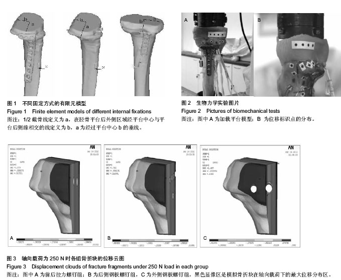
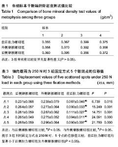
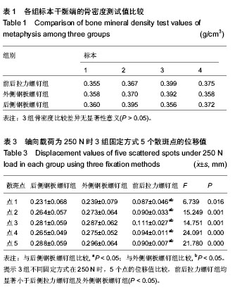
2.1 骨密度测定 3组标本干骺端的骨密度测试值见表1,前后拉力螺钉组的骨密度为(0.374±0.019) g/cm3,外侧钢板螺钉组的骨密度为(0.370±0.016) g/cm3,后侧钢板螺钉组的骨密度为(0.371±0.017) g/cm3,经过分析比较3组差异无显著性意义(P > 0.05)。 2.2 有限元法骨折块的轴向最大位移 3种模型在轴向载荷分别为250,500,1 000 N时,骨折块的轴向最大位移值依次变大。在250 N的轴向载荷下前后拉力螺钉组骨折块的轴向位移值最小,其次为后侧钢板螺钉组,外侧钢板螺钉组最大,位移值为0.138 200 mm。在500,1 000 N轴向载荷下,骨折块的轴向最大位移值情况同250 N情况类似。根据模拟出的骨折三维立体图像显示,在250 N时3组固定方式轴向最大位移值的分布区域都在骨折块的后外侧处,其余两种载荷下骨折块最大位移值的位置与250 N相似,见表2,图3。 2.3 实验生物力学结果 3组不同固定方式在250 N时,5个点在前后拉力螺钉组、后侧钢板螺钉组、外侧钢板螺钉组间所测量的位移值见表3。5个点分别经S-N-K法对3组进行两两间q 检验,前后拉力螺钉组与后侧拉力螺钉组比较,差异有显著性意义(P < 0.05);前后拉力螺钉组与外侧钢板螺钉组比较,差异有显著性意义(P < 0.05);后侧钢板螺钉组与外侧钢板螺钉组比较,两组间差异无显著性意义(P > 0.05)。每组在500,1 000 N时5个点在3组固定模型上的位移值趋势同250 N相似,随着轴向载荷力的增加,位移值相应增加,3组间两两比较的统计学结果同250 N一致,在3种固定方式的5个点中,点5的位移值最大,分布区域在骨折模型的后外侧。"
"

"
| [1] Piposar J, Fowler JR, Gaughan JP, et al. Race may not affect [correct] outcomes in operatively treated tibia fractures.Clin Orthop Relat Res. 2012;470(5):1513-1517. [2] Babis GC, Evangelopoulos DS, Kontovazenitis P, et al. High energy tibial plateau fractures treated with hybrid external fixation. J Orthop Surg Res. 2011;(14):35. [3] Zura RD, Browne JM, Black MD,et al.Current management of high-energy tibial plateau fractures. Curr Orthop. 2007;21(3): 229-235. [4] Papagelopoulos PJ, Partsinevelos AA, Themistocleous GS, et al. Complications after tibia plateau fracture surgery. Injury. 2006;37(6):475-484. [5] Stauffer KD, Tuttle TA, Elkins AD, et al. Complications associated with 696 tibial plateau leveling osteotomies (2001-2003). J Am Anim Hosp Assoc. 2006;42(1):44-50. [6] Cross WW 3rd, Levy BA, Morgan JA, et al. Periarticular raft constructs and fracture stability in split-depression tibial plateau fractures. Injury. 2013;44(6):796-801. [7] Heikkilä JT, Kukkonen J, Aho AJ, et al. Bioactive glass granules: a suitable bone substitute material in the operative treatment of depressed lateral tibialplateau fractures: a prospective, randomized 1 year follow-up study. J Mater Sci Mater Med. 2011;22(4):1073-1080. [8] Hsieh CH, Huang HT, Liu PC,et al. Anterior approach for posteromedial tibial plateau fractures. Kaohsiung J Med Sci. 2010;26(3):130-135. [9] Luo CF, Hu CF, Gao H, et al. Three-column classification for tibial plateau fractures. Chin J Orthop Trauma 2009;11(3): 201-205. [10] Luo CF, Sun H, Zhang B. Three-column fixation for complex tibial plateau fractures. J Orthop Trauma. 2010;24(11): 683-692. [11] Cooper HJ, Kummer FJ, Egol KA, et al.The effect of screw type on the fixation of depressed fragments in tibial plateau fractures. Bull Hosp Jt Dis. 2002;60:72-75. [12] Ali AM, Yang L, Hashmi M, et al. Bicondylar tibial plateau fractures managed with the Sheffield Hybrid Fixator. Biomechanicalstudy and operative technique. Injury. 2001;32 Suppl 4:SD86-91. [13] Ali AM, Saleh M, Bolongaro S, et al. The strength of different fixation techniques forbicondylar tibial plateau fractures-abiomechanical study. Clin Biomech. 2003; 18: 864-870. [14] Yushkevich PA,Piven J, Hazlett HC,et al.User- guided 3D active contour segmentation of anatomical structures: Significantly improved efficiency andreliabiity. Neurolmage. 2006;3:1116-1128. [15] Sonoda N, Chosa E, Totoribe K,et al. Biomechanical analysis for stress fractures of the anterior middle third of the tibia in athletes:nonlinear analysis using a three-dimensional finite element method. J Orthop Sci. 2003;4:505-513. [16] Lacroix D, Prendergast PJ. Three-dimensional simulation of fracture repair in the human tibia. Comput Methods Biomech Biomed Engin. 2002;5: 369-376. [17] 龚璐璐,杨铁毅,张岩,等.LISS钢板治疗胫骨远端骨折有限元建模及分析[J].生物医学工程进展,2010, 31(1):12-17. [18] 孙江波,廖怀章,蒋勇,等.胫骨平台后柱骨折钢板固定的三维有限元分析[J].临床医学研究,2012, 29(5):813-817. [19] Sonoda N, Chosa E, Totoribe K,et al. Biomechanical analysis for stress fractures of the anterior middle third of the tibia in athletes:nonlinear analysis using a three-dimensional finite element method. J Orthop Sci. 2003;8:505-513. [20] Meng L, Zhang Y, Lu Y. Three-dimensional finite element analysis of mini-external fixation and Kirschner wire internal fixation in Bennettfracture treatment. Orthop Traumatol Surg Res. 2013;99(1):21-29, [21] Kantardzi? I, Vasiljevi? D, Blazi? L,et al. Influence of cavity design preparation on stress values in maxillary premolar: a finite element analysis. Croat Med J. 2012;53(6):568-576. [22] Herrera A, Ibarz E, Cegoñino J, et al. Applications of finite element simulation in orthopedic and trauma surgery. World J Orthop. 2012; 3(4): 25-41. [23] Liu XS, Wang J, Zhou B,et al. Fast trabecular bone strength predictions of HR-pQCT and individual trabeculae segmentation-based plate and rod finite element model discriminate postmenopausal vertebral fractures. J Bone Miner Res. 2013;28(7):1666-1678. [24] Varga P, Schefzig P, Unger E, et al. Finite element based estimation of contact areas and pressures of the human scaphoid in various functional positions of the hand. J Biomech. 2013;46(5):984-990. [25] Nishiyama KK, Macdonald HM, Hanley DA, et al. Women with previous fragility fractures can be classified based on bone microarchitecture and finite element analysis measured with HR-pQCT. Osteoporos Int. 2013;24(5):1733-1740. [26] Miranda MA. Locking plate technology and its role in osteoporotic fractures. Injury.2007;38(9):Suppl 3:S35-39. [27] Will R, Englund R, Lubahn J, et al. Locking plates have increased torsionalstiffness compared to standard plates in a segmental defect model of claviclefracture. Arch Orthop Trauma Surg. 2011;131(6):841–847. [28] Jia YW, Cheng LM, Yu GR, et al. A finite element analysis of the pelvic reconstruction using fibular transplantation fixed with four different rod-screw systems after type I resection. Chin Med J. 2008;121(4):321-326. [29] Graeff C, Marin F, Petto H,et al. High resolution quantitative computed tomography-based assessment of trabecular microstructure and strength estimates by finite-element analysis of the spine, but not DXA, reflects vertebral fracture status in men with glucocorticoid-induced osteoporosis. Bone. 2013;52(2):568-577. [30] 张国柱,蒋协远,王满宜.CT扫描对胫骨平台骨折分型及治疗的影响[J].中华创伤骨科杂志,2006,8(4):326-329. [31] 谢祖宏,吕亮,胡汉敏,等.胫骨平台骨折后需早期康复锻炼但不宜过早负重锻炼[J].中国临床康复, 2002, 6(2): 238-239. [32] Howard NE, Phaff M, Aird J,et al. Does human immunodeficiency virus status affect early wound healing in open surgically stabilised tibial fractures?: A prospective study. Bone Joint J. 2013;95-B(12):1703-1707. [33] Uchiyama S, Itsubo T, Nakamura K,et al. Effect of early administration of alendronate after surgery for distal radial fragility fracture on radiological fracture healing time.Bone Joint J. 2013;95-B(11):1544-1550. [34] Matsunaga FT, Tamaoki MJ, Matsumoto MH, et al. Treatment of the humeral shaft fractures--minimally invasive osteosynthesis with bridge plate versus conservative treatment with functional brace: study protocol for a randomised controlled trial. Trials. 2013;14:246. [35] Urita A, Iwasaki N, Kondo M, et al. Effect of low-intensity pulsed ultrasound on bone healing at osteotomy sites after forearm bone shortening. J Hand Surg Am. 2013;38(3): 498-503. [36] Epari DR, Kassi JP, Schell H., et al. Timely fracture-healing requires optimization of axial fixation stability. J Bone Joint Surg Am. 2007:89(7):1575-1585. [37] Guo LX,Teo EC,Qiu TX.Prediction of biomechanical characteristics of intact and injured lower thoracic spine segment under different loads. J Musculoskel Res. 2004;8: 87-99. [38] 巴雪峰,孙改生,凯瑟尔.等. 胫骨平台骨折的治疗新进展[J].中国矫形外科杂志,2012,20(12):1104-1107. [39] 冯卫,郝廷,郝增涛,等.3种股骨近端髓内钉固定股骨转子间骨折的有限元分析[J].中国矫形外科杂志,2012,20(8):730-733. |
| [1] | Xu Feng, Kang Hui, Wei Tanjun, Xi Jintao. Biomechanical analysis of different fixation methods of pedicle screws for thoracolumbar fracture [J]. Chinese Journal of Tissue Engineering Research, 2021, 25(9): 1313-1317. |
| [2] | Chen Xinmin, Li Wenbiao, Xiong Kaikai, Xiong Xiaoyan, Zheng Liqin, Li Musheng, Zheng Yongze, Lin Ziling. Type A3.3 femoral intertrochanteric fracture with augmented proximal femoral nail anti-rotation in the elderly: finite element analysis of the optimal amount of bone cement [J]. Chinese Journal of Tissue Engineering Research, 2021, 25(9): 1404-1409. |
| [3] | Zhou Jihui, Li Xinzhi, Zhou You, Huang Wei, Chen Wenyao. Multiple problems in the selection of implants for patellar fracture [J]. Chinese Journal of Tissue Engineering Research, 2021, 25(9): 1440-1445. |
| [4] | Xu Yulin, Shen Shi, Zhuo Naiqiang, Yang Huilin, Yang Chao, Li Yang, Zhao Heng, Zhao Lu. Biomechanical comparison of three different plate fixation methods for acetabular posterior column fractures in standing and sitting positions [J]. Chinese Journal of Tissue Engineering Research, 2021, 25(6): 826-830. |
| [5] | Cai Qunbin, Zou Xia, Hu Jiantao, Chen Xinmin, Zheng Liqin, Huang Peizhen, Lin Ziling, Jiang Ziwei. Relationship between tip-apex distance and stability of intertrochanteric femoral fractures with proximal femoral anti-rotation nail: a finite element analysis [J]. Chinese Journal of Tissue Engineering Research, 2021, 25(6): 831-836. |
| [6] | Song Chengjie, Chang Hengrui, Shi Mingxin, Meng Xianzhong. Research progress in biomechanical stability of lateral lumbar interbody fusion [J]. Chinese Journal of Tissue Engineering Research, 2021, 25(6): 923-928. |
| [7] | Liu Zhao, Xu Xilin, Shen Yiwei, Zhang Xiaofeng, Lü Hang, Zhao Jun, Wang Zhengchun, Liu Xuzhuo, Wang Haitao. Guiding role and prospect of staging and classification combined collapse prediction method for osteonecrosis of femoral head [J]. Chinese Journal of Tissue Engineering Research, 2021, 25(6): 929-934. |
| [8] | Xie Chongxin, Zhang Lei. Comparison of knee degeneration after anterior cruciate ligament reconstruction with or without remnant preservation [J]. Chinese Journal of Tissue Engineering Research, 2021, 25(5): 735-740. |
| [9] | Nie Shaobo, Li Jiantao, Sun Jien, Zhao Zhe, Zhao Yanpeng, Zhang Licheng, Tang Peifu. Mechanical stability of medial support nail in treatment of severe osteoporotic intertrochanteric fracture [J]. Chinese Journal of Tissue Engineering Research, 2021, 25(3): 329-333. |
| [10] | Tan Jiachang, Yuan Zhenchao, Wu Zhenjie, Liu Bin, Zhao Jinmin. Biomechanical analysis of elastic nail combined with end caps and wire fixation for long oblique femoral shaft fractures [J]. Chinese Journal of Tissue Engineering Research, 2021, 25(3): 334-338. |
| [11] | Chen Lu, Zhang Jianguang, Deng Changgong, Yan Caiping, Zhang Wei, Zhang Yuan. Finite element analysis of locking screw assisted acetabular cup fixation [J]. Chinese Journal of Tissue Engineering Research, 2021, 25(3): 356-361. |
| [12] | Zhou Jihui, Li Xinzhi, Zhou You, Huang Wei, Chen Wenyao. Comparison of the advantages and disadvantages of multiple implants in treatment of traumatic dislocation of sternoclavicular joint [J]. Chinese Journal of Tissue Engineering Research, 2021, 25(3): 443-448. |
| [13] | Li Kun, Li Zhijun, Zhang Shaojie, Gao Shang, Sun Hao, Yang Xi, Wang Xing, Dai Lina . A 4-year-old child model of occipito-atlanto-axial joints established by finite element dynamic simulation [J]. Chinese Journal of Tissue Engineering Research, 2021, 25(24): 3773-3778. |
| [14] | Shu Qihang, Liao Yijia, Xue Jingbo, Yan Yiguo, Wang Cheng. Three-dimensional finite element analysis of a new three-dimensional printed porous fusion cage for cervical vertebra [J]. Chinese Journal of Tissue Engineering Research, 2021, 25(24): 3810-3815. |
| [15] | Sun Maji, Wang Qiuan, Zhang Xingchen, Guo Chong, Yuan Feng, Guo Kaijin. Development and biomechanical analysis of a new anterior cervical pedicle screw fixation system [J]. Chinese Journal of Tissue Engineering Research, 2021, 25(24): 3821-3825. |
| Viewed | ||||||
|
Full text |
|
|||||
|
Abstract |
|
|||||