Chinese Journal of Tissue Engineering Research ›› 2014, Vol. 18 ›› Issue (6): 880-887.doi: 10.3969/j.issn.2095-4344.2014.06.010
Previous Articles Next Articles
Proliferation of hematopoietic stem cells differentiated from embryonic stem cells via sustained Wnt pathway activation
Lin Fang1, 2, Jin Jing-jun2, Zhang Tao2, Ji Bin-feng1, Chen Yong2, 3
- 1School of Bioscience and Engineering, Fuzhou University, Fuzhou 350108, Fujian Province, China; 2Fujian Academy of Medical Sciences, Fuzhou 350001, Fujian Province, China; 3School of Life Science, Fujian Normal University, Fuzhou 350108, Fujian Province, China
-
Revised:2013-12-10Online:2014-02-05Published:2014-02-05 -
Contact:Jin Jing-jun, Investigator, Master’s supervisor, Fujian Academy of Medical Sciences, Fuzhou 350001, Fujian Province, China -
About author:Lin Fang, School of Bioscience and Engineering, Fuzhou University, Fuzhou 350108, Fujian Province, China; Fujian Academy of Medical Sciences, Fuzhou 350001, Fujian Province, China -
Supported by:the Natural Science Foundation of Fujian Province, No. 2012J01325; the Medical Innovative Subject of Fujian Province, No. 2012-cx-13; Fundamental Scientific Research Activities at Non-Profit Research Institutions of Fujian Province, No. 2010R1034-1
CLC Number:
Cite this article
Lin Fang, Jin Jing-jun, Zhang Tao, Ji Bin-feng, Chen Yong. Proliferation of hematopoietic stem cells differentiated from embryonic stem cells via sustained Wnt pathway activation[J]. Chinese Journal of Tissue Engineering Research, 2014, 18(6): 880-887.
share this article
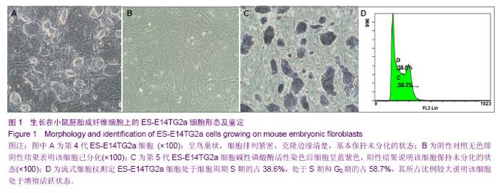
2.1 显微镜下观察生长在小鼠胚胎成纤维细胞上的ES-E14TG2a细胞的形态 复苏的ES-E14TG2a小鼠胚胎干细胞在小鼠胚胎成纤维细胞饲养层细胞上培养3 d时,可以看出该ES-E14TG2a细胞呈鸟巢状,细胞排列紧密,克隆边缘清楚,基本保持未分化的状态(图1A)。 2.2 AP染色 阴性对照为经丝裂霉素处理的P3代的小鼠胚胎成纤维细胞染色结果为无色即阴性结果表明该细胞已分化(图1B),实验组为P5代的ES-E14TG2a胚胎干细胞经碱性磷酸酶染色后细胞呈蓝紫色,阳性结果说明该细胞保持未分化的状态(图1C)。 2.3 ES-E14TG2a细胞的细胞周期 ES-E14TG2a细胞处于细胞周期S期的占38.6%,处于S期和G2期的占58.7%,S期和G2期均是DNA合成期,且S早期合成的DNA是调节细胞增殖和表达的DNA,其所占比例较大说明该细胞处于增殖活跃状态(图1D)。"
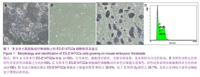
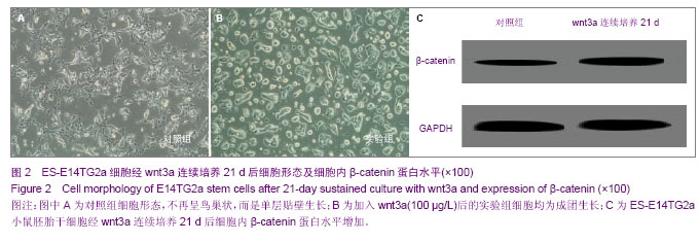
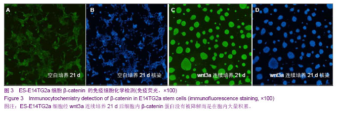
2.4 显微镜下观察ES-E14TG2a细胞经Wnt3a诱导后细胞集落的变化 将P5代的ES-E14TG2a细胞从小鼠胚胎成纤维细胞饲养层上分离后接种于经0.1%明胶包被的60mm培养皿上细胞形态不再呈鸟巢状,而是单层贴壁生长(图2A)。加入Wnt3a(100 µg/L)培养24 h后细胞又出现成团生长,连续培养21 d中细胞均为成团生长(图2B)。 2.5 β-catenin蛋白免疫荧光 对照组中的荧光强度较弱(图3A,B),而ES-E14TG2a小鼠胚胎干细胞经Wnt3a连续培养21 d后β-catenin蛋白的细胞荧光明显较强(图3C,D),说明ES-E14TG2a细胞经Wnt3a连续培养21 d后细胞内β-catenin蛋白没有被降解而是在胞内大量积累。 2.6 蛋白免疫印迹(Western Blot) B条带比A条带明显较粗说明ES-E14TG2a小鼠胚胎干细胞经Wnt3a连续培养 21 d后细胞内β-catenin蛋白水平增加(图2C)。"

"
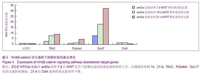
2.7 Wnt/β-Catenin信号通路下游靶标基因的表达 Pitx2、Frizzled、Sox17、Oct4是Wnt信号下游靶标基因,从ES-E14TG2a细胞经Wnt3a培养7 d后Wnt信号下游靶标基因的表达量均有所上升,且继续培养到14,21 d,Pitx2、Frizzled、Sox17的表达量持续增加,而连续培养21 d后Oct4基因的表达量有所下降(图4)。"
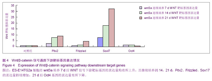

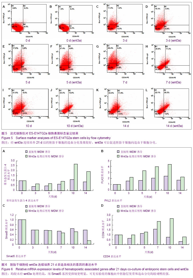
2.8 造血干细胞表面标记物检测 CD34和Sca-1是造血干细胞的两个重要表面标志[13-14],利用流式细胞仪检测发现直接将ES-E14TG2a细胞用拟胚体法诱导其向造血干细胞分化在3,5,7,10,14 d其双阳性的比率分别为1.2%,5.8%,6.2%,8.5%,11.9%;而先用Wnt3a连续培养21 d后的ES-E14TG2a细胞再通过单层贴壁培养法诱导其造血分化在3,5,7,10,14 d的双阳性比率为3.8%,6.8%,11.3%,15.9%,20.2%。可见经Wnt3a连续培养21 d后的胚胎干细胞的造血分化效果较好,Wnt3a可以促进胚胎干细胞向造血干细胞分化(图5)。2.9 与造血相关的基因的表达 胚胎干细胞经Wnt3a连续培养21 d后,再进行单层贴壁培养诱导其造血分化过程中BMP4、FLK2及CD34基因的表达量均较未经Wnt3a处理的高,而Smad5基因受抑制更明显,可见实验组的细胞向中胚胎层发育和造血分化均较对照组强(图6)。"
| [1] Keller G. Embryonic stem cell differentiation: emergence of a new era in biology and medicine. Genes Dev. 2005;19(10): 1129-1155. [2] Donovan PJ, Gearhart J.The end of the beginning for pluripotent stem cells. Nature. 2001;414(6859):92-97.[3] Keller G, Kennedy M, Papayannopoulou T, et al. Hematopoietic commitment during embryonic stem cell differentiation in culture. Mol Cell Biol. 1993;13(1):473-486.[4] McClanahan T, Dalrymple S, Barkett M, et al. Hematopoietic growth factor receptor genes as markers of lineage commitment during in vitro development of hematopoietic cells. Blood.1993;81(11):2903-2915.[5] Schmitt RM, Bruyns E, Snodgrass HR. Hematopoietic development of embryonic stem cells in vitro: cytokine and receptor gene expression. Genes Dev. 1991;5(5):728-740.[6] Johansson BM, Wiles MV. Evidence for involvement of activinA and bone morphogenetic protein 4 in mammalian mesoderm and hematopoietic development. Mol Cell Biol. 1995;15(1):141-151.[7] Sato N, Meijer L, Skaltsounis L, et al. Maintenance of pluripotency in human and mouse embryonic stem cells through activation of Wnt signaling by a pharmacological GSK-3-specific inhibitor. Nat Med. 2004;10(1):55-63.[8] Smith AG, Embryo-derived stem cells: of mice and men. Annu Rev Cell Dev Biol. 2001; 17:435-462.[9] Bejsove A. Wnt passway activation new relations and Iacations. Cell. 2005;120(1):11-24. [10] Willert K, Brown JD, Danenberg E, et al. Wnt proteins are lipid-modified and can act as stem cell growth factors. Nature. 2003;423(6938):448-452.[11] Lako M, Lindsay S, Lincoln J, et al.Characterisation of Wnt gene expression during the differentiation of murine embryonic stem cells in vitro : role of Wnt3a in enhancing haematopoietic differentiation. Mech De . 2001;103(2):49-59.[12] 徐兰,刘敏英.小鼠胚胎干细胞的培养-饲养层的制备[J].细胞生物学杂志,2009,32(2):291-292.[13] 田晨.小鼠造血干细胞表型及其分离纯化的研究进展[J].中国实验血液学杂志,2012,20(1):196-199.[14] Takamiya M,Haider KH,Ashraf M.Identification and characterization of a novel multipotent sub-population of scal-1 cardiac progenitor cells for myocardial regeneration. PLoS One.2011;6(9):252-256.[15] 赖永榕.造血干细胞移植的临床应用进展[J].广西医学.2005, 27(10):1499-1503.[16] Smith C,Storms B.Hematopoietic stem Cells.Clinical Orthopaedics Related Res.2000;379S:S91-S97. [17] 俞受程,叶建平,刘家赵.造血干细胞移植[J].中国医药科学,2011,1(21):9-12.[18] Manjiri MB, Aina H, Jamie C, et al. Generation of Multipotential Mesendodermal Progenitors from Mouse Embryonic Stem Cells via Sustained Wnt Pathway Activation. The Journal Of Biological Chemistry. 2007;282(43): 31703-31712.[19] Tamai K, Zeng X, Liu C, et al. A mechanism for Wnt coreceptor activation. Mol Cell. 2004;13(1):149-154.[20] Kikuchi A, Yamamoto H, Sato A. Selective activation mechanisms of Wnt signaling pathways. Trends Cell Biol. 2009;19(3):119-129.[21] Mulholland DJ,Dedhar S,Coetzee GA, et al. Interaction of Nuclear Receptor swith the Wnt/beta-catenin/Tcf Signaling axis: Wnt You Like to Know ? Endocr Rev.2005;26(7): 898-915.[22] Kikuchi A,Yamamoto H,Sato A.Selective activation mechanisms of Wnt signaling pathways.Trends CellBiol.2009; 19(3):119-129.[23] 刘娜,陆敏.胚胎干细胞自我更新相关信号转导途径及信号分子[J].科学通报,2005,50(7):623-628.[24] Niwa H,Miyazaki J,Smith AG.Quantitative expression of Oct-3/4 defines differentiation,dedifferentiation or self-renewal of ES cells.Nat Genet. 2000; 24(4):372-376. [25] Fei T,Xia K,Li Z,et al.Genome-wide mapping of SMAD target genes reveals the role of BMP signaling in embryonic stem cell fate determination.Genome Res.2010;20(4):36-44.[26] 王跃嗣,李建远,靳韶华.骨形态发生蛋白4在人胚胎卵黄囊造血过程中的表达[J].解剖学报.2008,39(5):723-727.[27] Wang Y,Umeda K,Nakayama N.Collaboration between WNT and BMP signaling promotes hemoangiogenic cell development from human fibroblast-derived iPS cells.Stem Cell.2010;4(3)223-231.[28] Heo J, Lee JS, Chu IS, et al. Spontaneous differentiation of mouse embryonic stem cells in vitro:Characterization by global gene expression profiles. Biochem Biophys Res Commun. 2005;332(4):1061-1069.[29] Kappel A,Schlaeger TM,Flamme I,et al.Role of SCL/Tel-1,GATA,and ets transcription factor binding sites for the regulation of flk-1 expression during murine vasular development.Blood.2000;96(9):3078-3085.[30] Zhang Y, Derynck R. Regulation of smad signalling by protein associations and signalling crosstalk.Trends Cell Biol. 1999; 9(7):274-279.[31] Kandyba E, Hazen VM, Kobielak A, et al. Smad1 and 5 but not smad8 establish stem cell quiescence which is critical to transform the premature hair follicle during morphogenesis toward the postnatal state. Stem Cells. 2014;32(2):534-547.[32] Tong KK, Kwan KM. Common partner Smad-independent canonical bone morphogenetic protein signaling in the specification process of the anterior rhombic lip during cerebellum development. Mol Cell Biol. 2013;33(10): 1925- 1937.[33] Chakravorty N, Hamlet S, Jaiprakash A, et al. Pro-osteogenic topographical cues promote early activation of osteoprogenitor differentiation via enhanced TGFβ, Wnt, and Notch signaling. Clin Oral Implants Res. 2013 .[34] Takamiya M, Haider KH, Ashraf M. Identification and characterization of a novel multipotent subpopulation of sca-1 cardiac progenitor cells for myocardial regeneration. PLoS One. 2011;6(9):252-265. |
| [1] | Li Cai, Zhao Ting, Tan Ge, Zheng Yulin, Zhang Ruonan, Wu Yan, Tang Junming. Platelet-derived growth factor-BB promotes proliferation, differentiation and migration of skeletal muscle myoblast [J]. Chinese Journal of Tissue Engineering Research, 2021, 25(7): 1050-1055. |
| [2] | Liu Cong, Liu Su. Molecular mechanism of miR-17-5p regulation of hypoxia inducible factor-1α mediated adipocyte differentiation and angiogenesis [J]. Chinese Journal of Tissue Engineering Research, 2021, 25(7): 1069-1074. |
| [3] | Ma Zetao, Zeng Hui, Wang Deli, Weng Jian, Feng Song. MicroRNA-138-5p regulates chondrocyte proliferation and autophagy [J]. Chinese Journal of Tissue Engineering Research, 2021, 25(5): 674-678. |
| [4] | Wang Yujiao, Liu Dan, Sun Song, Sun Yong. Biphasic calcium phosphate loaded with advanced platelet rich fibrin can promote the activity of rabbit bone marrow mesenchymal stem cells [J]. Chinese Journal of Tissue Engineering Research, 2021, 25(4): 504-509. |
| [5] | Zhou Jihui, Yao Meng, Wang Yansong, Li Xinzhi, Zhou You, Huang Wei, Chen Wenyao. Influence of novel nanoscaffolds on biological behaviors of neural stem cells and the related gene expression [J]. Chinese Journal of Tissue Engineering Research, 2021, 25(4): 532-536. |
| [6] | Chen Yang, Huang Denggao, Gao Yuanhui, Wang Shunlan, Cao Hui, Zheng Linlin, He Haowei, Luo Siqin, Xiao Jingchuan, Zhang Yingai, Zhang Shufang. Low-intensity pulsed ultrasound promotes the proliferation and adhesion of human adipose-derived mesenchymal stem cells [J]. Chinese Journal of Tissue Engineering Research, 2021, 25(25): 3949-3955. |
| [7] | Zhou Wu, Wang Binping, Wang Yawen, Cheng Yanan, Huang Xieshan. Transforming growth factor beta combined with bone morphogenetic protein-2 induces the proliferation and differentiation of mouse MC3T3-E1 cells [J]. Chinese Journal of Tissue Engineering Research, 2021, 25(23): 3630-3635. |
| [8] | Yu Chengshuai, Du Gang, Pang Shenning, Lao Shan. Chemerin, a pro-inflammatory adipokine, regulates chondrocyte proliferation and metabolism by increasing production of nitric oxide [J]. Chinese Journal of Tissue Engineering Research, 2021, 25(2): 258-263. |
| [9] | Dai Yaling, Chen Lewen, He Xiaojun, Lin Huawei, Jia Weiwei, Chen Lidian, Tao Jing, Liu Weilin. Construction of miR-146b overexpression lentiviral vector and the effect on the proliferation of hippocampal neural stem cells [J]. Chinese Journal of Tissue Engineering Research, 2021, 25(19): 3024-3030. |
| [10] | Ailimaierdan·Ainiwaer, Wang Ling, Gu Li, Dilidaer•Taxifulati, Wang Shan, Yin Hongbin. Effect of transforming growth factor-beta3 on the proliferation and osteogenic capability of osteoblasts [J]. Chinese Journal of Tissue Engineering Research, 2021, 25(17): 2664-2669. |
| [11] | Wang Renxian, Cao Jingjing, Wang Honggang, Wan Ben, Liu Weifeng. Effects of dispersants on aggregation, intracellular distribution and cell proliferation of nano-hydroxyapatite [J]. Chinese Journal of Tissue Engineering Research, 2021, 25(16): 2500-2505. |
| [12] | Jiang Tao, Wu Shuo, Li Zhiqiang, Shou Xi, Mayire·Nuermaimaiti, Ma Chuang, Wei Qin. Platelet-derived growth factor BB promotes the proliferation of bone marrow mesenchymal stem cells of Sprague-Dawley rats [J]. Chinese Journal of Tissue Engineering Research, 2021, 25(13): 1976-1981. |
| [13] | Yan Nan, Si Xiaofeng, Zeng Liang, Tian Wei, Shan Guangdong, Xiong Lishuo, Yang Weijie, Wang Zhengdong. Nerve growth factor interferes with proliferation and alpha-actin expression of skeletal muscle satellite cells in rats [J]. Chinese Journal of Tissue Engineering Research, 2021, 25(13): 2030-2035. |
| [14] | Wang Huili, Chen Zhijiang, Wu Bingyi. Glia maturation factor-gamma inhibits the proliferation of human colorectal cancer LoVo cells and affects the cytoskeletal motion of human umbilical vein endothelial cells [J]. Chinese Journal of Tissue Engineering Research, 2021, 25(13): 2055-2059. |
| [15] | Chen Qiang, Zhuo Hongwu, Xia Tian, Ye Zhewei . Toxic effects of different-concentration isoniazid on newborn rat osteoblasts in vitro [J]. Chinese Journal of Tissue Engineering Research, 2020, 24(8): 1162-1167. |
| Viewed | ||||||
|
Full text |
|
|||||
|
Abstract |
|
|||||