Chinese Journal of Tissue Engineering Research ›› 2014, Vol. 18 ›› Issue (4): 547-552.doi: 10.3969/j.issn.2095-4344.2014.04.010
Previous Articles Next Articles
Application of pedical screw visualization technique in screw placement of lumbar vertebrae fracture
Yu Hai-long, Liu Jun, Chen Yu, Wang Hong-wei, Wang Qi, Ma Jun-xiong, Ren Wei-jian, Meng Ling-zhi, Xiang Liang-bi
- Department of Orthopedics, General Hospital of Shenyang Military Area Command of Chinese PLA, Shenyang 110016, Liaoning Province, China
-
Revised:2013-10-16Online:2014-01-22Published:2014-01-22 -
Contact:Xiang Liang-bi, Chief physician, Department of Orthopedics, General Hospital of Shenyang Military Area Command of Chinese PLA, Shenyang 110016, Liaoning Province, China -
About author:Yu Hai-long, M.D., Associate chief physician, Department of Orthopedics, General Hospital of Shenyang Military Area Command of Chinese PLA, Shenyang 110016, Liaoning Province, China -
Supported by:a grant from Military Medicine Science and Technology Research during the Twelfth Five-Year Plan Period, No. CWS11J209; Science and Technology Program of Liaoning Province, No. 2012225019
CLC Number:
Cite this article
Yu Hai-long, Liu Jun, Chen Yu, Wang Hong-wei, Wang Qi, Ma Jun-xiong, Ren Wei-jian, Meng Ling-zhi, Xiang Liang-bi . Application of pedical screw visualization technique in screw placement of lumbar vertebrae fracture [J]. Chinese Journal of Tissue Engineering Research, 2014, 18(4): 547-552.
share this article
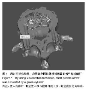
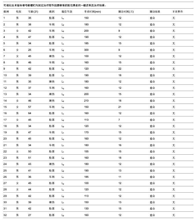
2.1 参与者数量分析 按意向性分析,纳入32例单一节段腰椎骨折患者,均进行有效随访,全部进入结果分析,无脱落。 2.2 椎弓根螺钉可视化技术对腰椎骨折伤椎置钉患者知情同意满意度的影响 通过椎弓根螺钉可视化技术,可以生动形象地向患者交代置钉方式、固定节段等相关的解剖学知识,通过图像向患者讲述可能出现的风险,比如术中由于脊柱严重畸形导致解剖结构复杂,可能会出现螺钉位置误置导致神经损伤,医生可以通过虚拟系统进行演示向患者讲述螺钉向椎管内置入损伤脊髓进而引起神经损伤,这样的术前告知便于患者及家属的理解,使其切实做到知情同意,本组患者的知情同意评分为非常满意的患者占100%。 2.3 椎弓根螺钉可视化技术对腰椎骨折伤椎置钉方式及内植物选择的影响 通过椎弓根螺钉的可视化技术,可以利用计算机辅助手术规划技术,利用获得的医学影像数据在计算机上建立患体的三维数字模型,进而制定最佳置钉方案。通过椎弓根螺钉的可视化技术,可以在术前拟置钉椎体的椎弓根做详细评估,尤其是对观察伤椎椎弓根的完整性上起着非常重要的作用,通过此技术作者判断出10例患者的伤椎单侧完好,但是另一侧破坏严重,无法置钉,这就为置钉方式选择提供了有力参考,节省了置钉时间。本组应用椎弓根螺钉可视化技术术前测量制定最优化参数如置钉的水平面、矢状面角度、螺钉的长度,术中按照术前计划进行螺钉置入,如图1测量的长度为33 mm,医师可选择35 mm×6.5 mm的短椎弓根螺钉置入伤椎,根据测量的角度将合适的椎弓根螺钉置入伤椎内,从而保证不会因椎弓根螺钉过长而影响骨折块的复位情况以及增加手术风险,显著提高了螺钉置入的准确性,本组中置钉后CT平扫螺钉完全位于椎弓根内的比例为95.1%。"

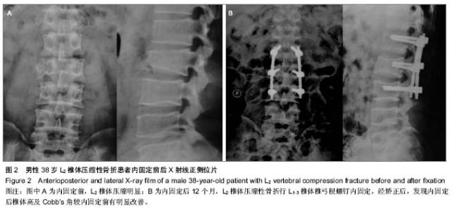
2.4 椎弓根螺钉可视化技术对腰椎骨折伤椎置钉手术指标的影响 通过椎弓根螺钉可视化技术,置钉前对置钉方式及内固定物的选择,显著节省了术中操作时间,提高了椎弓根螺钉置入的准确性。本组患者的手术时间为100-300 min,平均185 min;置钉后CT平扫173枚螺钉(95.1%)完全位于椎弓根内;术中失血165-1 950 mL,平均850 mL。 2.5 椎弓根螺钉可视化技术对腰椎骨折伤椎置钉效果的影响 患者随访7-22个月,所有患者骨折均达到临床骨性愈合。置钉后2周伤椎椎体前缘高度(96.6±3.4)%,较置钉前的(55.9±5.8)%明显升高(P < 0.05);置钉后2周Cobb’s角(4.7±1.3)°,较置钉前(26.4±3.2)°明显降低( P < 0.05);置钉后8个月伤椎椎体前缘高度(95.1±2.2)%,Cobb’s角(6.3±1.9)°,与置钉后2周差异无显著性意义(P > 0.05)。 2.6 典型病例 男性患者,38岁,诊断为L2椎体压缩性骨折,行腰椎后路切开复位椎弓根螺钉内固定植骨融合,内固定后12个月随访,骨折愈合良好(图2)。"

2.7 不良事件 置钉过程中未发生血管、神经损伤及椎弓根钉置入位置错误,无置钉后切口感染、深部感染、内固定松动、断裂等并发症。"
| [1] 贾水淼,董胜利,张凯,等.伤椎单侧椎弓根固定治疗胸腰椎骨折的临床探讨[J].中国矫形外科杂志,2008,16(20):1595-1596.[2] 陈国栋,张玉舰,沙俊峰,等. 伤椎斜交叉椎弓根固定联合传统内固定治疗胸腰椎骨折的实验研究[J].山东医药,2009,49(6):34-35.[3] 袁志峰,邵斌,曾景平.经伤椎置钉治疗胸腰椎骨折的临床运用及疗效分析[J].脊椎外科杂志,2013,11(1): 32-35.[4] 郑永宏,郝定均,吴起宁,等.伤椎固定在严重胸腰椎骨折中的应用[J].中国现代医药杂志,2009,11(9):37-39.[5] 俞阳,范海泉,陈铭.经伤椎和跨伤椎椎弓根钉棒系统内固定治疗胸腰椎骨折[J].脊柱外科杂志,2012,10(4): 228-231.[6] Gonzalvo A, Fitt G, Liew S, et al. The learning curve of pedicle screw placement: how many screws are enough? Spine (Phila Pa 1976). 2009;34:E761-765.[7] Gang C, Haibo L, Fancai L, et al. Learning curve of thoracic pedicle screw placement using the free-hand technique in scoliosis: how many screws needed for an apprentice? Eur Spine J. 2012;21:1151-1156.[8] Samdani AF, Ranade A, Sciubba DM, et al. Accuracy of free-hand placement of thoracic pedicle screws in adolescent idiopathic scoliosis: how much of a difference does surgeon experience make? Eur Spine J. 2010;19:91-95.[9] Samdani AF, Ranade A, Saldanha V, et al. Learning curve for placement of thoracic pedicle screws in the deformed spine. Neurosurgery. 2010;66:290-294; discussion 294-295.[10] Samdani AF, Ranade A, Sciubba DM, et al. Accuracy of free-hand placement of thoracic pedicle screws in adolescent idiopathic scoliosis: how much of a difference does surgeon experience make? Eur Spine J. 2010;19:91-95.[11] Klein S, Whyne CM, Rush R, et al. CT-based patient-specific simulation software for pedicle screw insertion. J Spinal Disord Tech. 2009;22:502-506.[12] Eftekhar B, Ghodsi M, Ketabchi E, et al. Surgical simulation software for insertion of pedicle screws. Neurosurgery. 2002; 50:222-224.[13] Richards PJ, Kurta IC, Jasani V, et al. Assessment of CAOS as a training model in spinal surgery: a randomized study. Eur Spine J. 2007;16:239-244.[14] Podolsky DJ, Martin AR, Whyne CM, et al. Exploring the role of 3-dimensional simulation in surgical training: feedback from a pilot study. J Spinal Disord Tech. 2010;23:e70-74.[15] Aubin CE, Labelle H, Chevrefils C, et al. Preoperative planning simulator for spinal deformity surgeries. Spine (Phila Pa 1976). 2008;33:2143-2152.[16] Majdouline Y, Aubin CE, Sangole A, et al. Computer simulation for the optimization of instrumentation strategies in adolescent idiopathic scoliosis. Med Biol Eng Comput. 2009; 47:1143-1154.[17] Aurouer N, Obeid I, Gille O, et al. Computerized preoperative planning for correction of sagittal deformity of the spine. Surg Radiol Anat. 2009;31:781-792.[18] 于海龙,项良碧,刘军,等.CT三维重建技术在手术治疗胸腰段椎体骨折中应用[J]. 中国矫形外科杂志,2010,18(2):157-159.[19] 徐凯,陈春,黄山东,等.腰椎椎弓根螺钉内固定术三维可视化设计[J].解放军医学杂志,2011,36(9):935-937.[20] Beck M, Mittlmeier T, Gierer P, et al. Benifil and accuracy of intraoperative 3D-imaging after pedicle screws placement: a prospective study in stabilizing thoracolumbar fractures. Eur Spine J. 2009;18(10):1469-1477. [21] Ershad M, Ahmadian A, Dadashi Serej N, et al. Minimization of target registration error for vertebra in image-guided spine surgery. Int J Comput Assist Radiol Surg. 2013. [22] Shin BJ, James AR, Njoku IU, et al. Pedicle screw navigation: a systematic review and meta-analysis of perforation risk for computer-navigated versus freehand insertion. J Neurosurg Spine. 2012;17(2):113-122.[23] 张宗宝.术前CT测量胸腰椎椎弓根相关参数在螺钉内固定术中的应用[J].实用医学影像杂志,2011,12(2):97-100.[24] Verma R, Krishan S, Haendlmayer K, et al. Functional outcome of computer-assisted spinal pedicle screw placement: a systematic review and meta-analysis of 23 studies including 5,992 pedicle screws. Eur Spine J. 2010; 19(3):370-375.[25] 李响,李春志,唐光才,等.多层螺旋CT腰椎三维成像在椎弓根螺钉置入术中的应用[J].中国医学影像学杂志,2011,19(8): 580-584.[26] 乔国勇,苗海敏,关凤英,等.椎弓根三维形态的CT测量在实施个体化置钉中的作用研究[J].临床医学工程,2013,20(2):139- 142.[27] 杨波,方世兵,尹飚,等.三维重建腰椎椎弓根螺钉置入的精确性[J].中国组织工程研究,2013,17(13):2333-2338.[28] 刘少喻,龙厚清,李佛保,等.单节段椎弓根螺钉骨折椎固定治疗新鲜创伤性胸腰椎骨折初步探讨[C]. 中华医学会骨科学分会脊柱外科学组.第七届全国脊柱外科学术会议论文集. 北京,2004:94-95.[29] Dick JC, Jones MP, Zdeblick TA, et al. A biomechanical comparison evaluation the use of intermediate screws and cross-linkage in lumbar pedicle fixation. J Spinal Discord. 1994;5:402-407. [30] Shen WJ, Liu TJ, Shen YS. Nonoperative treatment versus posterior fixation for thoracolumbar junction burst fractures without neurologic deficit. Spine. 2001;9: 1038-1045. [31] Weinstein JN, Rydevik BL, Rauschning W.Anatomic and technical considerations of pedicle screw fixation. Clin Orthop Relat Res. 1992;284:34-46. [32] Park P,Garton HJ,Gala VC,et al.Adjacent segment disease after lumbar or lumbosacral fusion:review of the literature.Spine(Phila Pa 1976). 2004;29(17):1938-1944.[33] Yang JY, Lee JK, Song HS. The impact of adjacent segment degeneration on the clinical outcome after lumbar spinal fusion.Spine(Phila Pa 1976). 2008;33(5):503-507. |
| [1] | Chen Qun-qun, Qiao Rong-qin, Duan Rui-qi, Hu Nian-hong, Li Zhao, Shao Min. Acu-Loc®2 volar distal radius bone plate system for repairing type C fracture of distal radius [J]. Chinese Journal of Tissue Engineering Research, 2017, 21(7): 1025-1030. |
| [2] | Li Peng, Li Hong-wei, Wang Shuang, Wang Hai-zhou. Digital anatomy of lumbar spinous process tilt angle of adults in northeast China: prodinding reference for pedicle screw insertion [J]. Chinese Journal of Tissue Engineering Research, 2017, 21(7): 1064-1068. |
| [3] | Ling Guan-han, Ou Zhi-xue, Yao Lan, Wen Li-chun, Wang Guo-xiang, Lin Heng-feng. Establishment of simulating three-dimensional model of China-Japan Friendship Hospital Classification for L type osteonecrosis of the femoral head [J]. Chinese Journal of Tissue Engineering Research, 2017, 21(7): 1074-1079. |
| [4] | Chen Lu-yao, Hu Shi-qiang, Wang Xiao-ping, Wu Wei-wei, Wei Zhan-tu, Huang Jian. Accuracy of digital orthopedic three-dimensional reconstruction for thoracolumbar pedicle screw placement [J]. Chinese Journal of Tissue Engineering Research, 2017, 21(3): 373-377. |
| [5] | Qiu Hao, Lu Min-peng, Dong Jing, Zhang Zhong-zu, Chu Tong-wei, Wang Qun-bo, Quan Zheng-xue, Jiang Dian-ming. Subtotal corpectomy and reconstruction with titanium mesh cage implantation and pedicle screw fixation through posterior approach in treatment of thoracolumbar burst fracture or thoracolumbar fracture dislocation [J]. Chinese Journal of Tissue Engineering Research, 2016, 20(53): 7932-7938. |
| [6] | Luo Li, Chi Peng-fei, Ma Ming, Zhao Yong, Sun Wu-dong, Huang Hai-yang. Design and analysis of hand rehabilitation training device based on three-dimensional modeling [J]. Chinese Journal of Tissue Engineering Research, 2016, 20(53): 7952-7958. |
| [7] | Abudunaibi•Aili, Zhang Hong-qi, Huang Wei-min, Li Lei, Tian Hui-zhong. Special formed titanium mesh cages for treating spinal tuberculosis via one-stage posterior approach [J]. Chinese Journal of Tissue Engineering Research, 2016, 20(48): 7192-7199. |
| [8] | Wang Gang, Li Xin-ying, Zhang Shu-quan, Li Ya-jun. Creep characteristics of tibial fractures with three internal and external fixation devices [J]. Chinese Journal of Tissue Engineering Research, 2016, 20(48): 7206-7211. |
| [9] | Zhang Wei, Tang Zai-xiang, Geng De-chun, Zhu Feng, Dong Han-qing, Wang Yi-jun, Xu Yao-zeng. Multiple linear regression analysis of hip function and vitamin D levels before and after hip arthroplasty [J]. Chinese Journal of Tissue Engineering Research, 2016, 20(44): 6557-6563. |
| [10] | Lian Zhi-ming, Yang Jing, Zhang Tai-liang, Ma Chuang, Liu Qiang, Yang Guang-zhong. Bridging external fixation combined with Kirschner-wire fixation versus volar locked plate fixation for unstable fractures of the distal radius [J]. Chinese Journal of Tissue Engineering Research, 2016, 20(44): 6590-6598. |
| [11] | Liu Hong-bin, Zhao Han-qing, Shi Yue, Hou Li-jun, Wang Zhong-xun. Establishment and volume estimation of three-dimensional digital model of osteoarthrosis of the femoral head [J]. Chinese Journal of Tissue Engineering Research, 2016, 20(44): 6629-6635. |
| [12] | Fu Feng, Zhao Ming-liang, Li Xiao-hong, Chen Chong, Wang Li-na, Sun Hong-tao, Tu Yue, Zhang Sai . Dynamic changes of brain cavity in rats after traumatic brain injury detected by MRI-based three-dimensional reconstruction [J]. Chinese Journal of Tissue Engineering Research, 2016, 20(40): 5946-5952. |
| [13] | Zhai Peng-fei, Liu Wei, Sun Zhi-ming, Zhang Xue-li. Adjacent segment degeneration after anterior cervical corpectomy and fusion [J]. Chinese Journal of Tissue Engineering Research, 2016, 20(35): 5216-5223. |
| [14] | Zhao Xue-zhai, Li Hai-jun, Meng Cai-yun, Li Yan, Zhang Shi-feng, Liu Ming-hui. Absorbable screw and metal screw fixation for ankle fractures: comparison of biocompatibility and ankle function [J]. Chinese Journal of Tissue Engineering Research, 2016, 20(31): 4687-4692. |
| [15] | Ni Peng-hui, Zhang Ying, Yang Jing, Xu Zi-ang, NuErBoLi, Cheng Kui, Liu Da-peng. Application of finite element analysis in orthopedics: new theory and new progress [J]. Chinese Journal of Tissue Engineering Research, 2016, 20(31): 4693-4699. |
| Viewed | ||||||
|
Full text |
|
|||||
|
Abstract |
|
|||||